Journal of Peking University (Health Sciences) ›› 2020, Vol. 52 ›› Issue (5): 924-930. doi: 10.19723/j.issn.1671-167X.2020.05.022
Previous Articles Next Articles
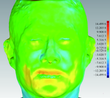
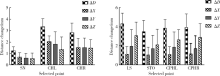
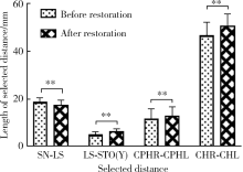
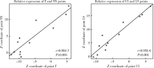
Validation of the digital integration technology for evaluating the nasolabial morphology variation after the cross-arch fixed restoration of maxillary implant-supported prostheses
Ke-yi HAO,Jia LUO,Ping DI,Hou-zuo GUO,Hui-dan SHEN,Yan-ping LIU,Yu ZHANG( ),Ye LIN
- Department of Oral Implantology, Peking University School and Hospital of Stomatology & National Clinical Research Center for Oral Diseases & National Engineering Laboratory for Digital and Material Technology of Stomatology & Beijing Key Laboratory of Digital Stomatology, Beijing 100081, China
CLC Number:
- R783


| [1] |
Kau CH, Richmond S, Zhurov A, et al. Use of 3-dimensional surface acquisition to study facial morphology in 5 populations[J]. Am J Orthod Dentofacial Orthop, 2010,137(4 Suppl):S56-57.
pmid: 20381762 |
| [2] |
Karatas OH, Toy E. Three-dimensional imaging techniques: A literature review[J]. Eur J Dent, 2014,8(1):132-140.
pmid: 24966761 |
| [3] |
Maló P, de Araújo Nobre M, Lopes A, et al. “All-on-4” imme-diate-function concept for completely edentulous maxillae: a clinical report on the medium (3 years) and long-term (5 years) outcomes[J]. Clin Implant Dent Relat Res, 2012,14(Suppl 1):e139-150.
doi: 10.1111/j.1708-8208.2011.00395.x |
| [4] |
Lopes A, Maló P, de Araújo Nobre M, et al. The NobelGuide® All-on-4® treatment concept for rehabilitation of edentulous jaws: a retrospective report on the 7-years clinical and 5-years radiographic outcomes [J]. Clin Implant Dent Relat Res, 2017,19(2):233-244.
pmid: 27758069 |
| [5] | 张宇, 林野, 刘洋, 等. 牙周炎晚期伴上颌牙槽骨前突畸形患者即刻种植全牙列固定修复的侧貌变化初探[J]. 中华口腔医学杂志, 2017,52(10):625-630. |
| [6] | 邸萍, 林野, 李健慧, 等. 单颌拔牙后即刻种植即刻修复的临床回顾研究[J]. 中华口腔医学杂志, 2013,48(4):216-222. |
| [7] | Holzinger D, Seemann R, Matoni N, et al. Effect of dental implants on bisphosphonate-related osteonecrosis of the jaws[J]. J Oral Maxillofac Surg, 2014, 72(10): 1937.e1-8. |
| [8] |
Tian K, Li Q, Wang X, et al. Reproducibility of natural head position in normal Chinese people[J]. Am J Orthod Dentofacial Orthop, 2015,148(3):503-510.
pmid: 26321348 |
| [9] |
Huang Y, Zhang X, Fan Y, et al. Reshaping 3D facial scans for facial appearance modeling and 3D facial expression analysis[J]. Image and Vision Computing, 2012,30(10):750-761.
doi: 10.1016/j.imavis.2011.12.008 |
| [10] |
Littlefield T, Kelly K, Cherney J, et al. Development of a new three-dimensional cranial imaging system[J]. J Craniofac Surg, 2004,15(1):175-181.
pmid: 14704586 |
| [11] |
Rangel FA, Maal TJ, Bergé SJ, et al. Integration of digital dental casts in 3-dimensional facial photographs[J]. Am J Orthod Dentofacial Orthop, 2008,134(6):820-826.
pmid: 19061810 |
| [12] |
Rosati R, De Menezes M, Rossetti A, et al. Digital dental cast placement in 3-dimensional, full-face reconstruction: a technical evaluation[J]. Am J Orthod Dentofacial Orthop, 2010,138(1):84-88.
pmid: 20620838 |
| [13] |
Avrampou M, Mericske-Stern R, Blatz MB, et al. Virtual implant planning in the edentulous maxilla: criteria for decision making of prosjournal design[J]. Clin Oral Implants Res, 2013,24(Suppl A100):152-159.
doi: 10.1111/j.1600-0501.2011.02407.x |
| [14] |
Mu CQ, Wang SQ, Liu Y, et al. Development of a facescan 3D facial reconstruction technology method for quantitative evaluation of cheilitis granulomatosa[J]. Sci Rep, 2017,7(1):1295.
pmid: 28465526 |
| [15] |
Kamashita Y, Kamada Y, Kawahata N, et al. Influence of lip support on the soft-tissue profile of complete denture wearers[J]. J Oral Rehabil, 2006,33(2):102-109.
doi: 10.1111/j.1365-2842.2006.01575.x pmid: 16457669 |
| [16] |
Fourie Z, Damstra J, Gerrits PO, et al. Evaluation of anthropometric accuracy and reliability using different three-dimensional scanning systems[J]. Forensic Sci Int, 2011,207(1-3):127-134.
doi: 10.1016/j.forsciint.2010.09.018 |
| [17] |
Metzger TE, Kula KS, Eckert GJ, et al. Orthodontic soft-tissue parameters: a comparison of cone-beam computed tomography and the 3dMD imaging system[J]. Am J Orthod Dentofacial Orthop, 2013,144(5):672-681.
pmid: 24182583 |
| [18] |
Desesa CR, Metzler P, Sawh-Martinez R, et al. Three-dimensional nasolabial morphologic alterations following Le Fort I[J]. Plast Reconstr Surg Glob Open, 2016,4(8):e848.
pmid: 27622116 |
| [19] |
Mccollum AGH, Dancaster JT, Evans WG, et al. Sagittal soft-tissue changes related to the surgical correction of maxillary-deficient Class III malocclusions[J]. Seminars in Orthodontics, 2009,15(3):172-184.
doi: 10.1053/j.sodo.2009.03.003 |
| [20] | 卫彦, 陈贵, 韩冰, 等. 三维照相定量评价总义齿修复前后面部软组织变化[J]. 北京大学学报(医学版), 2014,46(1):100-103. |
| [1] | Xiaoqiang LIU,Yin ZHOU. Risk factors of perioperative hypertension in dental implant surgeries with bone augmentation [J]. Journal of Peking University (Health Sciences), 2024, 56(1): 93-98. |
| [2] | Meng-en OU,Yun DING,Wei-feng TANG,Yong-sheng ZHOU. Three-dimensional finite element analysis of cement flow in abutment margin-crown platform switching [J]. Journal of Peking University (Health Sciences), 2023, 55(3): 548-552. |
| [3] | Ao-nan WEN,Wei LIU,Da-wei LIU,Yu-jia ZHU,Ning XIAO,Yong WANG,Yi-jiao ZHAO. Preliminary evaluation of the trueness of 5 chairside 3D facial scanning techniques [J]. Journal of Peking University (Health Sciences), 2023, 55(2): 343-350. |
| [4] | WANG Juan,YU Hua-jie,SUN Jing-de,QIU Li-xin. Application evaluation of prefabricated rigid connecting bar in implants immediate impression preparation of edentulous jaw [J]. Journal of Peking University (Health Sciences), 2022, 54(1): 187-192. |
| [5] | LI Xin-fei, PENG Yi-ji, YU Xiao-teng, XIONG Sheng-wei, CHENG Si-da, DING Guang-pu, YANG Kun-lin, TANG Qi, MI Yue, WU Jing-yun, ZHANG Peng, XIE Jia-xin, HAO Han, WANG He, QIU Jian-xing, YANG Jian, LI Xue-song, ZHOU Li-qun. Three dimensional nephrometry system for partial nephrectomy: Our initial exploration [J]. Journal of Peking University (Health Sciences), 2021, 53(3): 613-622. |
| [6] | LIU Xiao-qiang,YANG Yang,ZHOU Jian-feng,LIU Jian-zhang,TAN Jian-guo. Blood pressure and heart rate changes of 640 single dental implant surgeries [J]. Journal of Peking University (Health Sciences), 2021, 53(2): 390-395. |
| [7] | HUANG Xin-rui,LI Sha,GAO Song. Progress in filters for denoising cryo-electron microscopy images [J]. Journal of Peking University (Health Sciences), 2021, 53(2): 425-433. |
| [8] | Jia LUO,Yu ZHANG,Hong-yan CUI,Ning ZHU,Hui-dan SHEN,Ping DI,Ye LIN. Digital workflow coupling conic retention for the immediate restoration of adjacent posterior implants [J]. Journal of Peking University (Health Sciences), 2020, 52(5): 964-970. |
| [9] | Wei-ting LI,Peng LI,Mu-zi PIAO,Fang ZHANG,Jie DI. Study on bone volume harvested from the implant sites with different methods [J]. Journal of Peking University(Health Sciences), 2020, 52(1): 103-106. |
| [10] | Shun-ji WANG,Wen-bo ZHANG,Yao YU,Xiao-yan XIE,Hong-yu YANG,Xin PENG. Application of computer-assisted design for anterolateral thigh flap in oral and maxillofacial reconstruction [J]. Journal of Peking University(Health Sciences), 2020, 52(1): 119-123. |
| [11] | Qiang LUO,Qian DING,Lei ZHANG,Qiu-fei XIE. Quantitative analysis of occlusal changes in posterior partial fixed implant supported prostheses [J]. Journal of Peking University(Health Sciences), 2019, 51(6): 1119-1123. |
| [12] | Jian-hua ZHU,Jing WANG,Xiao-jing LIU,Chuan-bin GUO. Accuracy analysis of robotic assistant needle placement for trigeminal gasserian ganglion [J]. Journal of Peking University(Health Sciences), 2019, 51(5): 973-976. |
| [13] | Ye YAN,Hai-zhui XIA,Xu-sheng LI,Wei HE,Xue-hua ZHU,Zhi-ying ZHANG,Chun-lei XIAO,Yu-qing LIU,Hua HUANG,Liang-hua HE,Jian LU. Application of U-shaped convolutional neural network in auto segmentation and reconstruction of 3D prostate model in laparoscopic prostatectomy navigation [J]. Journal of Peking University(Health Sciences), 2019, 51(3): 596-601. |
| [14] | CHAI Jin-you, LIU Jian-zhang, WANG Bing, QU Jian, SUN Zhen, GAO Wen-hui, GUO Tian-hao, FENG Hai-lan, PAN Shao-xia. Evaluation of the fabrication deviation of a kind of milling digital implant surgical guides#br# [J]. Journal of Peking University(Health Sciences), 2018, 50(5): 892-898. |
| [15] | WU Min-jie, ZOU Li-dong, LIANG Feng. Clinical observation on soft and hard tissue changes of immediate implantation and immediate reconstruction in anterior region after loading 3 years [J]. Journal of Peking University(Health Sciences), 2018, 50(4): 694-699. |
|
||