Journal of Peking University (Health Sciences) ›› 2021, Vol. 53 ›› Issue (1): 120-125. doi: 10.19723/j.issn.1671-167X.2021.01.018
Previous Articles Next Articles
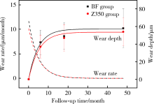
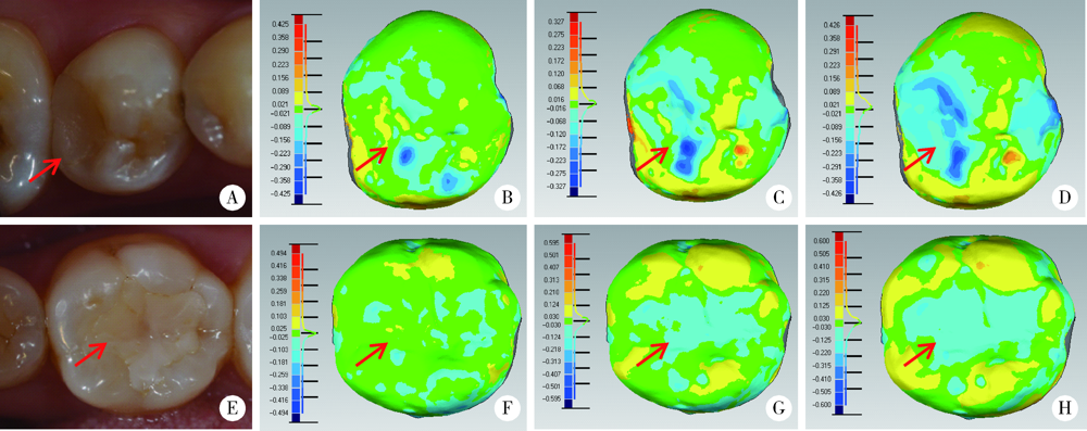
Evaluation of wear property of Giomer and universal composite in vivo
MU Hai-li1,2,TIAN Fu-cong3,WANG Xiao-yan1,Δ( ),GAO Xue-jun1
- 1. Department of Cariology and Endodontology, Peking University School and Hospital of Stomatology & National Clinical Research Center for Oral Diseases & National Engineering Laboratory for Digital and Material Technology of Stomatology & Beijing Key Laboratory of Digital Stomatology, Beijing 100081, China
2. First Clinical Division, Peking University School and Hospital of Stomatology, Beijing 100034, China
CLC Number:
- R783.1


| [1] | Gordan VV, Blaser PK, Watson RE, et al. A clinical evaluation of a giomer restorative system containing surface prereacted glass ionomer filler: results from a 13-year recall examination[J]. J Am Dent Assoc, 2014,145(10):1036-1043. |
| [2] |
Manhart J, Chen HY, Hickel R. Clinical evaluation of the poste-rior composite Quixfil in class Ⅰ and Ⅱ cavities: 4-year follow-up of a randomized controlled trial[J]. J Adhes Dent, 2010,12(3):237-243.
doi: 10.3290/j.jad.a17551 pmid: 20157663 |
| [3] |
Oz FD, Ergin E, Canatan S. Twenty-four-month clinical perfor-mance of different universal adhesives in etch-and-rinse, selective etching and self-etch application modes in NCCL: a randomized controlled clinical trial[J]. J Appl Oral Sci, 2019,27:e20180358.
doi: 10.1590/1678-7757-2018-0358 pmid: 30994773 |
| [4] |
Koc Vural U, Meral E, Ergin E, et al. Twenty-four-month clinical performance of a glass hybrid restorative in non-carious cervical lesions of patients with bruxism: a split-mouth, randomized clinical trial[J]. Clin Oral Investig, 2020,24(3):1229-1238.
doi: 10.1007/s00784-019-02986-x pmid: 31297658 |
| [5] |
Hayashi M, Wilson NH. Failure risk of posterior composites with post-operative sensitivity[J]. Oper Dent, 2003,28(6):681-688.
pmid: 14653280 |
| [6] |
Heintze SD. Clinical relevance of tests on bond strength, microleakage and marginal adaptation[J]. Dent Mater, 2013,29(1):59-84.
doi: 10.1016/j.dental.2012.07.158 |
| [7] |
Naoum S, Ellakwa A, Martin F, et al. Fluoride release, recharge and mechanical property stability of various fluoride-containing resin composites[J]. Oper Dent, 2011,36(4):422-432.
doi: 10.2341/10-414-L pmid: 21819201 |
| [8] |
Ikemura K, Tay FR, Endo T, et al. A review of chemical-approach and ultramorphological studies on the development of fluoride-releasing dental adhesives comprising new pre-reacted glass ionomer (PRG) fillers[J]. Dent Mater J, 2008,27(3):315-339.
doi: 10.4012/dmj.27.315 pmid: 18717159 |
| [9] |
Saku S, Kotake H, Scougall-Vilchis RJ, et al. Antibacterial acti-vity of composite resin with glass-ionomer filler particles[J]. Dent Mater J, 2010,29(2):193-198.
doi: 10.4012/dmj.2009-050 pmid: 20379030 |
| [10] |
Kitagawa H, Miki-Oka S, Mayanagi G, et al. Inhibitory effect of resin composite containing S-PRG filler on Streptococcus mutans glucose metabolism[J]. J Dent, 2018,70:92-96.
doi: 10.1016/j.jdent.2017.12.017 pmid: 29294301 |
| [11] |
Kakuta K, Wonglamsam A, Goto S, et al. Surface textures of composite resins after combined wear test simulating both occlusal wear and brushing wear[J]. Dent Mater J, 2012,31(1):61-67.
doi: 10.4012/dmj.2010-091 |
| [12] |
Ruivo MA, Pacheco RR, Sebold M, et al. Surface roughness and filler particles characterization of resin-based composites[J]. Microsc Res Tech, 2019,82(10):1756-1767.
doi: 10.1002/jemt.23342 pmid: 31313442 |
| [13] |
Condo R, Cerroni L, Pasquantonio G, et al. A deep morphological characterization and comparison of different dental restorative materials[J]. Biomed Res Int, 2017,2017:7346317.
doi: 10.1155/2017/7346317 pmid: 28752095 |
| [14] |
Heintze SD, Faouzi M, Rousson V, et al. Correlation of wear in vivo and six laboratory wear methods[J]. Dent Mater, 2012,28(9):961-973.
doi: 10.1016/j.dental.2012.04.006 |
| [15] |
Heintze SD, Ilie N, Hickel R, et al. Laboratory mechanical parameters of composite resins and their relation to fractures and wear in clinical trials: A systematic review[J]. Dent Mater, 2017,33(3):e101-e114.
doi: 10.1016/j.dental.2016.11.013 pmid: 27993372 |
| [16] |
Hickel R, Roulet JF, Bayne S, et al. Recommendations for conducting controlled clinical studies of dental restorative materials[J]. Clin Oral Investig, 2007,11(1):5-33.
doi: 10.1007/s00784-006-0095-7 pmid: 17262225 |
| [17] |
Leinfelder KF, Taylor DF, Barkmeier WW, et al. Quantitative wear measurement of posterior composite resins[J]. Dent Mater, 1986,2(5):198-201.
doi: 10.1016/S0109-5641(86)80013-6 pmid: 3468024 |
| [18] |
Mehl A, Gloger W, Kunzelmann KH, et al. A new optical 3-D device for the detection of wear[J]. J Dent Res, 1997,76(11):1799-1807.
doi: 10.1177/00220345970760111201 pmid: 9372798 |
| [19] |
Palotie U, Eronen AK, Vehkalahti K, et al. Longevity of 2- and 3-surface restorations in posterior teeth of 25- to 30-year-old attending Public Dental Service: A 13-year observation[J]. J Dent, 2017,62:13-17.
doi: 10.1016/j.jdent.2017.05.012 pmid: 28529175 |
| [20] | The American Dental Association. ADA acceptance program guidelines: resin based composites for posterior restorations [R]. Chicago: ADA Council on Scientific Affairs, 2001. |
| [21] |
Lempel E, Toth A, Fabian T, et al. Retrospective evaluation of posterior direct composite restorations: 10-year findings[J]. Dent Mater, 2015,31(2):115-122.
doi: 10.1016/j.dental.2014.11.001 pmid: 25480695 |
| [22] |
Demarco FF, Correa MB, Cenci MS, et al. Longevity of posterior composite restorations: not only a matter of materials[J]. Dent Mater, 2012,28(1):87-101.
doi: 10.1016/j.dental.2011.09.003 |
| [23] |
Hewlett ER, Orro ME, Clark GT. Accuracy testing of three-dimensional digitizing systems[J]. Dent Mater, 1992,8(1):49-53.
doi: 10.1016/0109-5641(92)90053-f pmid: 1521684 |
| [24] |
Thongthammachat S, Moore BK, Barco MT 2nd, et al. Dimensional accuracy of dental casts: influence of tray material, impression material, and time[J]. J Prosthodont, 2002,11(2):98-108.
pmid: 12087547 |
| [25] |
Palaniappan S, Bharadwaj D, Mattar DL, et al. Three-year randomized clinical trial to evaluate the clinical performance and wear of a nanocomposite versus a hybrid composite[J]. Dent Mater, 2009,25(11):1302-1314.
doi: 10.1016/j.dental.2009.06.001 |
| [26] |
Palaniappan S, Elsen L, Lijnen I, et al. Nanohybrid and microfilled hybrid versus conventional hybrid composite restorations: 5-year clinical wear performance[J]. Clin Oral Investig, 2012,16(1):181-190.
doi: 10.1007/s00784-010-0500-0 pmid: 21221678 |
| [27] |
Goldberg AJ, Rydinge E, Santucci EA, et al. Clinical evaluation methods for posterior composite restorations[J]. J Dent Res, 1984,63(12):1387-1391.
doi: 10.1177/00220345840630120901 pmid: 6239885 |
| [28] |
da Rosa Rodolpho PA, Cenci MS, Donassollo TA, et al. A clinical evaluation of posterior composite restorations: 17-year findings[J]. J Dent, 2006,34(7):427-435.
doi: 10.1016/j.jdent.2005.09.006 pmid: 16314023 |
| [29] |
Wilson NHF, Norman RD. Five-year findings of a multiclinical trial for posterior composite[J]. J Dent, 1991,19(3):153-159.
doi: 10.1016/0300-5712(91)90005-j pmid: 1939815 |
| [30] |
Satou N, Khan AM, Satou K, et al. In-vitro and in-vivo wear profile of composite resins[J]. J Oral Rehabil, 1992,19(1):31-37.
doi: 10.1111/j.1365-2842.1992.tb01588.x pmid: 1316435 |
| [31] |
Salgado VE, Cavalcante LM, Silikas N, et al. The influence of nanoscale inorganic content over optical and surface properties of model composites[J]. J Dent, 2013,41(Suppl 5):e45-53.
doi: 10.1016/j.jdent.2013.05.011 |
| [32] |
Lim BS, Ferracane JL, Condon JR, et al. Effect of filler fraction and filler surface treatment on wear of microfilled composites[J]. Dent Mater, 2002,18(1):1-11.
doi: 10.1016/S0109-5641(00)00103-2 |
| [33] |
Garoushi S, Vallittu PK, Lassila L. Characterization of fluoride releasing restorative dental materials[J]. Dent Mater J, 2018,37(2):293-300.
doi: 10.4012/dmj.2017-161 pmid: 29279547 |
| [34] |
Gonulol N, Ozer S, Sen Tunc E. Water sorption, solubility, and color stability of giomer restoratives[J]. J Esthet Restor Dent, 2015,27(5):300-306.
doi: 10.1111/jerd.12119 pmid: 25145876 |
| [35] |
Park CA, Hyun SH, Lee JH, et al. Evaluation of polymerization in fluoride-containing composite resins[J]. J Mater Sci Mater Med, 2007,18(8):1549-1556.
doi: 10.1007/s10856-007-3023-8 pmid: 17437069 |
| [1] | Yuxuan TIAN,Mingjian RUAN,Yi LIU,Derun LI,Jingyun WU,Qi SHEN,Yu FAN,Jie JIN. Predictive effect of the dual-parametric MRI modified maximum diameter of the lesions with PI-RADS 4 and 5 on the clinically significant prostate cancer [J]. Journal of Peking University (Health Sciences), 2024, 56(4): 567-574. |
| [2] | Liang LYU,Mingjin ZHANG,Aonan WEN,Yijiao ZHAO,Yong WANG,Jing LI,Gengchen YANG,Dawei LIU. Preliminary evaluation of chin symmetry with three dimentional soft tissue spatial angle wireframe template [J]. Journal of Peking University (Health Sciences), 2024, 56(1): 106-110. |
| [3] | Bochun MAO,Yajing TIAN,Xuedong WANG,Jing LI,Yanheng ZHOU. Soft and hard tissue changes of hyperdivergent class Ⅱ patients before and after orthodontic extraction treatment [J]. Journal of Peking University (Health Sciences), 2024, 56(1): 111-119. |
| [4] | Xiaotong LING,Liuyang QU,Danni ZHENG,Jing YANG,Xuebing YAN,Denggao LIU,Yan GAO. Three-dimensional radiographic features of calcifying odontogenic cyst and calcifying epithelial odontogenic tumor [J]. Journal of Peking University (Health Sciences), 2024, 56(1): 131-137. |
| [5] | Yi LIU,Chang-wei YUAN,Jing-yun WU,Qi SHEN,Jiang-xi XIAO,Zheng ZHAO,Xiao-ying WANG,Xue-song LI,Zhi-song HE,Li-qun ZHOU. Diagnostic efficacy of prostate cancer using targeted biopsy with 6-core systematic biopsy for patients with PI-RADS 5 [J]. Journal of Peking University (Health Sciences), 2023, 55(5): 812-817. |
| [6] | Chang-wei YUAN,De-run LI,Zhi-hua LI,Yi LIU,Gang-zhi SHAN,Xue-song LI,Li-qun ZHOU. Application of dynamic contrast enhanced status in multiparametric magnetic resonance imaging for prostatic cancer with PI-RADS 4 lesion [J]. Journal of Peking University (Health Sciences), 2023, 55(5): 838-842. |
| [7] | Zhan-yi ZHANG,Fan ZHANG,Ye YAN,Cai-guang CAO,Chang-jian LI,Shao-hui DENG,Yue-hao SUN,Tian-liang HUANG,Yun-he GUAN,Nan LI,Min LU,Zhen-hua HU,Shu-dong ZHANG. Near-infrared targeted probe designed for intraoperative imaging of prostatic neurovascular bundles [J]. Journal of Peking University (Health Sciences), 2023, 55(5): 843-850. |
| [8] | Zhuo-hua LIN,Ru-yi CAI,Yang SUN,Rong MU,Li-gang CUI. Methodology and clinical use of superb microvascular imaging in assessing micro-circulation changes of fingertips in systemic sclerosis [J]. Journal of Peking University (Health Sciences), 2023, 55(4): 636-640. |
| [9] | Ying LIU,Ran HUO,Hui-min XU,Zheng WANG,Tao WANG,Hui-shu YUAN. Correlations between plaque characteristics and cerebral blood flow in patients with moderate to severe carotid stenosis using magnetic resonance vessel wall imaging [J]. Journal of Peking University (Health Sciences), 2023, 55(4): 646-651. |
| [10] | Qiang FU,Guan-ying GAO,Yan XU,Zhuo-hua LIN,You-jing SUN,Li-gang CUI. Comparative study of ultrasound and magnetic resonance imaging in the diagnosis of asymptomatic anterosuperior acetabular labrum tears [J]. Journal of Peking University (Health Sciences), 2023, 55(4): 665-669. |
| [11] | Xiang LIU,Hui-hui XIE,Yu-feng XU,Xiao-dong ZHANG,Xiao-feng TAO,Lin LIU,Xiao-ying WANG. Value of artificial intelligence in the improvement of diagnostic consistency of radiology residents [J]. Journal of Peking University (Health Sciences), 2023, 55(4): 670-675. |
| [12] | Wen ZHANG,Xiao-jing LIU,Zi-li LI,Yi ZHANG. Effect of alar base cinch suture based on anatomic landmarks on the morphology of nasolabial region in patients after orthognathic surgery [J]. Journal of Peking University (Health Sciences), 2023, 55(4): 736-742. |
| [13] | Meng-en OU,Yun DING,Wei-feng TANG,Yong-sheng ZHOU. Three-dimensional finite element analysis of cement flow in abutment margin-crown platform switching [J]. Journal of Peking University (Health Sciences), 2023, 55(3): 548-552. |
| [14] | Li-jia MA,Pan-pan HU,Xiao-guang LIU. Spinal metastases combined with leptomeningeal metastasis: A case report [J]. Journal of Peking University (Health Sciences), 2023, 55(3): 563-566. |
| [15] | Ao-nan WEN,Wei LIU,Da-wei LIU,Yu-jia ZHU,Ning XIAO,Yong WANG,Yi-jiao ZHAO. Preliminary evaluation of the trueness of 5 chairside 3D facial scanning techniques [J]. Journal of Peking University (Health Sciences), 2023, 55(2): 343-350. |
|
||