Journal of Peking University (Health Sciences) ›› 2021, Vol. 53 ›› Issue (2): 355-363. doi: 10.19723/j.issn.1671-167X.2021.02.021

Previous Articles     Next Articles

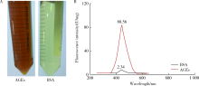
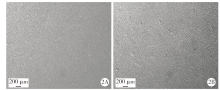
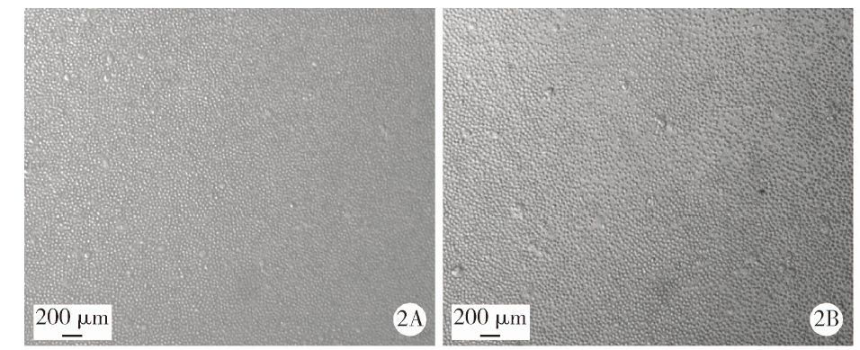
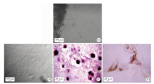
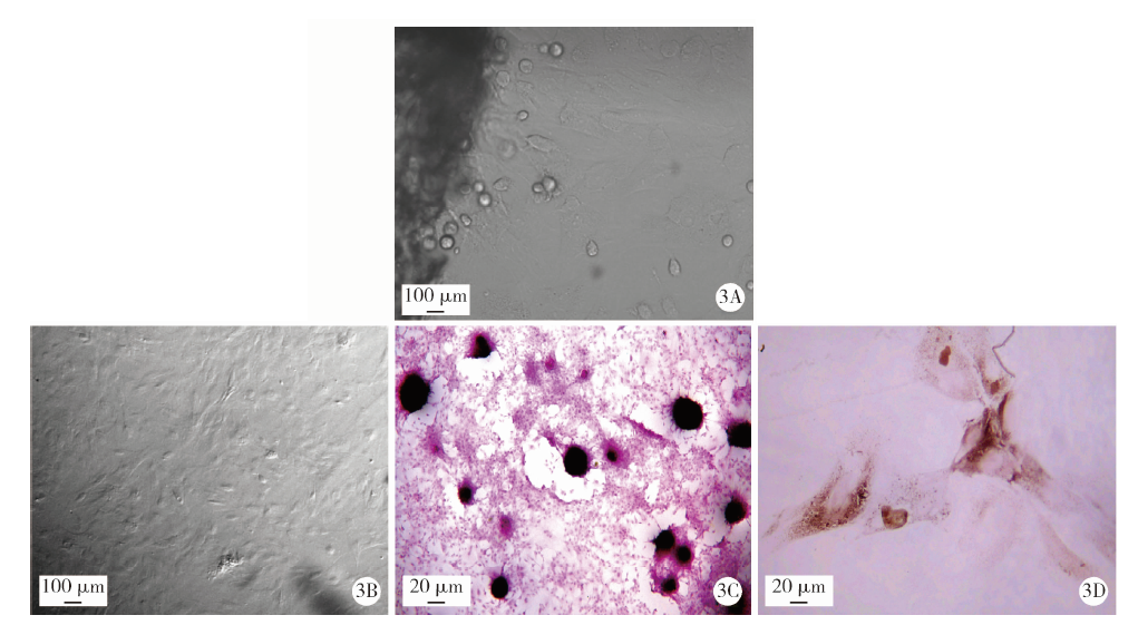
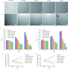
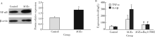
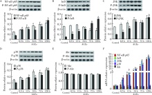
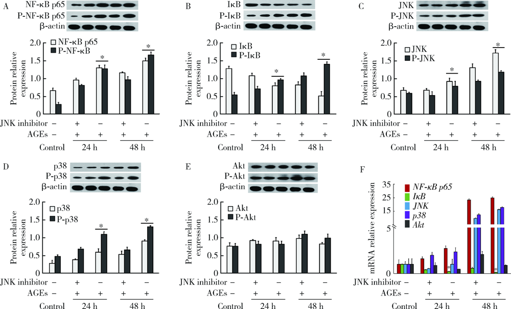
Mechanism of advanced glycation end products inhibiting the proliferation of peripheral blood mononuclear cells and osteoblasts in rats

LI Zheng1,WANG Xiao1,Δ(),HONG Tian-pei2,WANG Hao-jie1,GAO Zhan-yi1,WAN Meng1   

  1. 1. Department of Stomatology, Peking University Third Hospital, Beijing 100191, China
    2. Department of Endocrinology, Peking University Third Hospital, Beijing 100191, China
  • Received:2019-09-16 Online:2021-04-18 Published:2021-04-21
  • Contact: Xiao WANG E-mail:bysywangxiao@163.com
  • Supported by:
    Special Fund for the Study of the Relationship between Oral Diseases and Systemic Diseases of the Chinese Stomatological Association(CSA-Y2015-08)

Abstract:

Objective: To explore the mechanism of nuclear factor-kappa B (NF-κB), phosphatidylinositol 3-kinase (PI3K)/protein kinase B(PKB/Akt) and mitogen-activated protein kinase (MAPK) signaling pathways after intervention of advanced glycosylation end products (AGEs) in peripheral blood mononuclear cells (PBMCs) and osteoblasts (OB) in rats, so as to provide certain experimental basis and theoretical basis for further research on the clinical treatment of periodontal tissue inflammation caused by diabetes mellitus. Methods: AGEs were prepared, PBMCs and OB were isolated and cultured in vitro. CCK-8 was used to detect the cell viability intervened by different concentrations and time of AGEs. Western blot and qRT-PCR were used to detect the expression changes of genes related to NF-κB, PI3K/PKB and MAPK signaling pathways. Results: OB and PBMCs were successfully isolated and cultured in vitro. The activity of PBMCs and OB cells was significantly correlated with the concentration, time and interaction of AGEs. With the increase of AGEs concentration and time, the activity of PBMCs and OB cells significantly decreased (P<0.001). AGEs stimulation significantly increased the expression of NF-κB in PBMCs and the contents of tumor necrosis factor α(TNF-α), interleukin-1β(IL-1β) (P<0.01). TNF-α, IL-1β levels were significantly reduced after inhibition of NF-κB pathway (P<0.01). NF-κB p65, JNK, and p38 phosphorylated and non-phosphorylated proteins increased significantly after AGEs stimulation of OB (P<0.05). The phosphorylated protein expression of IκB was significantly increased, while the expression of non-phosphorylated protein was decreased (P<0.01).The expressions of NF-κB p65, JNK, and IκB were significantly increased at the mRNA levels, and the expressions of IκB mRNA were significantly decreased (P<0.05). There was no difference in the expression of Akt in either phosphorylated or non-phosphorylated proteins or at the mRNA level (P>0.05). With the addition of MAPK signaling pathway inhibitors, the phosphorylation and non-phosphorylated protein expressions of NF-κB p65, p38 and JNK were significantly reduced, and the phosphorylated protein of IκB was significantly decreased and the non-phosphorylated protein was significantly increased compared with the group with AGEs alone (P<0.05). The results of qRT-PCR showed that the expression of IκB increased significantly after the addition of the JNK pathway blocker (P<0.05), and the expression of NF-κB p65, p38 and JNK decreased, but the difference was not significant (P>0.05). While NF-κB p65, p38 and JNK were significantly decreased and IκB was significantly increased in the AGEs group after the addition of the p38 pathway blocker (P<0.05). At this time, there was still no significant change in the expression of Akt at the protein level and mRNA level (P>0.05). Conclusion: AGEs inhibit the proliferation of PBMCs and OB, and the NF-κB and MAPK pathways are likely involved in regulating this process, but not the PI3K/PKB pathway.

Key words: Glycation end products, advanced, Peripheral blood mononuclear cells, Osteoblasts, Signal transduction

CLC Number: 

  • R781.4

Table 1

Primer sequences used for qRT-PCR"

Gene Primer sequence Fragment length/bp
NF-κB p65 Forward: CGGATTGAAGAAAAACG
Reverse: TTGAAAAGGCATAGGGC
169
JNK Forward: TTAGATGAAAGGGAGCA
Reverse: TGACGCCATTCTTAGTT
91
p38 Forward: CAGATGCCGAAGATGAAC
Reverse: CCTCTTATCCGAGTCCAA
99
IκB Forward: CTGCGTGATGAAGATTGGA
Reverse: GCATCCCTGAGCAGTTGG
116
β-actin Forward: GGCACCACACCTTCTAC
Reverse: CTGGGTCATCTTTTCAC
107

Figure 1

The prepared AGEs and BSA reagents (A),analysis of BSA and AGEs by fluorescence spectrophotometer (B) AGEs, advanced glycation end products; BSA, bovine serum albumin."

Figure 2

The cultured PBMCs of the primary (A) and the third generation (B), observed by optical microscope"

Figure 3

The cultured OB of the primary (A) and the third generation (B) and the staining of alizarin red (C) and ALP (D)"

Figure 4

The AGEs stimulated cell morphology of PBMCs (A) and OB (B) observed by optical microscope for 48 h; the effects of AGEs at different concentrations for 24, 48, 72 h on cell PBMCs (C) and OB (D) viability and the interaction (E, F) between different concentrations and action times by CCK-8 test A, B, scale bar is 200 μm. *P<0.001, vs. control; AGEs, advanced glycation end products. E, PBMCs: Times, P=0.001; Concentrations, P<0.001; Time×Concentration, P<0.001 (GLM, F value: 14.1-1 851.4). F, OB: Times, P<0.001; Concentrations: P<0.001; Time×Concentration, P<0.001 (GLM, F: 64.4-373.1)."

Figure 5

NF-κB p65 expression examined by Western blot (A) and TNF-α and IL-1β level examined by ELISA (B) after AGEs treatment with PBMCs * P<0.01, vs. control; # P<0.01, vs. AGEs group. Bay117082, NF-κB signaling pathway inhibitor."

Figure 6

The related gene expression of NF-κB, PI3K/PKB, MAPK signaling pathways examined by Western blot (A-E) and qRT-PCR (F) after the AGEs stimulated OB P-NF-κB p65, P-IκB, P-p38, P-JNK, P-Akt are the phosphorylation products of NF-κB p65, IκB, p38, JNK, Akt, respectively. *P<0.05, #P<0.01, △P<0.001, vs. control respectively."

Figure 7

The related gene expression of NF-κB, PI3K/PKB, MAPK signaling pathways examined by Western blot (A-E) and qRT-PCR (F) after blocking by JNK inhibitor P-NF-κB p65, P-IκB, P-p38, P-JNK, P-Akt are the phosphorylation products of NF- κB p65, IκB, p38, JNK, Akt, respectively. *P<0.05, vs. JNK inhibitor at the same time."

Figure 8

The related gene expression of NF-κB, PI3K/PKB, MAPK signaling pathways examined by Western blot (A-E) and qRT-PCR (F) after blocking by p38 inhibitor P-NF-κB p65, P-IκB, P-p38, P-JNK, P-Akt are the phosphorylation products of NF-κB p65, IκB, p38, JNK, Akt, respectively. *P<0.05, vs. p38 inhibitor at the same time."

[1] Petersen PE, Ogawa H. The global burden of periodontal disease: Towards integration with chronic disease prevention and control[J]. Periodontol 2000, 2012,60(1):15-39.
doi: 10.1111/j.1600-0757.2011.00425.x pmid: 22909104
[2] 李峥, 沙月琴, 张博学, 等. 参加社区慢性病管理的糖尿病患者牙周健康状况调查及相关因素分析[J]. 中华口腔医学杂志, 2007: 42(2):100-101.
[3] Papapanou PN. Periodontal disease: Epidemiology[J]. Annals of Periodontol, 1996,1(1):11-36.
[4] Khader YS, Dauod AS, El-Qaderi SS, et al. Periodontal status of diabetics compared with nondiabetics: A meta-analysis[J]. J Diabetes Complications, 2006,20(1):59-68.
pmid: 16389170
[5] Taylor JJ, Preshaw PM, Lalla E. A review of the evidence for pathogenic mechanisms that may link periodontitis and diabetes[J]. J Periodontol, 2013,84(4 Suppl.):S113-S134.
pmid: 23631573
[6] Taiyeb-Ali TB, Cheta Raman RP, Vaithilingam RD. Relationship between periodontal disease and diabetes mellitus: An Asian perspective[J]. Periodontol 2000, 2011,56(1):258-268.
pmid: 21501247
[7] Ulrich P, Cerami A. Protein glycation, diabetes, and aging[J]. Recent Prog Horm Res, 2001,56(1):1-21.
[8] Ramasamy R, Vannucci SJ, Yan SS, et al. Advanced glycation end products and RAGE: A common thread in aging, diabetes, neurodegeneration, and inflammation[J]. Glycobiology, 2005,15(7):16R-28R.
[9] Neeper M, Schmidt AM, Brett J, et al. Cloning and expression of a cell surface receptor for advanced glycosylation end products of proteins[J]. J Biol Chem, 1992,267(21):14998-15004.
pmid: 1378843
[10] Southerlan JH, Taylor GW, Moss K, et al. Commonality in chro-nic inflammatory diseases: Periodontitis, diabetes, and coronary artery disease[J]. Periodontol 2000, 2006,40(1):130-143.
[11] Lin L. RAGE on the Toll Road?[J]. Cell Mol Immunol, 2006,3(5):351-358.
pmid: 17092432
[12] Schmidt A, Yan S, Brett J, et al. Regulation of human mononuclear phagocyte migration by cell surface-binding proteins for advanced glycation end products[J]. J Clin Invest, 1993,91(5):2155-2168.
doi: 10.1172/JCI116442 pmid: 8387541
[13] Anfelika B, Stephan S, Markus S, et al. Diabetes-associated sustained activation of the transcription factor nuclear factor-kappa B[J]. Diabetes, 2001,50(12):2792-2808.
pmid: 11723063
[14] Chen Q, Dong L, Wang L, et al. Advanced glycation end pro-ducts impair function of late endothelial progenitor cells through effects on protein kinase Akt and cyclooxygenase-2[J]. Biochem Biophys Res Commun, 2009,381(2):192-197.
pmid: 19232321
[1] WANG Zheng,DING Qian,GAO Yuan,MA Quan-quan,ZHANG Lei,GE Xi-yuan,SUN Yu-chun,XIE Qiu-fei. Effect of porous zirconia ceramics on proliferation and differentiation of osteoblasts [J]. Journal of Peking University (Health Sciences), 2022, 54(1): 31-39.
[2] XIAO Ruo-tao,LIU Cheng,XU Chu-xiao,HE Wei,MA Lu-lin. Prognostic value of preoperative platelet parameters in locally advanced renal cell carcinoma [J]. Journal of Peking University (Health Sciences), 2021, 53(4): 647-652.
[3] Jia-yu WANG,Mei-ping ZHAO. Fluorescence assay for the detection of apurinic/apyrimidinic endonuclease 1 (APE1) activity in human blood samples [J]. Journal of Peking University(Health Sciences), 2019, 51(3): 487-492.
[4] GAN Hong-quan, WANG Qian, ZHANG Hui, LIU Xin, DENG Hua-min, SONG Hui-ping, WANG ZHi-qiang, LI Qi-jia. Effects of RGD peptides-grafted porous tantalum on morphological change of MG63 osteoblasts-tantalum conjunctive interface and expression of osteogenesis factors [J]. Journal of Peking University(Health Sciences), 2018, 50(1): 176-182.
[5] GUO Qian, CHEN Xu-yong, SU Yin. Interleukin-2 signaling pathway regulating molecules in systemic lupus erythematosus [J]. Journal of Peking University(Health Sciences), 2016, 48(6): 1100-1104.
[6] LING Long, ZHAO Yu-ming, GE Li-hong. Impact of different degree pulpitis on cell proliferation and osteoblastic differentiation of dental pulp stem cell in Beagle immature premolars [J]. Journal of Peking University(Health Sciences), 2016, 48(5): 878-883.
[7] XIAO Yang, DU Yao-yao, GAO Cheng, KONG Wei. Dynamic alteration of microRNA in high phosphorus induced calcification of vascular smooth muscle cell [J]. Journal of Peking University(Health Sciences), 2016, 48(5): 756-765.
[8] CAO Pei, JIANG Xue-jun, XI Zhi-jun. Sunitinib induces autophagy via suppressing Akt/mTOR pathway in renal cell carcinoma [J]. Journal of Peking University(Health Sciences), 2016, 48(4): 584-589.
[9] WANG Zhu-Qing, WANG Ping, WU-CHOU Yah-huei, YE Xiao-Qian, HUANG Shang-Zhi, SHI Bing, WANG Ke, YUAN Yuan, LIU Dong-Jing, WU Tao, WANG Hong, Terri H. Beaty. Association study between candidate genes on transforming growth factor-β signaling pathway and the risk of non-syndromic cleft lip with or without cleft palate in Chinese populations [J]. Journal of Peking University(Health Sciences), 2015, 47(3): 384-389.
[10] Li-Jing-Jin, CHEN Hong, REN Jing-Yi. Targeted effect of microRNA on nerve growth factor pathway and its functional network in patients with unstable angina [J]. Journal of Peking University(Health Sciences), 2014, 46(6): 868-874.
[11] CHANG Si-jia, ZOU Xiao-ying, ZHUANG Heng, YUE Lin, GAO Xue-jun. Notch activation delayed ageing of human dental pulp cells [J]. Journal of Peking University(Health Sciences), 2014, 46(1): 5-11.
Viewed
Full text


Abstract

Cited

  Shared   
  Discussed   
[1] Author. English Title Test[J]. Journal of Peking University(Health Sciences), 2010, 42(1): 1 -10 .
[2] . [J]. Journal of Peking University(Health Sciences), 2009, 41(2): 188 -191 .
[3] . [J]. Journal of Peking University(Health Sciences), 2009, 41(3): 376 -379 .
[4] . [J]. Journal of Peking University(Health Sciences), 2009, 41(4): 459 -462 .
[5] . [J]. Journal of Peking University(Health Sciences), 2010, 42(1): 82 -84 .
[6] . [J]. Journal of Peking University(Health Sciences), 2007, 39(3): 319 -322 .
[7] . [J]. Journal of Peking University(Health Sciences), 2007, 39(3): 333 -336 .
[8] . [J]. Journal of Peking University(Health Sciences), 2007, 39(3): 337 -340 .
[9] . [J]. Journal of Peking University(Health Sciences), 2007, 39(3): 225 -328 .
[10] . [J]. Journal of Peking University(Health Sciences), 2007, 39(4): 346 -350 .