Journal of Peking University (Health Sciences) ›› 2021, Vol. 53 ›› Issue (5): 896-901. doi: 10.19723/j.issn.1671-167X.2021.05.014
Previous Articles Next Articles
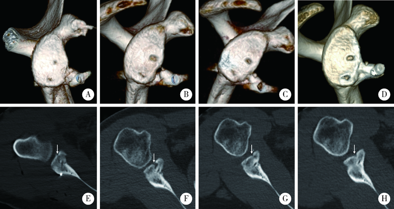
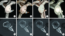
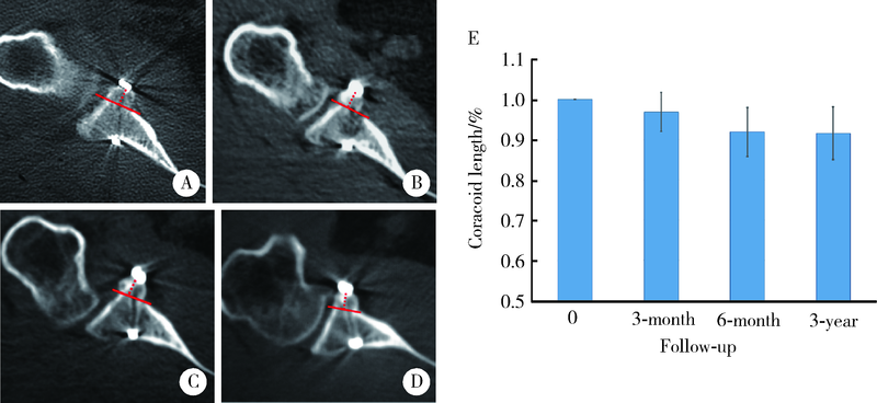
An arthroscopic “inlay” Bristow procedure with suture button fixation: Surgical technique and radiology evaluation
SHAO Zhen-xing1,SONG Qing-fa1,ZHAO Yu-qing2,CUI Guo-qing1,△( )
)
- 1. Department of Sports Medicine, Peking University Third Hospital, Institute of Sports Medicine of Peking University, Beijing Key Laboratory of Sports Injuries, Beijing 100191, China
2. Department of Radiology, Peking University Third Hospital, Beijing 100191, China
CLC Number:
- R684.73


| [1] |
Cowling PD, Akhtar MA, Liow RY. What is a Bristow-Latarjet procedure? A review of the described operative techniques and outcomes [J]. Bone Joint J, 2016, 98-B(9):1208-1214.
doi: 10.1302/0301-620X.98B9.37948 pmid: 27587522 |
| [2] |
Bhatia S, Frank RM, Ghodadra NS, et al. The outcomes and surgical techniques of the latarjet procedure [J]. Arthroscopy, 2014, 30(2):227-235.
doi: 10.1016/j.arthro.2013.10.013 |
| [3] |
Griesser MJ, Harris JD, McCoy BW, et al. Complications and re-operations after Bristow-Latarjet shoulder stabilization: A systema-tic review [J]. J Shoulder Elbow Surg, 2013, 22(2):286-292.
doi: 10.1016/j.jse.2012.09.009 pmid: 23352473 |
| [4] |
van der Linde JA, van Kampen DA, Terwee CB, et al. Long-term results after arthroscopic shoulder stabilization using suture anchors: An 8- to 10-year follow-up [J]. Am J Sports Med, 2011, 39(11):2396-2403.
doi: 10.1177/0363546511415657 pmid: 21803980 |
| [5] |
Casabianca L, Gerometta A, Massein A, et al. Graft position and fusion rate following arthroscopic Latarjet [J]. Knee Surg Sports Traumatol Arthrosc, 2016, 24(2):507-512.
doi: 10.1007/s00167-015-3551-6 |
| [6] |
Kany J, Flamand O, Grimberg J, et al. Arthroscopic Latarjet procedure: Is optimal positioning of the bone block and screws possible? A prospective computed tomography scan analysis [J]. J Shoulder Elbow Surg, 2016, 25(1):69-77.
doi: 10.1016/j.jse.2015.06.010 |
| [7] |
Allain J, Goutallier D, Glorion C. Long-term results of the Latarjet procedure for the treatment of anterior instability of the shoulder [J]. J Bone Joint Surg Am, 1998, 80(6):841-852.
pmid: 9655102 |
| [8] |
Bessiere C, Trojani C, Pelegri C, et al. Coracoid bone block versus arthroscopic Bankart repair: A comparative paired study with 5-year follow-up [J]. Orthop Traumatol Surg Res, 2013, 99(2):123-130.
doi: 10.1016/j.otsr.2012.12.010 |
| [9] | Lafosse L, Lejeune E, Bouchard A, et al. The arthroscopic Latarjet procedure for the treatment of anterior shoulder instability [J]. Arthroscopy, 2007, 23(11):1241-1245. |
| [10] |
Boileau P, Gendre P, Baba M, et al. A guided surgical approach and novel fixation method for arthroscopic Latarjet [J]. J Shoulder Elbow Surg, 2016, 25(1):78-89.
doi: 10.1016/j.jse.2015.06.001 pmid: 26256014 |
| [11] |
Butt U, Charalambous CP. Complications associated with open coracoid transfer procedures for shoulder instability [J]. J Shoulder Elbow Surg, 2012, 21(8):1110-1119.
doi: 10.1016/j.jse.2012.02.008 |
| [12] |
Boileau P, Hardy MB, McClelland WB, et al. Arthroscopic posterior bone block procedure: A new technique using suture anchor fixation [J]. Arthrosc Tech, 2013, 2(4):473-477.
doi: 10.1016/j.eats.2013.07.004 pmid: 24892011 |
| [13] |
Boileau P, Saliken D, Gendre P, et al. Arthroscopic Latarjet: Suture-button fixation is a safe and reliable alternative to screw fixation [J]. Arthroscopy, 2019, 35(4):1050-1061.
doi: S0749-8063(18)31066-1 pmid: 30857907 |
| [14] |
Giles JW, Degen RM, Johnson JA, et al. The Bristow and Latarjet procedures: Why these techniques should not be considered synonymous [J]. J Bone Joint Surg A, 2014, 96(16):1340-1348.
doi: 10.2106/JBJS.M.00627 |
| [15] |
van der Linde JA, van Wijngaarden R, Somford MP, et al. The Bristow-Latarjet procedure, a historical note on a technique in comeback [J]. Knee Surg Sports Traumatol Arthrosc, 2016, 24(2):470-478.
doi: 10.1007/s00167-015-3704-7 |
| [16] |
Boileau P, Thelu CE, Mercier N, et al. Arthroscopic Bristow-Latarjet combined with bankart repair restores shoulder stability in patients with glenoid bone loss [J]. Clin Orthop Relat Res, 2014, 472(8):2413-2424.
doi: 10.1007/s11999-014-3691-x |
| [17] |
Shao Z, Song Q, Cheng X, et al. An arthroscopic “Inlay” Bristow procedure with suture button fixation for the treatment of recurrent anterior glenohumeral instability: 3-year follow-up [J]. Am J Sports Med, 2020, 48(11):2638-2649.
doi: 10.1177/0363546520943633 |
| [18] |
Nourissat G, Delaroche C, Bouillet B, et al. Optimization of bone-block positioning in the Bristow-Latarjet procedure: A biomechanical study [J]. Orthop Traumatol Surg Res, 2014, 100(5):509-513.
doi: 10.1016/j.otsr.2014.03.023 |
| [19] |
Hovelius L, Sandstrom B, Olofsson A, et al. The effect of capsular repair, bone block healing, and position on the results of the Bristow-Latarjet procedure (study Ⅲ): Long-term follow-up in 319 shoulders [J]. J Shoulder Elbow Surg, 2012, 21(5):647-660.
doi: 10.1016/j.jse.2011.03.020 pmid: 21719316 |
| [20] |
Kee YM, Kim JY, Kim HJ, et al. Fate of coracoid grafts after the Latarjet procedure: Will be analogous to the original glenoid by remodelling [J]. Knee Surg Sports Traumatol Arthrosc, 2018, 26(3):926-932.
doi: 10.1007/s00167-017-4808-z |
| [21] |
Samilson RL, Prieto V. Dislocation arthropathy of the shoulder [J]. J Bone Joint Surg, 1983, 65(4):456-460.
doi: 10.2106/00004623-198365040-00005 |
| [22] | Garcia JC, do Amaral FM, Belchior RJ, et al. Comparative systematic review of fixation methods of the coracoid and conjoined tendon in the anterior glenoid to treat anterior shoulder instability [J]. Orthop J Sports Med, 2019, 7(1):2325-2328. |
| [23] |
Xu J, Liu H, Lu W, et al. Modified arthroscopic Latarjet procedure: Suture-button fixation achieves excellent remodeling at 3-year follow-up [J]. Am J Sports Med, 2019, 48(1):39-47.
doi: 10.1177/0363546519887959 |
| [1] | Jia-peng ZHENG,Qi XIAO,Hui-yun DENG,Qing-quan WU,Wen-liang ZHAI,Da-sheng LIN. Arthroscopic classification and management for the popliteal hiatus of the lateral meniscus tears [J]. Journal of Peking University (Health Sciences), 2021, 53(5): 891-895. |
| [2] | HOU Zong-chen,AO Ying-fang,HU Yue-lin,JIAO Chen,GUO Qin-wei,HUANG Hong-shi,REN Shuang,ZHANG Si,XIE Xing,CHEN Lin-xin,ZHAO Feng,PI Yan-bin,LI Nan,JIANG Dong. Characteristics and related factors of plantar pressure in the chronic ankle instability individuals [J]. Journal of Peking University (Health Sciences), 2021, 53(2): 279-285. |
| [3] | Zhong-di LIU,Ting-min XU,Yu DANG,Dian-ying ZHANG,Zhong-guo FU. A mid-term clinical follow-up study on repair of the meniscus tears by a modified arthroscopic outside-in puncture suture technique [J]. Journal of Peking University (Health Sciences), 2020, 52(5): 870-874. |
| [4] | Dong JIANG,Yue-lin HU,Chen JIAO,Qin-wei GUO,Xing XIE,Lin-xin CHEN,Feng ZHAO,Yan-bin PI. Mid-to-long term outcomes and influence factors of postoperative concurrent chronic ankle instability and posterior ankle impingement [J]. Journal of Peking University(Health Sciences), 2019, 51(3): 505-509. |
| [5] | Cui-ping ZHANG,Pei-pei LIU,Qiang FU,Guan-ying GAO,Li-gang CUI,Yan XU,Jian-quan WANG. Application of ultrasound-guided hip joint drug injection in the postoperative rehabilitation of arthroscopie repair of acetabular labral tears [J]. Journal of Peking University(Health Sciences), 2019, 51(2): 265-267. |
| [6] | RONG Yan-bo, TIAN Guang-lei, CHEN Shan-lin. Biomechanical analysis of the deep radioulnar ligaments stabilizing the distal radioulnar joint [J]. Journal of Peking University(Health Sciences), 2017, 49(3): 518-521. |
| [7] | ZHANG Hui, LIU Xin, HONG Lei, GENG Xiang-su, FENG Hua. Arthroscopic all-inside reconstruction for posterior cruciate ligament and popliteus tendon compared with popliteofibular ligament reconstruction: clinical outcome of minimum 2-year follow-up [J]. Journal of Peking University(Health Sciences), 2016, 48(2): 237-243. |
| [8] | LIU Bo, CHEN Shan-lin, ZHU Jin, WANG Zhi-xin, YANG Chen, SHEN Jie, TIAN Guan-lei. Arthroscopic management of lesser arc perilunate injuries [J]. Journal of Peking University(Health Sciences), 2016, 48(2): 234-236. |
| [9] | WU Guan, JIANG Chun-Yan, LU Yi, ZHU Yi-Ming, LI Feng-Long, LI Xu. Modified arthroscopic Latarjet procedure for the treatment of anterior shoulder instability [J]. Journal of Peking University(Health Sciences), 2015, 47(2): 321-325. |
| [10] | LI Feng-Long, JIANG Chun-Yan, LU Yi, ZHU Yi-Ming, LI Xu. Arthroscopic coracoclavicular ligament reconstruction versus open modified Weaver-Dunn procedure for acromioclavicular joint dislocations:comparison of curative effect [J]. Journal of Peking University(Health Sciences), 2015, 47(2): 253-257. |
| [11] | ZHU Yi-Ming, JIANG Chun-Yan, LU Yi, LI Feng-Long, LI Xu, LI Yue. Clinical follow-up study after open Latarjet procedure in patients with recurrent anterior shoulder dislocation [J]. Journal of Peking University(Health Sciences), 2015, 47(2): 226-231. |
|
||