Journal of Peking University (Health Sciences) ›› 2021, Vol. 53 ›› Issue (5): 891-895. doi: 10.19723/j.issn.1671-167X.2021.05.013
Previous Articles Next Articles
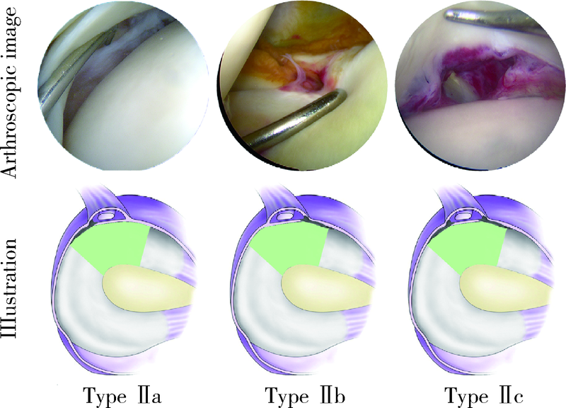
Arthroscopic classification and management for the popliteal hiatus of the lateral meniscus tears
ZHENG Jia-peng,XIAO Qi,DENG Hui-yun,WU Qing-quan,ZHAI Wen-liang,LIN Da-sheng( )
)
- Department of Orthopaedic Surgery, Xiamen University Affiliated Southeast Hospital, Zhangzhou 363000, Fujian, China
CLC Number:
- R684.7


| [1] |
Cohn AK, Mains DB. Popliteal hiatus of the lateral meniscus. Anatomy and measurement at dissection of 10 specimens [J]. Am J Sports Med, 1979, 7(4):221-226.
doi: 10.1177/036354657900700402 |
| [2] |
Stäubli HU, Birrer S. The popliteus tendon and its fascicles at the popliteal hiatus: Gross anatomy and functional arthroscopic evaluation with and without anterior cruciate ligament deficiency [J]. Arthroscopy, 1990, 6(3):209-220.
pmid: 2206184 |
| [3] |
LaPrade RF, Wozniczka JK, Stellmaker MP, et al. Analysis of the static function of the popliteus tendon and evaluation of an anatomicreconstruction: the “fifth ligament” of the knee [J]. Am J Sports Med, 2010, 38(3):543-549.
doi: 10.1177/0363546509349493 pmid: 20042547 |
| [4] |
Guimaraes JB, Facchetti L, Schwaiger BJ, et al. Natural evolution of popliteomeniscal fascicle tears over 2 years and its association with lateral articular knee cartilage degeneration in patients with traumatic anterior cruciate ligament tear [J]. Eur Radiol, 2018, 28(8):3542-3549.
doi: 10.1007/s00330-017-5279-9 pmid: 29476215 |
| [5] | Snyder RL, Jansson KA. Technical note: peripheral meniscus repairs anterior to the popliteal hiatus [J]. Arthroscopy, 2000, 16(8):E19. |
| [6] |
Ouanezar H, Blakeney WG, Latrobe C, et al. The popliteus tendon provides a safe and reliable location for all-inside meniscal repair device placement [J]. Knee Surg Sports Traumatol Arthrosc, 2018, 26(12):3611-3619.
doi: 10.1007/s00167-018-4889-3 |
| [7] |
Peduto AJ, Nguyen A, Trudell DJ, et al. Popliteomeniscal fascicles: Anatomic considerations using MR arthrography in cadavers [J]. AJR Am J Roentgenol, 2008, 190(2):442-448.
doi: 10.2214/AJR.07.2643 |
| [8] |
Aman ZS, DePhillipo NN, Storaci HW, et al. Quantitative and qualitative assessment of posterolateral meniscal anatomy: Defining the popliteal hiatus, popliteomeniscal fascicles, and the lateral meniscotibial ligament [J]. Am J Sports Med, 2019, 47(8):1797-1803.
doi: 10.1177/0363546519849933 |
| [9] |
Lee DW, Jang HW, Lee SR, et al. Clinical, radiological, and morphological evaluations of posterior horn tears of the lateral meniscus left in situ during anterior cruciate ligament reconstruction [J]. Am J Sports Med, 2014, 42(2):327-335.
doi: 10.1177/0363546513508374 |
| [10] |
Shin HK, Lee HS, Lee YK, et al. Popliteomeniscal fascicle tear: Diagnosis and operative technique [J]. Arthrosc Tech, 2012, 1(1):e101-e106.
doi: 10.1016/j.eats.2012.04.004 |
| [11] |
Fang CH, Liu H, Di ZL, et al. Arthroscopic all-inside repair with suture hook for horizontal tear of the lateral meniscus at the popli-teal hiatus region: A preliminary report [J]. BMC Musculoskelet Disord, 2020, 21(1):52.
doi: 10.1186/s12891-020-3066-2 |
| [12] |
Leafblad ND, Leland DP, Camp CL, et al. Arthroscopic repair of double radial tears of the lateral meniscus [J]. Arthrosc Tech, 2019, 8(6):e541-e547.
doi: 10.1016/j.eats.2019.01.015 |
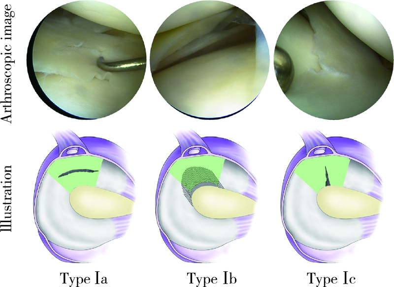
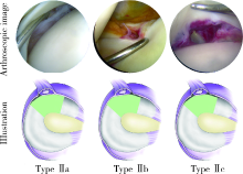
| [13] | Zheng J, Xiao Q, Wu Q, et al. Tears of the popliteomeniscal fascicles of the lateral meniscus: an arthroscopic classification [J]. Cartilage, 2020, 12(2020-12-08)[2021-03-08]. https://doi.org/10.1177/1947603520980156 . |
| [14] |
Pasque C, Noyes FR, Gibbons M, et al. The role of the popliteofibular ligament and the tendon of popliteus in providing stability in the human knee [J]. J Bone Joint Surg Br, 2003, 85(2):292-298.
pmid: 12678372 |
| [15] |
Yang JH, Jeong HI, Kim TS, et al. The management of the popliteus hiatus during lateral meniscal transplantation [J]. Knee, 2012, 19(6):959-961.
doi: 10.1016/j.knee.2012.04.007 |
| [16] |
Kimura M, Shirakura K, Hasegawa A, et al. Anatomy and pathophysiology of the popliteal tendon area in the lateral meniscus: 1. Arthroscopic and anatomical investigation [J]. Arthroscopy, 1992, 8(4):419-423.
pmid: 1466698 |
| [17] |
Kimura M, Shirakura K, Hasegawa A, et al. Anatomy and pathophysiology of the popliteal tendon area in the lateral meniscus: 2. Clinical investigation [J]. Arthroscopy, 1992, 8(4):424-427.
pmid: 1466699 |
| [18] |
van Steyn MO, Mariscalco MW, Pedroza AD, et al. The hypermobile lateral meniscus: A retrospective review of presentation, imaging, treatment, and results [J]. Knee Surg Sports Traumatol Arthrosc, 2016, 24(5):1555-1559.
doi: 10.1007/s00167-014-3497-0 |
| [19] |
Ahn JH, Lee SH, Kim KI, et al. Arthroscopic meniscus repair for recurrent subluxation of the lateral meniscus [J]. Knee Surg Sports Traumatol Arthrosc, 2018, 26(3):787-792.
doi: 10.1007/s00167-017-4420-2 |
| [20] | 冯华, 洪雷, 耿向苏, 等. 外侧半月板腘肌腱区损伤的缝合方法 [J]. 中国运动医学杂志, 2007, 26(2):159-163. |
| [1] | Zhenlong LIU, Zhenchen HOU, Xiaoqing HU, Shuang REN, Qinwei GUO, Yan XU, Xi GONG, Yingfang AO. Arthroscopic tissue engineering scaffold repair for cartilage injuries [J]. Journal of Peking University (Health Sciences), 2025, 57(2): 384-387. |
| [2] | Zhen-xing SHAO,Qing-fa SONG,Yu-qing ZHAO,Guo-qing CUI. An arthroscopic “inlay” Bristow procedure with suture button fixation: Surgical technique and radiology evaluation [J]. Journal of Peking University (Health Sciences), 2021, 53(5): 896-901. |
| [3] | Zhong-di LIU,Ting-min XU,Yu DANG,Dian-ying ZHANG,Zhong-guo FU. A mid-term clinical follow-up study on repair of the meniscus tears by a modified arthroscopic outside-in puncture suture technique [J]. Journal of Peking University (Health Sciences), 2020, 52(5): 870-874. |
| [4] | Dong JIANG,Yue-lin HU,Chen JIAO,Qin-wei GUO,Xing XIE,Lin-xin CHEN,Feng ZHAO,Yan-bin PI. Mid-to-long term outcomes and influence factors of postoperative concurrent chronic ankle instability and posterior ankle impingement [J]. Journal of Peking University(Health Sciences), 2019, 51(3): 505-509. |
| [5] | Cui-ping ZHANG,Pei-pei LIU,Qiang FU,Guan-ying GAO,Li-gang CUI,Yan XU,Jian-quan WANG. Application of ultrasound-guided hip joint drug injection in the postoperative rehabilitation of arthroscopie repair of acetabular labral tears [J]. Journal of Peking University(Health Sciences), 2019, 51(2): 265-267. |
| [6] | LIU Bo, CHEN Shan-lin, ZHU Jin, WANG Zhi-xin, YANG Chen, SHEN Jie, TIAN Guan-lei. Arthroscopic management of lesser arc perilunate injuries [J]. Journal of Peking University(Health Sciences), 2016, 48(2): 234-236. |
| [7] | WU Guan, JIANG Chun-Yan, LU Yi, ZHU Yi-Ming, LI Feng-Long, LI Xu. Modified arthroscopic Latarjet procedure for the treatment of anterior shoulder instability [J]. Journal of Peking University(Health Sciences), 2015, 47(2): 321-325. |
| [8] | LI Feng-Long, JIANG Chun-Yan, LU Yi, ZHU Yi-Ming, LI Xu. Arthroscopic coracoclavicular ligament reconstruction versus open modified Weaver-Dunn procedure for acromioclavicular joint dislocations:comparison of curative effect [J]. Journal of Peking University(Health Sciences), 2015, 47(2): 253-257. |
|
||