Journal of Peking University (Health Sciences) ›› 2026, Vol. 58 ›› Issue (1): 89-98. doi: 10.19723/j.issn.1671-167X.2026.01.012

Previous Articles     Next Articles

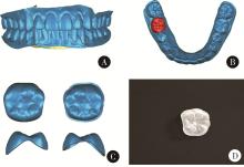
Development of a surface electromyography index system for orofacial muscles and validation of a discriminant model in unilateral molar occlusal interference

Wenbo LI1, Yufeng SHEN2, Yongtao YANG1, Shenyao SHAN1, Zixiang GAO3, Aonan WEN3, Xiangyi SHANG1, Yuwen TIAN1, Shuwei GUO3, Yizhen WANG1, Yong WANG3,*(), Yijiao ZHAO1,*()   

  1. 1. Institute of Medical Technology, Peking University Health Science Center, Beijing 100191, China
    2. Department of Stomatology, The First Affiliated Hospital of Shihezi University, Shihezi 832008, Xinjiang, China
    3. Center for Digital Dentistry, Peking University School and Hospital of Stomatology & National Center for Stomatology & National Clinical Research Center for Oral Diseases & National Engineering Research Center of Oral Biomaterials and Digital Medical Devices & NHC Key Laboratory of Digital Stomatology & Beijing Key Laboratory of Digital Stomatology, Beijing 100081, China
  • Received:2025-10-16 Online:2026-02-18 Published:2025-12-17
  • Contact: Yong WANG, Yijiao ZHAO
  • Supported by:
    the Beijing Natural Science Foundation(L232100); the Beijing Natural Science Foundation(L242132); the National Natural Science Foundation of China(82271039); the Young and Middle-Aged Backbone Talent in Science and Technology Innovation Project under the Shihezi City (Eighth Division) Financial Science and Technology Plan(2025RC04); the Youth Science Project under the Xinjiang Production and Construction Corps Science and Technology Plan(2025DB040); the National Key Research and Development Program of China(2022YFC2405401)

RICH HTML


Abstract:

Objective: To construct a standardized unilateral molar occlusal interference model, to establish a comprehensive surface electromyography (sEMG)-based index system for orofacial muscle function, and to develop an accurate discriminant model, thereby providing an objective electrophysiological basis for occlusal interference diagnosis. Methods: Twenty-six healthy adult volunteers were recruited and provided written informed consent. Utilizing advanced digital dental technology, including intraoral scanning, computer-aided design (CAD), and additive manufacturing, a standardized occlusal inter-ference patch with a precise thickness was fabricated. This patch was adhesively bonded to the occlusal surface of the mandibular first molar to create a reversible unilateral occlusal interference model. A self-developed, multi-channel wireless sEMG system was employed to collect high-fidelity electromyographic signals from key bilateral masticatory muscles: the anterior temporal muscles, masseters, and the anterior bellies of the digastric muscles. Data were recorded during 10 standardized mandibular functional activities both before (baseline) and after the induction of interference. From the raw sEMG signals, a multi-dimensional index system comprising 56 distinct indicators across time, frequency, and complexity domains was constructed. Sophisticated statistical analyses, including paired-sample t-tests (or Wilcoxon signed-rank tests), principal component analysis (PCA) for dimensionality reduction, and stepwise Logistic regression analysis, were applied to screen for the most significant feature variables and to build the optimal discriminant model. Results: Forty valid interference models were successfully established. Statistical analysis revealed 253 sEMG indicators showed significant differences following interference induction (P < 0.05), with lateral-movement-related parameters demonstrating particular sensitivity. PCA extracted 19 principal components (PCs) explaining 85.5% of cumulative variance, where PC1 (muscle fatigue level) and PC2 (functional movement amplitude) represented the primary explanatory components. The optimal Logistic regression model incorporated 3 principal components. Cross-validation showed the model achieved a mean accuracy of 0.840, with mean sensitivity and specificity of 0.851 and 0.828, respectively, and a mean area under the curve (AUC) of 0.923. Conclusion: The Logistic regression discriminant model for unilateral molar occlusal interference constructed in this study can effectively identify the occlusal interference state under the experimental conditions, demonstrating promising diagnostic potential.

Key words: Occlusal interference, Surface electromyography, Orofacial muscles, Regression model

CLC Number: 

  • R78

Figure 1

Design and fabrication process of the standardized occlusal interference patch A, three-dimensional morphological data of the maxillary and mandibular dentition; B, tissue surface morphology of the occlusal interference patch; C, views of the occlusal surface, tissue surface, mesial surface, and distal surface of the three-dimensional digital model of the occlusal interference patch; D, additively manufactured resin occlusal interference patch."

Table 1

sEMG parameter system for orofacial muscles established in this study"

Category Indicator name Data source Formula
Common sEMG indicators for muscle function assessment in oral medicine Percentage overlapping coeffificient,POC Normalized RMS data $\frac{1}{n}\sum\limits_{i = 1}^n {\left( {1 - \frac{{NI{S_i} - I{S_i}}}{{NI{S_i} + I{S_i}}}} \right)} \times 100\% {\rm{ }} $
Modified asymme- try index,mAI Normalized RMS data $ \frac{1}{n}\sum\limits_{i = 1}^n {\frac{{M{M_{NIS - i}} + T{A_{NIS - i}} + D{A_{NIS - i}} - M{M_{IS - i}} - T{A_{IS - i}} - D{A_{IS - i}}}}{{M{M_{NIS - i}} + T{A_{NIS - i}} + D{A_{NIS - i}} + M{M_{IS - i}} + T{A_{IS - i}} + D{A_{IS - i}}}}} \times 100\% $
Activity index,Ac Normalized RMS data $\frac{1}{n}\sum\limits_{i = 1}^n {\frac{{M{M_{NS - i}} + M{M_{IS - i}} - T{A_{NIS - i}} - T{A_{IS - i}}}}{{M{M_{NIS - i}} + M{M_{IS - i}} + T{A_{NIS - i}} + T{A_{IS - i}}}}} \times 100\% $
Torque coeffifi- cient,TC Normalized RMS data $ \frac{1}{n}\sum\limits_{i = 1}^n {\frac{{\left| {T{A_{NIS}} - T{A_{IS}}} \right| - \left| {M{M_{NIS}} - M{M_{IS}}} \right|}}{{\left( {M{M_{NIS}} + T{A_{NIS}} + M{M_{IS}} + T{A_{IS}}} \right)}}} \times 100\% $
Time-frequency domain sEMG indicators Average rectified value, ARV Filtered data ${\rm{ For\; channel }}j, \frac{1}{n}\sum\limits_{i = 1}^n {\left| {{x_{ij}}} \right|} $
Median frequency, MF Filtered data $ \int_0^{f_{\text {median }}} P(f) \mathrm{d} f=\frac{1}{2} \int_0^{f_{\text {max }}} P(f) \mathrm{d} f$
Mean power fre- quency, MPF Filtered data $ \frac{\int_0^{f_{\max }} f \cdot P(f) \mathrm{d} f}{\int_0^{f_{\max }} P(f) \mathrm{d} f}$
Novel sEMG indicators Coefficient of variation, CV Normalized RMS data $\begin{array}{*{20}{c}}{{\rm{For \;channel }}j{\rm{, }}}\\{C{V_j} = \frac{{\sqrt {\frac{1}{{n - 1}}\sum\limits_{i = 1}^n {{{\left( {{x_{ij}} - {{\bar x}_j}} \right)}^2}} } }}{{\frac{1}{n}\sum\limits_{i = 1}^n {{x_{ij}}} }} \times 100\% }\end{array} $
Muscle synergy in- dex, MSI Normalized RMS data Calculation of the Pearson correlation matrix from six-channel RMS data, extraction of the fifteen off-diagonal elements from its upper triangular part, and computation of the arithmetic mean of their absolute values.

Figure 2

Distribution of the number of surface electromyography (sEMG) parameters showing statistically significant differences before and after interference across mandibular movements IS, interference side; NIS, non-interference side; ICP, intercuspal position. The full names of all sEMG features have been provided in Table 1."

Figure 3

Scree plot and cumulative variance contribution rate of principal component analysis for orofacial muscle surface electromyography (sEMG) parameters Cum, cumulative variance; Exp, explained variance ratio."

Table 2

Representative factor loadings and characteristic interpretation of principal components"

Variable Primary feature High-loading indicators Loading
PC1 Muscle fatigue level MF_IS (mouth opening-closing movement) 0.70
MF_mean (mouth opening-closing movement) 0.67
MF_IS-DA (IS chewing) 0.66
MF_DA (IS lateral movement) 0.65
PC2 Functional movement amplitude ARV_NIS-TA (mouth opening-closing movement) 0.78
ARV_IS-MM (IS lateral movement) 0.77
ARV_NIS-MM (NIS lateral movement) 0.77
ARV_NIS-TA (protrusive movement) 0.74

Figure 4

Score scatter plot of interference and non-interference groups in the principal component (PC) space"

Table 3

Characteristics of principal components (PC) included in the Logistic regression model and their representative variables"

Principal component Primary feature High-loading indicators Loading
PC2 Functional movement amplitude ARV_NIS-TA (mouth opening-closing movement) 0.78
ARV_IS-MM (IS lateral movement) 0.77
ARV_NIS-MM (NIS lateral movement) 0.77
ARV_NIS-TA (protrusive movement) 0.74
PC8 Static activity stability MF_NIS-DA (rest position) 0.58
ARV_NIS-DA (clenching) 0.48
CV_IS (rest position) 0.41
PC9 Fine-motor control CV_MM (ICP light contact) 0.68
CV_IS-MM (ICP light contact) 0.59
MF_IS-DA (rest position) 0.49

Figure 5

Results of Logistic regression analysis based on principal components (PC)"

Figure 6

Boxplot of cross-validation performance metrics for the Logistic regression model developed in this study IQR, interquartile range; AUC, area under the receiver operating characteristic curve."

1
Nalamliang N , Sumonsiri P , Thongudomporn U . Are occlusal contact area asymmetry and masticatory muscle activity asymmetry related in adults with normal dentition[J]. Cranio, 2022, 40 (5): 409- 417.

doi: 10.1080/08869634.2020.1764270
2
Li J , Jiang T , Feng H , et al. The electromyographic activity of masseter and anterior temporalis during orofacial symptoms induced by experimental occlusal highspot[J]. J Oral Rehabil, 2008, 35 (2): 79- 87.

doi: 10.1111/j.1365-2842.2007.01750.x
3
Pativetpinyo D , Supronsinchai W , Changsiripun C . Immediate effects of temporary bite-raising with light-cured orthodontic band cement on the electromyographic response of masticatory muscles[J]. J Appl Oral Sci, 2018, 26, e20170214.
4
Riise C , Sheikholeslam A . Influence of experimental interfering occlusal contacts on the activity of the anterior temporal and masseter muscles during mastication[J]. J Oral Rehabil, 1984, 11 (4): 325- 333.

doi: 10.1111/j.1365-2842.1984.tb00583.x
5
Sheikholeslam A , Riise C . Influence of experimental interfering occlusal contacts on the activity of the anterior temporal and masseter muscles during submaximal and maximal bite in the intercuspal position[J]. J Oral Rehabil, 1983, 10 (3): 207- 214.

doi: 10.1111/j.1365-2842.1983.tb00114.x
6
张磊, 谢秋菲. 牙体解剖与口腔生理学[M]. 3版 北京大学医学出版社, 2022: 206- 207.
7
丁其川, 熊安斌, 赵新刚, 等. 基于表面肌电的运动意图识别方法研究及应用综述[J]. 自动化学报, 2016, 42 (1): 13- 25.
8
李文博, 朱玉佳, 秦庆钊, 等. 自主研发无线表面肌电系统对咀嚼肌功能活动的评价研究[J]. 华西口腔医学杂志, 2025, 43 (3): 346- 353.
9
Ferrario VF , Tartaglia GM , Galletta A , et al. The influence of occlusion on jaw and neck muscle activity: A surface EMG study in healthy young adults[J]. J Oral Rehabil, 2006, 33 (5): 341- 348.

doi: 10.1111/j.1365-2842.2005.01558.x
10
Ferrario VF , Sforza C , Colombo A , et al. An electromyographic investigation of masticatory muscles symmetry in normo-occlusion subjects[J]. J Oral Rehabil, 2000, 27 (1): 33- 40.

doi: 10.1046/j.1365-2842.2000.00490.x
11
Naeije M , McCarroll RS , Weijs WA . Electromyographic activity of the human masticatory muscles during submaximal clenching in the inter-cuspal position[J]. J Oral Rehabil, 1989, 16 (1): 63- 70.

doi: 10.1111/j.1365-2842.1989.tb01318.x
12
Sid'El Moctar SM , Rida I , Boudaoud S . Comprehensive review of feature extraction techniques for sEMG signal classification: From handcrafted features to deep learning approaches[J]. Irbm, 2024, 45 (6): 100866.

doi: 10.1016/j.irbm.2024.100866
13
王富, 牛丽娜, 陈吉华. 数字化咬合分析的方案与效能[J]. 中华口腔医学杂志, 2025, 60 (8): 822- 828.
14
孙欣荣, 冯玥, 刘伟才. 多模态数据融合的可视化技术在咬合重建中的应用[J]. 华西口腔医学杂志, 2022, 40 (4): 468- 475.
15
Baba K , Ai M , Mizutani H , et al. Influence of experimental occlusal discrepancy on masticatory muscle activity during clenching[J]. J Oral Rehabil, 1996, 23 (1): 55- 60.

doi: 10.1111/j.1365-2842.1996.tb00812.x
16
Michelotti A , Farella M , Gallo LM , et al. Effect of occlusal interference on habitual activity of human masseter[J]. J Dent Res, 2005, 84 (7): 644- 648.

doi: 10.1177/154405910508400712
17
Christensen LV , Rassouli NM . Experimental occlusal interferences. Part Ⅰ. A review[J]. J Oral Rehabil, 1995, 22 (7): 515- 520.

doi: 10.1111/j.1365-2842.1995.tb01197.x
18
Suvinen TI , Kemppainen P . Review of clinical EMG studies related to muscle and occlusal factors in healthy and TMD subjects[J]. J Oral Rehabil, 2007, 34 (9): 631- 644.

doi: 10.1111/j.1365-2842.2007.01769.x
19
Walton TR , Layton DM . Mediotrusive occlusal contacts: Best evidence consensus statement[J]. J Prosthodont, 2021, 30 (Suppl 1): 43- 51.
20
李宝勇, 周丽娟. TMD患者单侧下颌第三磨牙伸长咬合干扰与升颌肌肌电关系的研究[J/OL]. 实用口腔医学杂志, 2025(2025-10-15)[2025-10-16]. https://link.cnki.net/urlid/61.1062.R.20251015.1040.002.
21
李雪姣, 徐啸翔, 谢秋菲. 干扰与颞下颌关节紊乱病的复杂关系: 动物实验和临床研究的启示[J]. 实用口腔医学杂志, 2013, 29 (2): 266- 274.
[1] FAN Ying-ying,LIU Yun,CAO Ye,XIE Qiu-fei. Hippocampus is involved in 17β-estradiol exacerbating experimental occlusal inter-ference-induced chronic masseter hyperalgesia in ovariectomized rats [J]. Journal of Peking University (Health Sciences), 2022, 54(1): 40-47.
[2] Shu-dong YAN,Guang-ju YANG,Si-yi MO,Yun LIU,Qiu-fei XIE. Effect of long-term resistance exercise on masseter muscle mechanical hyperalgesia in rats [J]. Journal of Peking University(Health Sciences), 2019, 51(1): 21-27.
Viewed
Full text


Abstract

Cited

  Shared   
  Discussed   
No Suggested Reading articles found!