Journal of Peking University (Health Sciences) ›› 2026, Vol. 58 ›› Issue (1): 175-183. doi: 10.19723/j.issn.1671-167X.2026.01.023

Previous Articles     Next Articles

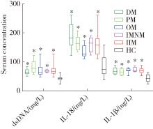
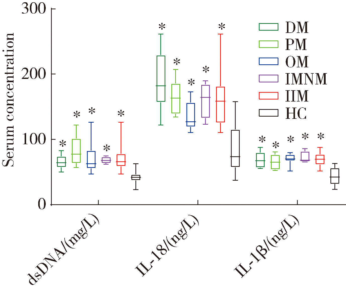
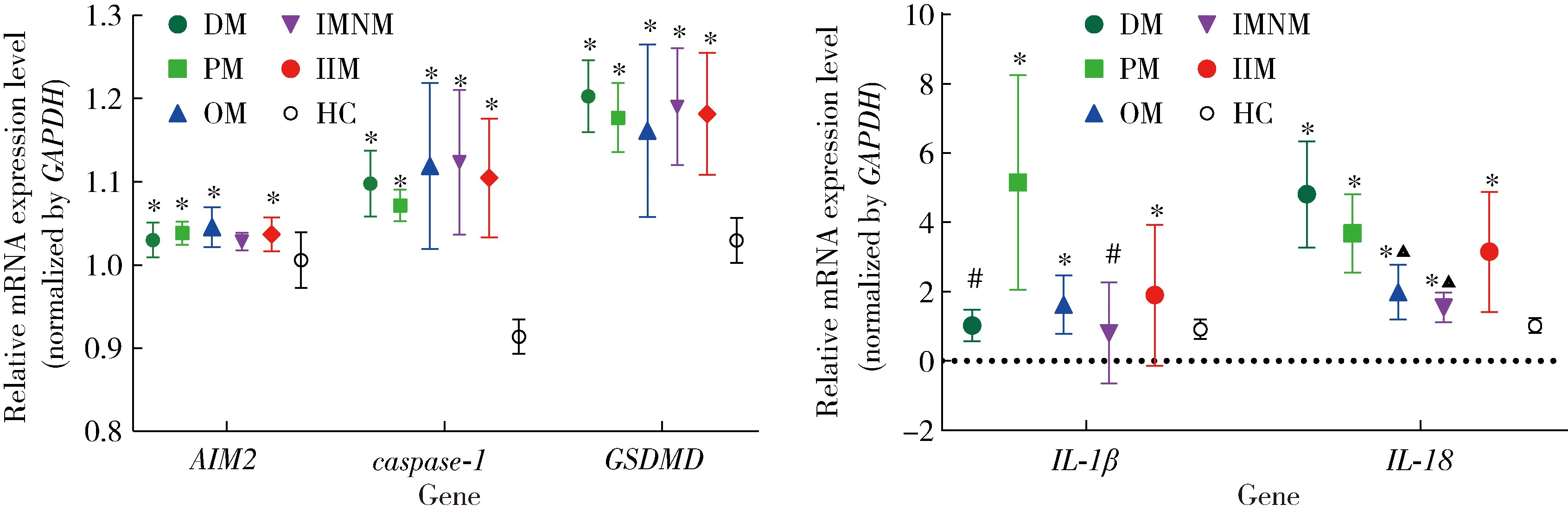
Expression of the melanoma 2-mediated pyroptosis pathway in peripheral blood mononuclear cells of patients with idiopathic inflammatory myopathies

Jiyan CHU1,2, Ping LI1,*(), Jing TIAN3, Diyu FU1,2, Lin GUO1, Rui SUN1, Yadi LI1   

  1. 1. Department of Rheumatology and Immunology, General Hospital of Northern Theater Command, Shenyang 110001, China
    2. Graduate School, Dalian Medical University, Dalian 116044, Liaoning, China
    3. Department of Orthopedics, General Hospital of Northern Theater Command, Shenyang 110001, China
  • Received:2023-07-29 Online:2026-02-18 Published:2026-01-05
  • Contact: Ping LI
  • Supported by:
    Natural Science Foundation of Liaoning Province(2019-MS-350); Key Project of Joint Logistic Support Force(19LBJ1003B)

RICH HTML


Abstract:

Objective: To detect the expression levels of absence in melanoma 2 (AIM2), cysteine aspartate-specific protease-1 (caspase-1), and gasdermin D (GSDMD) in peripheral blood mononuclear cell (PBMC) of patients with idiopathic inflammatory myopathy (IIM) and to explore their role in the pathogenesis of IIM. Methods: A total of 30 IIM patients (IIM group) who visited the Department of Rheumatology and Immunology, General Hospital of Northern Theater Command from May 2020 to June 2022 were recruited. Concurrently, 30 healthy volunteers matched by gender and age were recruited from the hospital's Health Examination Center. Clinical information, biochemical and immunological mar-kers, and venous blood samples were collected from the study subjects. Serum double-stranded DNA (dsDNA) levels were detected by fluorescence quantitative method, and the mRNA expression levels of AIM2, caspase-1, GSDMD, interleukin 1β (IL-1β), and IL-18 in PBMC were detected by reverse transcription quantitative real-time PCR (RT-qPCR). The protein expression levels of AIM2, caspase-1, GSDMD, IL-1β, and IL-18 in PBMC were detected using the Western blot (WB) method, and the serum levels of IL-1β and IL-18 were detected by enzyme-linked immunosorbent assay (ELISA). Results: The IIM group included 10 cases of dermatomyositis (DM), 5 cases of polymyositis (PM), 11 cases of overlap syndrome (OM), and 4 cases of immune-mediated necrotizing myopathy (IMNM). Compared with the healthy control group, the serum levels of dsDNA, IL-1β, and IL-18 were significantly increased in the IIM group and its subgroups (P < 0.05). Except for the fact that there was no statistically significant difference in AIM2 mRNA levels in PBMC of the IMNM subgroup compared to the healthy control group, the expression of AIM2, caspase-1, and GSDMD mRNA was significantly increased in the IIM group and other subgroups (P < 0.05); Except for the comparison of IL-1β mRNA levels in PBMC of the IMNM and OM subgroups with the healthy control group showing no statistical difference, the expression of IL-1β and IL-18 mRNA was significantly increased in the IIM group and other subgroups (P < 0.05); Comparisons between subgroups indicated that the expression of IL-1β mRNA in the DM subgroup was significantly higher than that in the OM and IMNM subgroups, and the expression of IL-18 mRNA in the PM subgroup was significantly higher than that in the DM and OM subgroups (P < 0.05). The expression levels of AIM2, caspase-1, GSDMD, IL-1β, and IL-18 proteins in PBMC of the IIM group and its subgroups were significantly higher than those in the healthy control group (P < 0.05); Comparisons among subgroups revealed that the expression of IL-18 protein in the OM subgroup was significantly higher than that in the PM subgroup (P < 0.05). In the IIM group, the mRNA of caspase-1, GSDMD, and IL-18 showed a positive correlation with AIM2 mRNA, and the protein expression of caspase-1, GSDMD, IL-1β, and IL-18 also showed a positive correlation with AIM2 protein expression. Conclusion: The AIM2 inflammasome-mediated pyroptosis pathway may be involved in the pathogenesis of IIM, providing a theoretical basis for further research on the etiology of IIM and the development of new therapies.

Key words: Idiopathic inflammatory myopathy, Absent in melanoma 2, Pyroptosis, Caspase-1, Gasdermin D

CLC Number: 

  • R593.26

Table 1

The primer sequences for AIM2 inflammasome pathway genes"

Gene Forward primer Reverse primer
GAPDH 5′-ACAACTTTGGTATCGTGGAAGG-3′ 5′-GCCATCACGCCACAGTTTC-3′
AIM2 5′-AGCAAGATATTATCGGCACAGTG-3′ 5′-GTTCAGCGGGACATTAACCTT-3′
caspase-1 5′-TTTCCGCAAGGTTCGATTTTCA-3′ 5′-GGCATCTGCGCTCTACCATC-3′
GSDMD 5′-GTGTGTCAACCTGTCTATCAAGG-3′ 5′-CATGGCATCGTAGAAGTGGAAG-3′
IL-1β 5′-TGAGCTCGCCAGTGAAATGAT-3′ 5′-TGCTGTAGTGGTGGTCGGAG-3′
IL-18 5′-TTCAAGACCAGCCTGACCAAC-3′ 5′-GCTCACCACAACCTCTACCTCC-3′

Table 2

Comparison of clinical data and laboratory indicators among the health control group, IIM group and its subgroups"

Items IIM group
DM subgroup (n = 10) PM subgroup (n = 5) OM subgroup (n = 11) IMNM subgroup (n = 4) Total (n = 30) HC group (n = 30)
Demographic characteristics
  Female 9 (90.0) 3 (60.0) 8 (72.7) 2 (50.0) 22 (73.3) 20 (66.7)
  Age/years 59.3 ± 11.7 52.6 ± 3.9 55.3 ± 15.5 65.5 ± 13.5 55.0 ± 15.1 53.1 ± 8.4
  Duration/months 5.5 (3.5 – 12.3) 24.0 (8.5 – 66.0) 12.0 (1.0 – 180.0) 14.0 (5.0 – 14.8)
Assessment of disease severity and activity
  VAS score 76.8 ± 9.4 75.7 ± 5.1 69.7 ± 21.5 68.3 ± 38.8
  MMT score 65.1 ± 14.6 75.0 ± 3.0 66.0 ± 17.4 68.7 ± 16.3
  MYOACT score 3.2 ± 0.9 2.6 ± 1.2 4.4 ± 1.2 3.9 ± 0.7
  MDAAT score 7.0 (3.8 – 8.3) 6.0 (4.3 – 8.5) 6.0 (4.5 – 8.0) 4.0 (3.3 – 6.3)
  MDI score 3.0 (2.0 – 7.5) 5.0 (3.0 – 7.5) 9.0 (4.0 – 19.0) 2.5 (1.3 – 6.0)
Laboratory indicators
  WBC/(×109/L) 6.6 (6.0 – 7.4) 7.0 (4.7 – 11.3) 10.1 (5.5 – 12.4) 10.1 (5.5 – 12.4) 6.9 (6.1 – 8.7)* 5.9 (5.0 – 6.5)
  LY/(×109/L) 1.1 (0.5 – 28.2)** 0.8 (0.7 – 2.1) 1.5 (0.7 – 1.8) 1.6 (1.0 – 1.7) 1.2 (0.5 – 28.2) 1.4 (0.3 – 3.5)
  Hb/(g/L) 119.3 ± 17.1* 136.2 ± 15.3 126.9 ± 18.4 130.3 ± 9.4 126.4 ± 16.9* 140.0 ± 12.7
  ALT/(U/L) 50.3 (9.0 – 290.4)** 123.0 (47.0 – 340.0)* 31.0 (11.0 – 340.0) 81.0 (80.0 – 410.9)** 55.5 (9.0 – 410.9)* 25.5 (10.0 – 89.0)
  AST/(U/L) 104.0 (12.0 – 441.7)** 107.0 (27.0 – 266.0)* 25.0 (10.0 – 136.0)△ 106.0 (73.0 – 589.1)** 76.0 (10.0 – 441.7)* 20.5 (13.0 – 55.0)
  LDH/(U/L) 513.5 (169.0 – 808.0)** 543.0 (184.0 – 1335.0)** 255.0 (162.0 – 1595.0)** 1858.5 (891.0 – 1957.0)** 551.0 (162.0 – 1957.0)* 174.0 (98.0 – 278.0)
  CK/(U/L) 442.0 (27.0 – 716.2)** 2 554.0 (55.0 – 7690.0)** 149.0 (36.0 – 1031.0)** 3428.5 (131.0 – 1679.0)** 488.0 (27.0 – 1639.0)* 90.0 (21.0 – 178.0)
  CK-MB/(U/L) 54.0 (13.1 – 573.5)** 60.8 (9.8 – 432.3)** 12.1 (9.0 – 54.1)** 200.0 (181.4 – 899.2)** 31.35 (9.0 – 899.0)* 12.0 (3.0 – 23.4)
  TNT/(μg/L) 27.0 (5.0 – 650.0)** 200.0 (5.0 – 2820.0)** 61.0 (5.0 – 279.0)** 611.0 (383.0 – 2700.0)** 92.0 (5.0 – 650.0)* 0.0 (0.0 – 10.0)
  ESR/(mm/h) 19.0 (2.0 – 58.0)** 15.0 (2.0 – 35.0)** 11.0 (2.0 – 106.0)** 13.0 (10.0 – 46.0)** 13.5 (2.0 – 106.0)* 2.0 (2.0 – 12.0)
  CRP/(mg/L) 4.9 (2.9 – 59.3)** 2.9 (1.3 – 11.0) 4.4 (1.3 – 81.5)** 9.1 (0.6 – 9.2) 4.0 (0.6 – 81.5)* 0.9 (0.2 – 2.5)
  SF/(mg/L) 187.5 (30.6 – 578.2)** 255.5 (30.62 – 1632.0)** 463.1 (142.0 – 1433.0)** 626.3 (503.7 – 1345.0)** 364.8 (30.6 – 1632.0)* 67.4 (15.8 – 176.5)
  IgG/(g/L) 16.9 ± 4.4* 11.1 ± 4.6* 11.6 ± 4.9* 14.0 ± 0.6 13.6 ± 4.9* 11.3 ± 3.6
  IgA/(g/L) 3.3 (2.0 – 5.0)** 1.8 (1.0 – 2.6)** 2.2 (0.9 – 3.4) 2.3 (1.9 – 3.2) 2.3 (0.6 – 6.6) 2.0 (0.8 – 4.1)
  IgM/(g/L) 1.3 (0.8 – 1.7) 1.1 (0.3 – 2.3) 1.0 (0.7 – 1.3) 1.5 (1.2 – 1.8)** 1.1 (0.8 – 1.5) 1.0 (0.8 – 1.4)
  ANA positive 6 (60) 4 (80) 9 (82) 2 (50) 21 (70) 0

Figure 1

Comparison of serum dsDNA, IL-1β and IL-18 concentrations among the healthy control group, IIM group and its subgroups (DM, PM, OM, IMNM) *P < 0.05, vs. HC. dsDNA, double-stranded DNA; Other abbreviations as in Table 1 and Table 2."

Figure 2

The relative expression of mRNA in PBMC of the health control group, IIM group and its subgroups (DM, PM, OM, IMNM) *P < 0.05, vs. HC; #P < 0.05, vs. PM; ▲P < 0.05, vs. DM. PBMC, peripheral blood mononuclear cell; Other abbreviations as in Table 1 and Table 2."

Figure 3

The expression of protein in PBMC of the health control group, IIM group and its subgroups (DM, PM, OM, IMNM) *P < 0.05 vs. HC; #P < 0.05 vs. PM. PBMC, peripheral blood mononuclear cell; Other abbreviations as in Table 1 and Table 2."

Figure 4

Correlation analysis of AIM2 with mRNA and protein in PBMC and serum inflammatory factors of IIM and health control groups PBMC, peripheral blood mononuclear cell; AIM2, absent in melanoma 2; GSDMD, gasdermin D; IL-1β, interleukin 1β; IL-18, interleukin 18."

1
Dobloug C , Garen T , Bitter H , et al. Prevalence and clinical characteristics of adult polymyositis and dermatomyositis; data from a large and unselected Norwegian cohort[J]. Ann Rheum Dis, 2015, 74 (8): 1551- 1556.

doi: 10.1136/annrheumdis-2013-205127
2
Tan JA , Roberts-Thomson PJ , Blumbergs P , et al. Incidence and prevalence of idiopathic inflammatory myopathies in South Australia: A 30-year epidemiologic study of histology-proven cases[J]. Int J Rheum Dis, 2013, 16 (3): 331- 338.

doi: 10.1111/j.1756-185X.2011.01669.x
3
Selva-O'Callaghan A , Pinal-Fernandez I , Trallero-Araguás E , et al. Classification and management of adult inflammatory myopathies[J]. Lancet Neurol, 2018, 17 (9): 816- 828.

doi: 10.1016/S1474-4422(18)30254-0
4
Lundberg IE , Tjärnlund A , Bottai M , et al. 2017 European League Against Rheumatism/American College of Rheumatology classification criteria for adult and juvenile idiopathic inflammatory myopathies and their major subgroups[J]. Arthritis Rheumatol, 2017, 69 (12): 2271- 2282.

doi: 10.1002/art.40320
5
Sharma BR , Karki R , Kanneganti TD . Role of AIM2 inflammasome in inflammatory diseases, cancer and infection[J]. Eur J Immunol, 2019, 49 (11): 1998- 2011.

doi: 10.1002/eji.201848070
6
Choubey D , Panchanathan R . Absent in melanoma 2 proteins in SLE[J]. Clin Immunol, 2017, 176, 42- 48.

doi: 10.1016/j.clim.2016.12.011
7
Méndez-Frausto G , Medina-Rosales MN , Uresti-Rivera EE , et al. Expression and activity of AIM2-inflammasome in rheumatoid arthritis patients[J]. Immunobiology, 2020, 225 (2): 151880.

doi: 10.1016/j.imbio.2019.11.015
8
Vakrakou AG , Svolaki IP , Evangelou K , et al. Cell-autonomous epithelial activation of AIM2 (absent in melanoma-2) inflammasome by cytoplasmic DNA accumulations in primary Sjögren' s syndrome[J]. J Autoimmun, 2020, 108, 102381.

doi: 10.1016/j.jaut.2019.102381
9
Loell I , Raouf J , Chen YW , et al. Effects on muscle tissue remodeling and lipid metabolism in muscle tissue from adult patients with polymyositis or dermatomyositis treated with immunosuppressive agents[J]. Arthritis Res Ther, 2016, 18 (1): 136.

doi: 10.1186/s13075-016-1033-y
10
Allenbach Y , Mammen AL , Benveniste O , et al. 224th ENMC International Workshop: : Clinico-sero-pathological classification of immune-mediated necrotizing myopathies Zandvoort, The Netherlands, 14-16 October 2016[J]. Neuromuscul Disord, 2018, 28 (1): 87- 99.

doi: 10.1016/j.nmd.2017.09.016
11
刘宁, 吴婵媛, 王迁, 等. 特发性炎性肌病核心评估指标[J]. 中华临床免疫和变态反应杂志, 2019, 13 (4): 318- 321.
12
潘蕾, 谢娟, 李嘉欣, 等. 特发性炎性肌病的评估与监测[J]. 临床内科杂志, 2023, 40 (3): 148- 151.
13
陈梦雅, 郑捷, 曹华. 皮肌炎皮损评分方法的临床应用[J]. 中华皮肤科杂志, 2017, 50 (1): 70- 72.
14
Nicholson LT , Haemel A . Outcome measures in dermatomyositis: Quality of life and the patient perspective[J]. Br J Dermatol, 2020, 182 (4): 830- 831.

doi: 10.1111/bjd.18600
15
Liu D , Xiao Y , Zhou B , et al. PKM2-dependent glycolysis promotes skeletal muscle cell pyroptosis by activating the NLRP3 inflammasome in dermatomyositis/polymyositis[J]. Rheumatology, 2021, 60 (5): 2177- 2189.

doi: 10.1093/rheumatology/keaa473
16
Yin X , Han GC , Jiang XW , et al. Increased expression of the NOD-like receptor family, pyrin domain containing 3 inflammasome in dermatomyositis and polymyositis is a potential contributor to their pathogenesis[J]. Chin Med J, 2016, 129 (9): 1047- 1052.

doi: 10.4103/0366-6999.180528
17
Seto N , Torres-Ruiz JJ , Carmona-Rivera C , et al. Neutrophil dysregulation is pathogenic in idiopathic inflammatory myopathies[J]. JCI Insight, 2020, 5 (3): e134189.

doi: 10.1172/jci.insight.134189
18
Duvvuri B , Pachman LM , Morgan G , et al. Neutrophil extracellular traps in tissue and periphery in juvenile dermatomyositis[J]. Arthritis Rheumatol, 2020, 72 (2): 348- 358.

doi: 10.1002/art.41078
19
Jin TC , Perry A , Jiang JS , et al. Structures of the HIN domain: DNA complexes reveal ligand binding and activation mechanisms of the AIM2 inflammasome and IFI16 receptor[J]. Immunity, 2012, 36 (4): 561- 571.

doi: 10.1016/j.immuni.2012.02.014
20
Hornung V , Ablasser A , Charrel-Dennis M , et al. AIM2 recognizes cytosolic dsDNA and forms a caspase-1-activating inflammasome with ASC[J]. Nature, 2009, 458 (7237): 514- 518.

doi: 10.1038/nature07725
21
Sun Q , Scott MJ . caspase-1 as a multifunctional inflammatory mediator: Noncytokine maturation roles[J]. J Leukoc Biol, 2016, 100 (5): 961- 967.

doi: 10.1189/jlb.3MR0516-224R
22
Aguilar-Vazquez A , Chavarria-Avila E , Salazar-Paramo M , et al. Impaired muscle strength is associated with ultrastructure damage in myositis[J]. Sci Rep, 2022, 12 (1): 17671.

doi: 10.1038/s41598-022-22754-4
23
Papadopoulou C , Hong Y , Krol P , et al. The vasculopathy of juvenile dermatomyositis: Endothelial injury, hypercoagulability, and increased arterial stiffness[J]. Arthritis Rheumatol, 2021, 73 (7): 1253- 1266.

doi: 10.1002/art.41639
24
Tucci M , Quatraro C , Dammacco F , et al. Interleukin-18 over-expression as a hallmark of the activity of autoimmune inflammatory myopathies[J]. Clin Exp Immunol, 2006, 146 (1): 21- 31.

doi: 10.1111/j.1365-2249.2006.03180.x
25
Helmers SB , Bruton M , Loell I , et al. Expression of interleukin-18 in muscle tissue of patients with polymyositis or dermatomyositis and effects of conventional immunosuppressive treatment[J]. Rheumatology, 2018, 57 (12): 2149- 2157.

doi: 10.1093/rheumatology/key222
26
Tsoi LC , Gharaee-Kermani M , Berthier CC , et al. IL18-containing 5-gene signature distinguishes histologically identical dermatomyositis and lupus erythematosus skin lesions[J]. JCI Insight, 2020, 5 (16): e139558.

doi: 10.1172/jci.insight.139558
27
Svensson J , Holmqvist M , Tjärnlund A , et al. Use of biologic agents in idiopathic inflammatory myopathies in Sweden: A descriptive study of real life treatment[J]. Clin Exp Rheumatol, 2017, 35 (3): 512- 515.
28
Broz P , Pelegrín P , Shao F . The gasdermins, a protein family executing cell death and inflammation[J]. Nat Rev Immunol, 2020, 20 (3): 143- 157.

doi: 10.1038/s41577-019-0228-2
29
Ma M , Chai K , Deng R . Study of the correlation between the noncanonical pathway of pyroptosis and idiopathic inflammatory myopathy[J]. Int Immunopharmacol, 2021, 98, 107810.

doi: 10.1016/j.intimp.2021.107810
30
邓蕊, 柴克霞. 细胞焦亡非经典途径蛋白质在皮肌炎/多发性肌炎患者肌肉组织中的表达及意义[J]. 中华微生物学和免疫学杂志, 2021, 41 (10): 771- 777.
[1] Yirui LIAN, Jingxuan LIU, Liang ZHAO, Jing ZHAO, Sitian ZANG, Yuhui LI. Rheumatic disease spectrum and immunological profile of anti-PM/Scl antibodies in idiopathic inflammatory myopathies [J]. Journal of Peking University (Health Sciences), 2025, 57(6): 1018-1023.
[2] YI Wen-xia,WEI Cui-jie,WU Ye,BAO Xin-hua,XIONG Hui,CHANG Xing-zhi. Long-term rituximab treatment of refractory idiopathic inflammatory myopathy: A report of 3 cases [J]. Journal of Peking University (Health Sciences), 2021, 53(6): 1191-1195.
[3] XIAO Yun-shu,ZHU Feng-yun-zhi,LUO Lan,XING Xiao-yan,LI Yu-hui,ZHANG Xue-wu,SHEN Dan-hua. Clinical and immunological characteristics of 88 cases of overlap myositis [J]. Journal of Peking University (Health Sciences), 2021, 53(6): 1088-1093.
Viewed
Full text


Abstract

Cited

  Shared   
  Discussed   
No Suggested Reading articles found!