Journal of Peking University (Health Sciences) ›› 2026, Vol. 58 ›› Issue (1): 208-213. doi: 10.19723/j.issn.1671-167X.2026.01.028

Previous Articles     Next Articles

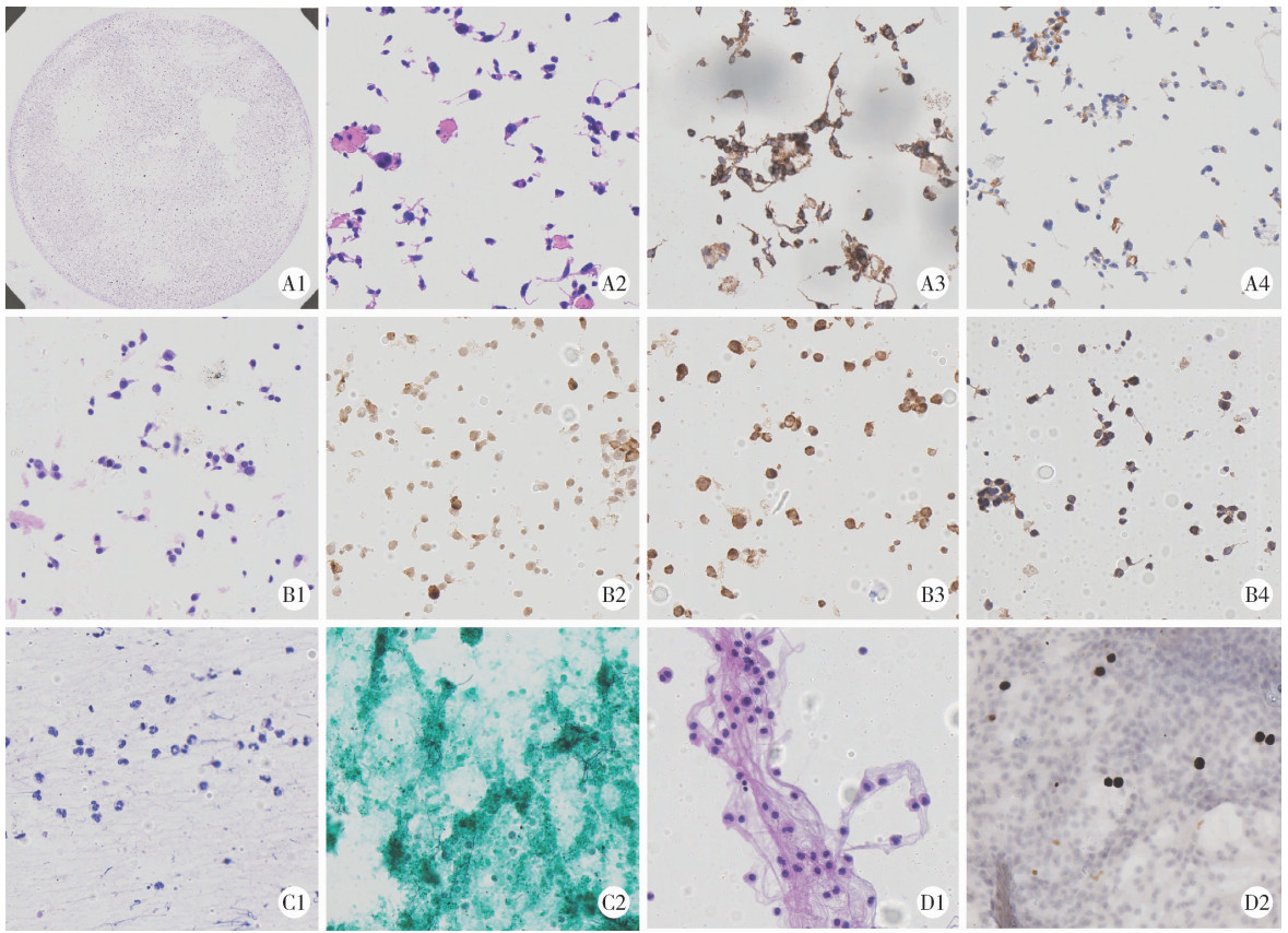
Application of cell transfer technology in pathological diagnosis of micro-volume cell fluid

Ye ZHAO, Xiaoli DIAO, Yan XIONG*()   

  1. Department of Pathology, Peking University First Hospital, Beijing 100034, China
  • Received:2025-07-04 Online:2026-02-18 Published:2026-01-04
  • Contact: Yan XIONG

RICH HTML


Abstract:

Objective: To explore the key technical points and value of cell transfer technology in the diagnosis of micro-volume cell fluid. Methods: In the study, 32 micro-volume cell fluid samples with the diagnosis of tumor or atypical cells in the Department of Pathology, Peking University First Hospital were collected from September 2024 to June 2025. The cells on the ThinPrep cytology test (TCT) slides were divided into several sections and transferred to corresponding slides for immunocytochemistry (ICC) and special staining. Hematoxylin-eosin (HE) staining slides before and after transfer were compared to evaluate the performance of cell transfer technology in maintaining the consistency of cell morphology. The re-diagnosis referring to the results of ICC and special staining of transfer slides were made. The diagnosis before and after cell transfer was compared to evaluate the value of technology in improving the differential diagnostic accuracy. Results: A total of 140 cell transfer slides were prepared from the 32 samples. Among them, 32 HE-stained slides were consistent with the original TCT slides in terms of staining quality, cell morphology and arrangement, with a success rate of 100%; 99 transfer slides were immuno-stained, of which 91 had accurate color and position of positivity and clear background of negativity, with a success rate of 91.91%; 9 special-stained slides had sharp color contrast and clear background, with a success rate of 100%. With the help of ICC and special staining results of transfer slides, 26 of the 32 samples were accurately diagnosed, including 18 cases of malignant tumors and 8 cases of non-neoplastic lesions; 6 cases remained undiagnosed, including four due to ICC staining failure and two due to too few cells. Compared with the original cytological diagnosis, a definitive differential diagnosis was obtained in 81.25% of cases after cell transfer. Conclusion: The application of cell transfer technology in TCT samples is feasible in clinical practice and is suitable for cases requiring ICC and special staining for auxiliary diagnosis. It can significantly improve the differential diagnostic accuracy for the micro-volume cell fluid samples, which is invaluable for the special cases which pathological diagnosis can only be made based on the micro-volume cell fluid samples because no more tissue sample is available.

Key words: Cytodiagnosis, Specimen handling, Immunohistochemistry, Cell-transfer technique, Micro-volume cell fluid

CLC Number: 

  • R446.8

Figure 1

Micro-volume cell fluid: Cerebrospinal fluid, bronchial washing, bronchoalveolar lavage fluid, thyroid fine-needle aspiration fluid (from left to right)"

Figure 2

The operation process of cell transfer technology A, divide the area and mark the directions; B, remove the cover glass and set it aside for later use, add Mount Quick gel; C, soaking in warm water; D, remove the gel film, place in the same direction as the cover glass; E, cut the gel film and make the marking copy; F, transfer to slides for different staining projects."

Figure 3

The staining images before and after cell transfer A, medullary thyroid carcinoma in fine needle aspiration fluid (A1, before, HE ×10; A2, after, HE ×200; A3, CgA+ + +, ICC ×200; A4, calcitonin+, ICC ×200); B, malignant melanoma in left inguinal lymph nodes fluid (B1, after, HE ×200; B2, HMB45+ +, ICC ×200; B3, malen-A+ +, ICC ×200; B4, S100+ +, ICC ×200); C, pulmonary infection in bronchoalveolar lavage fluid (C1, after, HE ×200; C2, GMS+, specific stain, ×200); D, brain metastasis of lung adenocarcinoma in cerebrospinal fluid (D1, after, HE ×200; D2, TTF-1+ + +, ICC ×200). HE, hematoxyllin-eosin; ICC, immunocytochemistry; CgA, chromogranin A; HMB45, human melanoma black 45; GMS, gomori methenamine silver; TTF-1, thyroid transcription factor-1."

Table 1

Enhancement of diagnostic results by cell transfer technology"

Diagnostic results before cell transfer Diagnostic results after cell transfer Sample n Marker
Consider tumor cells; Individual epithelial cells with atypia Adenocarcinoma of lung CSF, BALF 9 CKPan (AE1/AE3), TTF-1, Pax-8, TFE3, p40
A large number of cell nuclei with irregular shapes T-cell lymphoblastic lymphoma CSF 2 TdT, CD34, CD68, CKPan (AE1/AE3), CD3, CD20, Olig2, GFAP, Ki67
Malignant cell Malignant melanoma Left inguinal lymph node FNA fluid 1 CKPan (AE1/AE3), S-100, LCA/CD45, vimentin, HMB45, melan-A
Scattered atypical cells are distributed, and the nucleoli can be seen Cardia cancer CSF 1 CEA, CKPan (AE1/AE3), LCA/CD45
There are a large number of small lymphocytes and monocytes, as well as atypical cells with large nuclei and immature morphology B lymphoblastic leukemia CSF 2 TdT, Pax-5
Clustered atypical glandular cells, with mucous present in the cytoplasm and prominent nucleoli Endometrial cancer Peritoneal wash 1 Pax-8
A large number of mycelia were detected Pneumocystis pneumonia; Pulmonary infection Ebus needle aspiration fluid, BALF 2 GMS, PAS, AFB, WAFB
Malignant cell Adenocarcinoma of stomach Ascites (a little) 1 TTF-1, Pax-8, WT1, CK7, CK20, CDX-2
Consider cells with unclear pathological significance that exhibit atypical changes (TBSRTC Ⅲ) Medullary thyroid carcinoma Thyroid FNA fluid 1 Congo red, calcitonin, Syn, CgA
Cells are scattered and distributed in clusters, with enlarged nuclei and an increased nuclear-cytoplasmic ratio; Some epithelial cells show atypia Non-tumoral lesion BALF, bronchial washing, CSF 8 GATA3, CEA, Ki67, TdT, CD34, CD20, CD117, MPO, CD68, TTF-1, SALL-4, Syn, CD1a, Olig2
1
李春海, 李克勤. 肿瘤微血管生成的机制与肿瘤侵袭和转移[J]. 中华肿瘤杂志, 2000, 22 (3): 181- 183.
2
潘秦镜, 李凌, 张询, 等. 液基细胞学筛查宫颈癌的研究[J]. 中华肿瘤杂志, 2001, 23 (4): 309- 312.
3
Miller RT . Avoiding pitfalls in diagnostic immunohistochemistry: Important technical aspects that every pathologist should know[J]. Semin Diagn Pathol, 2019, 36 (5): 312- 335.

doi: 10.1053/j.semdp.2019.05.002
4
Wen CH , Lin CH , Ko PL , et al. Cell transfer technique for constructing cytological microarrays for immunocytochemical analysis[J]. Cytopathology, 2017, 28 (2): 157- 163.

doi: 10.1111/cyt.12360
5
王一凡, 张兰兰, 杨巧, 等. 2种免疫细胞化学染色方法在脑脊液转移性肺腺癌诊断中的应用[J]. 广西医科大学学报, 2024, 41 (8): 1171- 1175.
6
崔娣, 陈争先, 刘龙腾, 等. 细胞转移技术在细针穿刺细胞学诊断中的应用[J]. 中华病理学杂志, 2021, 50 (6): 615- 619.
7
Gong Y , Joseph T , Sneige N . Validation of commonly used immunostains on cell-transferred cytologic specimens[J]. Cancer, 2005, 105 (3): 158- 164.

doi: 10.1002/cncr.21063
8
Jimenez-Joseph D , Gangi MD . Application of Diatex compound in cytology: Use in preparing multiple slides from a single routine smear[J]. Acta Cytol, 1986, 30 (4): 446- 447.
9
石远凯, 孙燕, 于金明, 等. 中国肺癌脑转移诊治专家共识(2017年版)[J]. 中国肺癌杂志, 2017, 20 (1): 1- 12.
[1] YU Yan-fei,HE Shi-ming,WU Yu-cai,XIONG Sheng-wei,SHEN Qi,LI Yan-yan,YANG Feng,HE Qun,LI Xue-song. Clinicopathological features and prognosis of fumarate hydratase deficient renal cell carcinoma [J]. Journal of Peking University (Health Sciences), 2021, 53(4): 640-646.
[2] CHI Yan-ting,ZHANG Yan-ping,ZHANG Qiu-lu,LIU Cui-ling,LI bin-bin. Clinicopathological analysis of mucosa associated lymphoid tissue lymphoma secondary to Sjögren’s syndrome in salivary gland [J]. Journal of Peking University (Health Sciences), 2021, 53(1): 40-45.
[3] Ru MA,Xin-bao LI,Feng-cai YAN,Yu-lin LIN,Yan LI. Clinical evaluation of tumor-stroma ratio in pseudomyxoma peritonei from the appendix [J]. Journal of Peking University (Health Sciences), 2020, 52(2): 240-246.
[4] Xiao-peng ZHANG,Wei-yu ZHANG,Fei HUO,Hao HU,Qi WANG,Ke-xin XU. Outcome of surgical management and pathogenesis of female primary bladder neck obstruction [J]. Journal of Peking University(Health Sciences), 2019, 51(6): 1052-1055.
[5] Chun-feng ZHANG,Yun LIU,Min LU,Xiao-juan DU. Expression of hUTP14a in non-small cell lung cancer [J]. Journal of Peking University(Health Sciences), 2019, 51(1): 145-150.
[6] Lei LIU,Li-hua WANG,Yu-bo REN,Xiao-song RAO,Shao-min YANG. Clinicopathological analysis of aggressive angiomyxoma of soft tissue in abdomino-pelvic cavity [J]. Journal of Peking University(Health Sciences), 2018, 50(6): 1098-1101.
[7] MEI Fang, ZHAO Ting-ting, GAO Fei, ZHENG Jie. A rare pulmonary benign bi-phasic tumor: a case report of pulmonary adenofibroma and literature review [J]. Journal of Peking University(Health Sciences), 2017, 49(6): 1076-1080.
[8] LIU Chang, CUI Li-gang, WANG Hong-lei. Renal Ewing’s sarcoma/primitive neuroectodermal tumor: a case report and literature review [J]. Journal of Peking University(Health Sciences), 2017, 49(5): 919-923.
[9] XI Chen-guang, FAN Yu, YANG Xin-yu, LIU Li-bo, WANG Jing-hua, HU Shuai, LI Yan-yan, HE Qun. Clinicopathological features and differential diagnosis of metanephric adenoma: a report of sixteen cases [J]. Journal of Peking University(Health Sciences), 2016, 48(4): 598-602.
[10] SI Jing-wen, WANG Li, BA Xiao-jun, ZHANG Xu, DONG Ying, ZHANG Ji-xin, LI Wen-ting, LI Ting. Clinicopathological screening of Lynch syndrome: a report of 2 cases and literature review [J]. Journal of Peking University(Health Sciences), 2015, 47(5): 858-864.
[11] GONG Bei, HU Hui-Hui, ZHANG Man. Expression of apolipoprotein A-Ⅰ in eight histological types of renal neoplasms [J]. Journal of Peking University(Health Sciences), 2015, 47(1): 155-159.
[12] ZHANG Meng-Xue-1, PEI Fei-1△, WANG Tian-Li-2, HAN Xiang-3, YOU Jiang-Feng-1, ZOU Peng-Cheng-1, WANG Yue-Qi-1, LI Xu-Wen-1, LIU Xin-1, ZHONG Gao-Gao-1, LIU Yan-1, WANG Yu-Xiang-1, WANG Hua-1, ZHANG Bo-1. Anaplastic lymphoma kinase fusion gene expression, clinical pathological characteristics and prognosis in 95 Chinese patients with non-small cell lung cancer [J]. Journal of Peking University(Health Sciences), 2014, 46(4): 582-588.
[13] MA Rui-qiong, CHENG Hong-yan, YE Xue, CHEN Jun, CUI Heng, WEI Li-hui, CHANG Xiao-hong. Expression and significance of tumor necrosis factor receptor associated protein 1 in epithelial ovarian cancer [J]. Journal of Peking University(Health Sciences), 2014, 46(1): 120-124.
Viewed
Full text


Abstract

Cited

  Shared   
  Discussed   
No Suggested Reading articles found!