Journal of Peking University(Health Sciences) ›› 2019, Vol. 51 ›› Issue (3): 596-601. doi: 10.19723/j.issn.1671-167X.2019.03.033
Previous Articles Next Articles
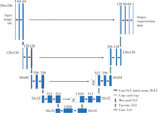
Application of U-shaped convolutional neural network in auto segmentation and reconstruction of 3D prostate model in laparoscopic prostatectomy navigation
Ye YAN1,*,Hai-zhui XIA1,*,Xu-sheng LI2,Wei HE3,Xue-hua ZHU1,Zhi-ying ZHANG1,Chun-lei XIAO1,Yu-qing LIU1,Hua HUANG4,Liang-hua HE2,Jian LU1△( )
)
CLC Number:
- R737.25


| [1] |
Siegel RL, Miller KD, Jemal A.Cancer statistics, 2018[J]. CA Cancer J Clin, 2018, 68(1): 7-30.
doi: 10.3322/caac.21442 |
| [2] |
Simmons MN, Stephenson AJ, Klein EA.Natural history of biochemical recurrence after radical prostatectomy: risk assessment for secondary therapy[J]. Eur Urol, 2007, 51(5): 1175-1184.
doi: 10.1016/j.eururo.2007.01.015 |
| [3] | Van den Broeck T, van den Bergh R, Arfi N, et al. Prognostic value of biochemical recurrence following treatment with curative intent for prostate cancer: A systematic review [J/OL]. Eur Urol,(2018-10-17) [2019-02-15]. https://doi.org/10.1016/j.eururo.2018.10.011. |
| [4] |
Ukimura O, Aron M, Nakamoto M, et al.Three-dimensional surgical navigation model with TilePro display during robot-assisted radical prostatectomy[J]. J Endourol, 2014, 28(6): 625-630.
doi: 10.1089/end.2013.0749 |
| [5] |
Hughes-Hallett A, Mayer EK, Marcus HJ, et al.Augmented rea-lity partial nephrectomy: examining the current status and future perspectives[J]. Urology, 2014, 83(2): 266-273.
doi: 10.1016/j.urology.2013.08.049 |
| [6] | 王燕, 高旭, 阳青松, 等. 3D打印技术辅助认知融合在前列腺穿刺活检术中的应用[J]. 临床泌尿外科杂志, 2016, 31(2): 104-107. |
| [7] | 邵叶秦, 杨新. 基于随机森林的CT前列腺分割[J]. CT理论与应用研究, 2015, 24(5): 647-655. |
| [8] | Ronneberger O, Fischer P, Brox T.U-net: Convolutional networks for biomedical image segmentation[C]. International Conference on Medical image computing and computer-assisted intervention. Cham: Springer, 2015: 234-241. |
| [9] | 詹曙, 梁植程, 谢栋栋. 前列腺磁共振图像分割的反卷积神经网络方法[J]. 中国图象图形学报, 2017, 22(4): 516-522. |
| [10] | Neher PF, Stieltjes B, Reisert M, et al.MITK global tractography[C]. Proceedings of SPIE: The International Society for Optical Engineering, 2012: 83144D. doi: 10.1117/12.911215. |
| [11] |
Lecun Y, Bottou L, Bengio Y, et al.Gradient-based learning applied to document recognition[J]. Proceedings of the IEEE, 1998, 86(11): 2278-2324.
doi: 10.1109/5.726791 |
| [12] |
Mahapatra D, Buhmann JM.Prostate MRI segmentation using learned semantic knowledge and graph cuts[J]. IEEE Transactions on Biomedical Engineering, 2014, 61(3): 756-764.
doi: 10.1109/TBME.2013.2289306 |
| [13] | Korez R, Likar B, Pernuš F, et al.Model-based segmentation of vertebral bodies from MR images with 3D CNNs[C]. International Conference on Medical Image Computing and Computer-Assisted Intervention. Cham: Springer, 2016: 433-441. |
| [14] |
Brosch T, Tang LY, Yoo Y, et al.Deep 3D convolutional encoder networks with shortcuts for multiscale feature integration applied to multiple sclerosis lesion segmentation[J]. IEEE Transactions on Medical Imaging, 2016, 35(5): 1229-1239.
doi: 10.1109/TMI.2016.2528821 |
| [15] |
Martínez F, Romero E, Dréan G, et al.Segmentation of pelvic structures for planning CT using a geometrical shape model tuned by a multi-scale edge detector[J]. Phys Med Biol, 2014, 59(6): 1471-1484.
doi: 10.1088/0031-9155/59/6/1471 |
| [16] | 凌彤, 杨琬琪, 杨明. 利用多模态U形网络的CT图像前列腺分割[J]. 智能系统学报, 2018, 13(6): 981-988. |
| [17] | Ebbing J, Jäderling F, Collins JW, et al.Comparison of 3D printed prostate models with standard radiological information to aid understanding of the precise location of prostate cancer: A construct validation study[J]. PLoS One, 2018, 13(6): e199477. |
| [18] |
Volonté F, Pugin F, Bucher P, et al.Augmented reality and image overlay navigation with OsiriX in laparoscopic and robotic surgery: not only a matter of fashion[J]. J Hepatobiliary Pancreat Sci, 2011, 18(4): 506-509.
doi: 10.1007/s00534-011-0385-6 |
| [19] |
Teber D, Guven S, Simpfendorfer T, et al.Augmented reality: a new tool to improve surgical accuracy during laparoscopic partial nephrectomy? Preliminary in vitro and in vivo results[J]. Eur Urol, 2009, 56(2): 332-338.
doi: 10.1016/j.eururo.2009.05.017 |
| [20] |
Porpiglia F, Fiori C, Checcucci E, et al.Augmented reality robot-assisted radical prostatectomy: Preliminary experience[J]. Urology, 2018, 115(5): 184.
doi: 10.1016/j.urology.2018.01.028 |
| [21] | Porpiglia F, Checcucci E, Amparore D, et al.Augmented-reality robot-assisted radical prostatectomy using hyper-accuracy three-dimensional reconstruction (HA 3DTM) technology: a radiological and pathological study[J]. BJU international, 2018, 123(5): 834-845. |
| [1] | Xiaoqiang BAI, Zhiruo YUAN, Yongsheng ZHOU, Longwei LV. Dynamic stretching promotes osteogenic differentiation of human bone marrow mesenchymal stem cells in three-dimensional culture [J]. Journal of Peking University (Health Sciences), 2026, 58(3): 641-649. |
| [2] | Zhaode BU, Mengyu FENG, Ke JI. Practice and reflection on sentinel lymph node navigation surgery for early gastric cancer [J]. Journal of Peking University (Health Sciences), 2026, 58(2): 239-243. |
| [3] | Wen DU, Wenbo ZHANG, Yao YU, Shuo LIU, Huiyu SU, Leihao HU, Zunan TANG, Binzhang WU, Zhen CHEN, Jiaqi LI, Hao WANG, Xin PENG. Exploration and clinical application of the "digital and intelligent surgery" diagnosis and treatment workflow for oral and maxillofacial tumors [J]. Journal of Peking University (Health Sciences), 2026, 58(2): 278-284. |
| [4] | Jingheng WU, Yunhao XUE, Shanlin CHEN, Yintao GUO, Yuntao LIU, Wei ZHANG. Super microsurgical lymphaticovenular anastomosis for limb lymphedema: An outcome analysis based on clinical stage and indocyanine green pattern [J]. Journal of Peking University (Health Sciences), 2026, 58(2): 359-364. |
| [5] | Aonan WEN, Xiaohui ZHANG, Yongtao YANG, Zixiang GAO, Wenbo LI, Shenyao SHAN, Xiangyi SHANG, Yuwen TIAN, Shuwei GUO, Yizhen WANG, Yong WANG, Yijiao ZHAO. Method of constructing 3D facial smile simulation sequence data based on non-rigid registration [J]. Journal of Peking University (Health Sciences), 2026, 58(1): 139-144. |
| [6] | Lu YU, Ling WU, Xiaojing LIU, Zili LI. Feasibility study of a surgical planning protocol for orthognathic surgery utilizing similarity retrieval from database: A randomized controlled trial [J]. Journal of Peking University (Health Sciences), 2026, 58(1): 145-152. |
| [7] | Yuting YANG, Liuyang QU, Danni ZHENG, Xiaotong LING, Xiaoyun XU, Denggao LIU. Demographic characteristic and clinical features in 1 812 patients with salivary gland stones [J]. Journal of Peking University (Health Sciences), 2026, 58(1): 153-159. |
| [8] | Rentao TANG, Liuchang YANG, Jie NIE, Xiaoyan WANG. Microbial communities in extraradicular infections of post-treatment apical periodontitis without or with sinus tracts [J]. Journal of Peking University (Health Sciences), 2026, 58(1): 43-49. |
| [9] | Liang SHAO, Wenjie MA, Ying CHEN, Qian DING, Lei ZHANG. Digital measurement and analysis of anatomical characteristics of protrusive and intercuspal position occlusal contacts in maxillary incisors [J]. Journal of Peking University (Health Sciences), 2026, 58(1): 99-106. |
| [10] | Cuiping WANG, Zhe CHEN, Yongjing CHENG. Correlation study of superb microvascular imaging on knee osteoarthritis [J]. Journal of Peking University (Health Sciences), 2025, 57(6): 1096-1100. |
| [11] | Yanhua LIU, Min LU, Xuyang ZHAO, Kuan'gen ZHANG, Rui WU, Fang MEI, Zhihao DAI, Jiangfeng YOU, Fei PEI. Effect of dephosphorylation of tumor metastasis suppressor gene LASS2 on vacuolar ATPase activity and invasiveness of prostate cancer [J]. Journal of Peking University (Health Sciences), 2025, 57(6): 1113-1123. |
| [12] | Zhemin LI, Jiafu JI, Guoxin LI, Ziyu LI, Zhaode BU, Xiangyu GAO, Di DONG, Lei TANG, Xiaofang XING, Shuqin JIA, Ting GUO, Lianhai ZHANG, Fei SHAN, Xin JI, Anqiang WANG. Development and dissemination of precision medicine approaches in gastric cancer management [J]. Journal of Peking University (Health Sciences), 2025, 57(5): 864-867. |
| [13] | Bowen LI, Qiang ZHANG, Yixin SUN. Establishment and validation of a risk prediction model for scoliosis after Nuss procedure in children and young adults with pectus excavatum [J]. Journal of Peking University (Health Sciences), 2025, 57(5): 941-946. |
| [14] | Xiaoyong YANG, Fan ZHANG, Lulin MA, Cheng LIU. Clinical characteristics and influencing factors of extraglandular invasion of prostatic ductal adenocarcinoma [J]. Journal of Peking University (Health Sciences), 2025, 57(5): 956-960. |
| [15] | Yujia XIAO, Bochun MAO, Yanheng ZHOU. Three-dimensional morphological analysis of posed smile [J]. Journal of Peking University (Health Sciences), 2025, 57(5): 989-995. |
|
||