Journal of Peking University (Health Sciences) ›› 2022, Vol. 54 ›› Issue (6): 1219-1223. doi: 10.19723/j.issn.1671-167X.2022.06.027

Previous Articles     Next Articles

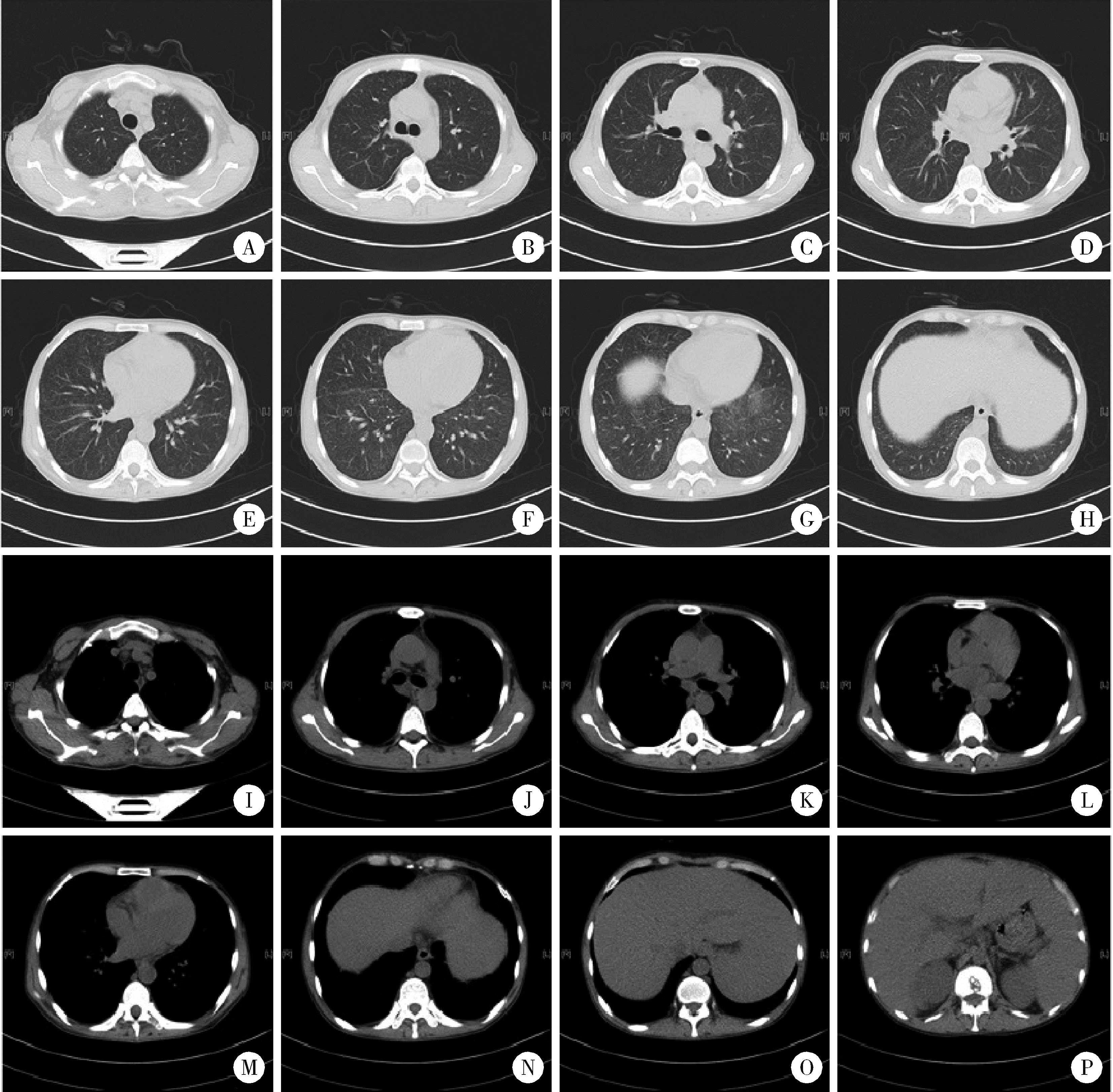
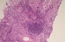
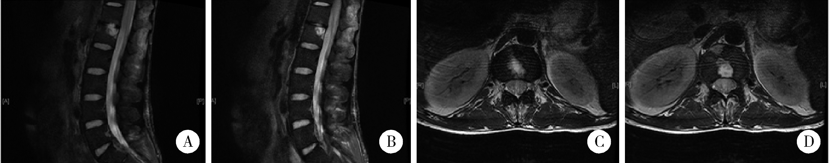
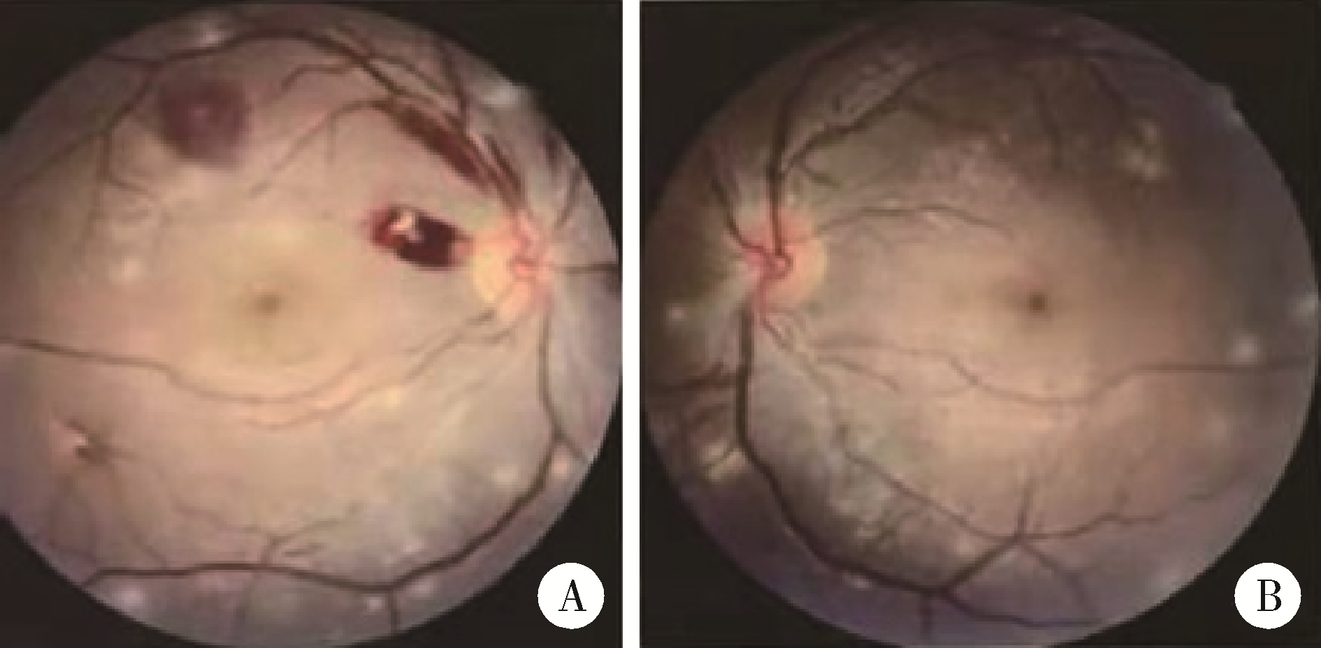
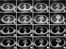
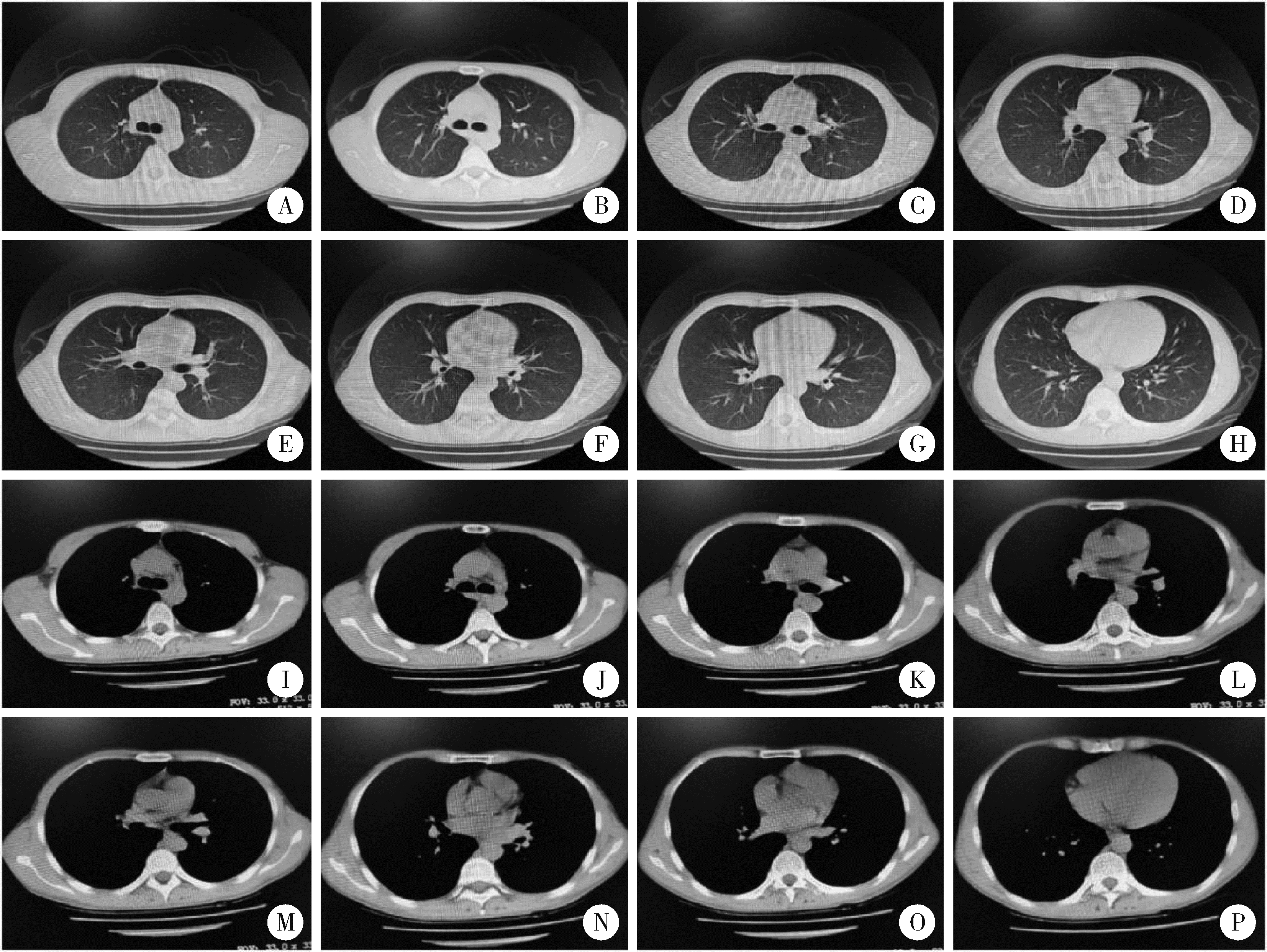
Hemophagocytic lymphohistiocytosis caused by hematogenous disseminated pulmonary tuberculosis: A case report

Qiu-yu LI1,Ying LIANG1,Ni-ni DAI1,Yu-xiang WANG2,Bo-tao ZHU1,Rui WU1,Hong ZHU1,*(),Yong-chang SUN1   

  1. 1. Department of Respiratory and Critical Care Medicine, Peking University Third Hospital, Beijing 100191, China
    2. Department of Pathology, Peking University Third Hospital, Beijing 100191, China
  • Received:2020-06-22 Online:2022-12-18 Published:2022-12-19
  • Contact: Hong ZHU E-mail:zhuhong_bysy@163.com

Abstract:

Hemophagocytic lymphohistiocytosis (HLH) was a life-threatening syndrome due to the uncontrolled immune activation of cytotoxic T lymphocytes, natural killer (NK) cells, and macrophages. HLH is characterized by primary and secondary causes, the early diagnosis and treatment of patients are closely related to the prognosis and clinical outcome of patients. The clinical presentation is variable but mostly includes prolonged fever, splenomegaly, coagulopathy, hypertriglyceridemia, and hemophagocytosis, none of them is specific and particular for HLH. Tuberculosis (TB) infection is one of the causes of HLH. HLH caused by TB is very rare clinically, but it has a high mortality. For patients with fever of unknown origin, HLH-related clinical manifestations sometimes present before the final diagnosis of TB, and HLH is associated with the most significant mortality rate. This article is mainly about a 28-year-old patient with HLH who suffered from severe TB infection. The patient attended a hospital with a history of 2 months of prolonged fever, 10 days booger and subcutaneous hemorrhage in lower limbs. Before this, he was in good health and denied any history of tuberculosis exposure. Combined with relevant laboratory test results (such as splenomegaly, hemoglobin, platelet count, and hypertriglyceridemia) and clinical manifestations (e.g. fever), the patient was diagnosed with hemophagocytic lymphohistiocytosis, but the etiology of HLH remained to be determined. To confirm the etiology, the patient was asked about the relevant medical history (intermittent low back pain) and was performed chest CT scan, bone marrow biopsy, and fundus photography. Finally, he was diagnosed with hemophagocytic lymphohistiocytosis caused by hematogenous disseminated pulmonary tuberculosis. In response to this, intravenous methylprednisolone and anti-tuberculosis treatment (isoniazid, pyrazinamide, moxifloxacin, and amikacin) were administered to the patient. After more than a month of treatment, the patient recovered from HLH caused by severe TB infection. Therefore, this case suggests that we should be vigilant to the patient who admitted to the hospital with fever for unknown reasons, to diagnose HLH as early as possible and clarify its cause, then perform interventions and treatment, especially HLH secondary to tuberculosis. Also, cases of atypical TB and severe TB should be carefully monitored to achieve early diagnosis and early intervention.

Key words: Hematogenous disseminated pulmonary tuberculosis, Hemophagocytic lymphohistiocytosis, Fever, Pulmonary nodule

CLC Number: 

  • R521.1

Figure 1

The chest computed tomography of the patient A-O, bronchovascular bundles of both lungs were enlarged and disordered, tree bud sign and diffuse dot-like shadow were seen, small ground glass density nodules were seen in the upper right lobe and the lower left lobe, trachea and main bronchus were smooth, no enlarged lymph nodes were seen in hilum and mediastinum, and no signs of hydrothorax were found on both sides; P, diffuse decrease of liver density, splenomegaly and bone destruction in L1 vertebra."

Figure 2

Biopsy of supraclavicular lymph node Reactive proliferation of lymphoid tissue, especially the proliferation of histiocytes, the formation of suspected small granuloma, no exact neoplastic lesions."

Figure 3

The lumbar magnetic resonance imaging of the patient A, B, lumbar curvature is straight and smooth, there are patchy short T1 long T2 signal shadows in T12 to L1 vertebral bodies; C, D, considering infectious lesions, the intervertebral space becomes narrow, no swelling or protrusion of the intervertebral discs and no obvious stenosis of the spinal canal is observed."

Figure 4

multiple nodules in fundoscopy Multiple small nodules can be seen, considering tuberculous lesions. A, right eye; B, left eye."

Figure 5

The chest computed tomography of the recovery patient A-H, thorax is symmetrical, the mediastinum is in the middle, the miliary nodular shadow is diffused in both lungs and between the lobes, the size is uniform, the distribution is generally uniform, and it is absorbed compared with the former, there is no air containing area under the pleura in the upper lobes, middle lobes and lower lobes of both lungs; I-P, opening of each lobe and segment of bronchus is smooth, and the 7 areas of mediastinum are slightly enlarged lymph nodes."

1 中华医学会放射学分会传染病放射学专业委员会. 肺结核影像学及分级诊断专家共识[J]. 新发传染病电子杂志, 2018, 3 (2): 118- 127.
doi: 10.3877/j.issn.2096-2738.2018.02.017
2 Schram AM , Berliner N . How I treat hemophagocytic lymphohis-tiocytosis in the adult patient[J]. Blood, 2015, 125 (19): 2908- 2914.
doi: 10.1182/blood-2015-01-551622
3 Henter JI , Home A , Arico M , et al. HLH-2004: diagnostic and therapeutic guidelines for hemophagocytic lymphohistiocytosis[J]. Pediatr Blood Cancer, 2007, 48 (2): 124- 131.
doi: 10.1002/pbc.21039
4 Darmawan G , Salido EO , Concepcion ML , et al. Hemophagocytic lymphohistiocytosis: "A dreadful mimic"[J]. Int J Rheum Dis, 2015, 18 (7): 810- 812.
doi: 10.1111/1756-185X.12506
5 Zhang Y , Liang G , Qin H , et al. Tuberculosis-associated hemophagocytic lymphohistiocytosis with initial presentation of fever of unknown origin in a general hospital: An analysis of 8 clinical cases[J]. Medicine, 2017, 96 (16): e6575.
doi: 10.1097/MD.0000000000006575
6 Asaji M , Tobino K , Murakami K , et al. Miliary tuberculosis in a young woman with hemophagocytic syndrome: A case report and literature review[J]. Intern Med, 2017, 56 (12): 1591- 1596.
doi: 10.2169/internalmedicine.56.8025
7 Shea YF , Chan JF , Kwok WC , et al. Haemophagocytic lymphohistiocytosis: an uncommon clinical presentation of tuberculosis[J]. Hong Kong Med J, 2012, 18 (6): 517- 525.
[1] Wei-bo GAO,Mao-jing SHI,Hai-yan ZHANG,Chun-bo WU,Ji-hong ZHU. Relationship between marked hyperferritinemia and hemophagocytic lymphohistiocytosis [J]. Journal of Peking University (Health Sciences), 2021, 53(5): 921-927.
[2] ZHANG Jia-wei, HAN Pei-en, YANG Li. Spatial accessibility of fever clinics for multi-tiered prevention and control on COVID-19 in Beijing [J]. Journal of Peking University (Health Sciences), 2021, 53(3): 543-548.
[3] Yu-qing OUYANG,Lian-fang NI,Xin-min LIU. Prognosis factors analysis of patients with malignant solitary pulmonary nodules [J]. Journal of Peking University(Health Sciences), 2020, 52(1): 158-162.
[4] WANG He, HU Zhao-heng, CHEN Ling2, PAN Yu3. Influence of history of oral bisphosphonates on the incidence rate of fever after intravenous injection of zoledronic acid in patients with osteoporosis [J]. Journal of Peking University(Health Sciences), 2016, 48(4): 680-682.
[5] KANG Lei, XU Xiao-Jie, FAN Yan, WANG Rong-Fu, MA Chao, FU Zhan-Li, ZHANG Jian-Hua, ZHANG Xu-Chu. Diagnostic value of fluorine-18 fluorodeoxyglucose positron emission tomography/computed tomography in fever of unknown origin [J]. Journal of Peking University(Health Sciences), 2015, 47(1): 175-180.
[6] NI Lian-Fang, LIU Xin-Min. Diagnostic value of serum tumor markers in differentiating malignant from benign solitary pulmonary nodules [J]. Journal of Peking University(Health Sciences), 2014, 46(5): 707-710.
Viewed
Full text


Abstract

Cited

  Shared   
  Discussed   
[1] . [J]. Journal of Peking University(Health Sciences), 2007, 39(4): 434 -436 .
[2] . [J]. Journal of Peking University(Health Sciences), 2001, 33(3): 288 -289 .
[3] . [J]. Journal of Peking University(Health Sciences), 2002, 34(2): 97 -98 .
[4] . [J]. Journal of Peking University(Health Sciences), 2002, 34(2): 112 -116 .
[5] . [J]. Journal of Peking University(Health Sciences), 2011, 43(1): 29 -33 .
[6] . [J]. Journal of Peking University(Health Sciences), 2011, 43(2): 179 -182 .
[7] . [J]. Journal of Peking University(Health Sciences), 2001, 33(1): 50 -53 .
[8] . [J]. Journal of Peking University(Health Sciences), 2002, 34(2): 140 -143 .
[9] . [J]. Journal of Peking University(Health Sciences), 2010, 42(4): 476 -479 .
[10] . [J]. Journal of Peking University(Health Sciences), 2008, 40(2): 208 -210 .