Journal of Peking University(Health Sciences) ›› 2019, Vol. 51 ›› Issue (1): 120-130. doi: 10.19723/j.issn.1671-167X.2019.01.022
Previous Articles Next Articles
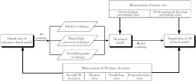
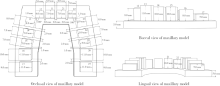
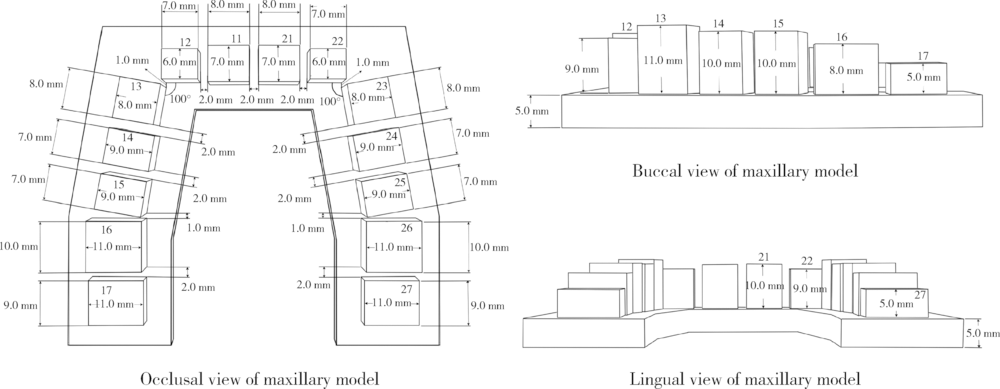
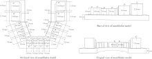
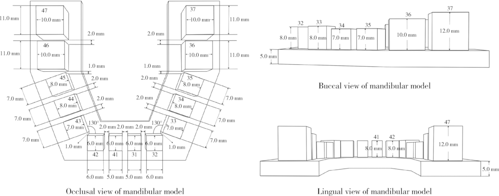
A method to evaluate the trueness of reconstructed dental models made with photo-curing 3D printing technologies
Ning XIAO,Yu-chun SUN,Yi-jiao ZHAO( ),Yong WANG()
- Center for Digital Dentistry, Department of Prosthodontics, Peking University School and Hospital of Stomatology & National Clinical Research Center for Oral Diseases & National Engineering Laboratory for Digital and Material Technology of Stomatology & Beijing Key Laboratory of Digital Stomatology, Beijing 100081, China
CLC Number:
- R783.1


| [1] |
Bell A, Ayoub AF, Siebert P . Assessment of the accuracy of a three-dimensionalimaging system for archiving dental study models[J]. J Orthod, 2003,30(3):219-223.
doi: 10.1093/ortho/30.3.219 pmid: 14530419 |
| [2] |
雷东辉, 杨建军, 安世昌 , 等. 四种超硬石膏模型精度及抗弯强度的比较[J]. 中国组织工程研究与临床康复, 2011,15(21):3979-3982.
doi: 10.3969/j.issn.1673-8225.2011.21.043 |
| [3] |
Santoro M, Galkin S, Teredesai M , et al. Comparison of measurements made on digital and plaster models[J]. Am J Orthod Den-tofacial Orthop, 2003,124(1):101-105.
doi: 10.1016/S0889-5406(03)00152-5 pmid: 12867904 |
| [4] |
杨慧芳, 赵建江, 王勇 . 3D打印技术在口腔医学领域中的应用[J]. 中国医疗设备, 2015,30(5):63-65.
doi: 10.3969/j.issn.1674-1633.2015.05.019 |
| [5] |
Dawood A, Marti MB, Sauret-Jackson V , et al. 3D printing in dentistry[J]. Br Dent J, 2015,219(11):521-529.
doi: 10.1038/sj.bdj.2015.914 |
| [6] |
Barazanchi A, Li KC, Al-Amleh B , et al. Additive technology: update on current materials and applications in dentistry[J]. J Prosthodont, 2017,26(2):156-163.
doi: 10.1111/jopr.12510 pmid: 27662423 |
| [7] |
孙玉春, 李榕, 周永胜 , 等. 三维打印在口腔修复领域中的应用[J]. 中华口腔医学杂志, 2017,52(6):381-385.
doi: 10.3760/cma.j.issn.1002-0098.2017.06.013 |
| [8] | 王惠芸 . 我国人牙的测量和统计[J]. 中华口腔科杂志, 1959,3(7):149-155. |
| [9] |
翁希里, 于世宾, 赵守亮 , 等. 关中地区1000个汉族人恒牙解剖形态测量[J]. 牙体牙髓牙周病学杂志, 2007,17(2):75-77.
doi: 10.3969/j.issn.1005-2593.2007.02.004 |
| [10] |
涂玲, 刘良奎, 胡延佳 . 湖南地区正常恒牙牙合牙及牙弓的测量研究[J]. 中国现代医学杂志, 2003,13(24):62-64.
doi: 10.3969/j.issn.1005-8982.2003.24.019 |
| [11] |
彭惠, 卢海燕, 王昕 . 汉族青年恒牙牙冠的测量研究[J]. 现代口腔医学杂志, 2003,17(1):57-59.
doi: 10.3969/j.issn.1003-7632.2003.01.020 |
| [12] |
于跃, 许天民 . Spee曲线相关研究的回顾[J]. 中华口腔正畸学杂志, 2013,20(4):214-217.
doi: 10.3760/cma.j.issn.1674-5760.2013.04.008 |
| [13] |
Kasparova M, Grafova L, Dvorak P , et al. Possibility of reconstruction of dental plaster cast from 3D digital study models[J]. Biomed Eng Online, 2013,12:49.
doi: 10.1186/1475-925X-12-49 pmid: 23721330 |
| [14] |
Rebong RE, Stewart KT, Utreja A , et al. Accuracy of three-dimensional dental resin models created by fused deposition mo-deling, stereolithography, and Polyjet prototype technologies: A comparative study[J]. Angle Orthod, 2018,88(3):363-369.
doi: 10.2319/071117-460.1 pmid: 29509023 |
| [15] |
Keating AP, Knox J, Bibb R , et al. A comparison of plaster, digital and reconstructed study model accuracy[J]. J Orthod, 2008,35(3):191-201.
doi: 10.1179/146531207225022626 |
| [16] |
Hazeveld A, Huddleston SJ, Ren Y . Accuracy and reproducibility of dental replica models reconstructed by different rapid prototyping techniques[J]. Am J Orthod Dentofacial Orthop, 2014,145(1):108-115.
doi: 10.1016/j.ajodo.2013.05.011 pmid: 24373661 |
| [17] |
Saleh WK, Ariffin E, Sherriff M , et al. Accuracy and reprodu-cibility of linear measurements of resin, plaster, digital and printed study-models[J]. J Orthod, 2015,42(4):301-306.
doi: 10.1179/1465313315Y.0000000016 pmid: 26216658 |
| [18] |
Hwang YC, Park YS, Kim HK , et al. The evaluation of working casts prepared from digital impressions[J]. Oper Dent, 2013,38(6):655-662.
doi: 10.2341/12-352-l pmid: 23570301 |
| [19] |
Wan HW, Yusoff Y, Mardi NA . Comparison of reconstructed rapid prototyping models produced by 3-dimensional printing and conventional stone models with different degrees of crowding[J]. Am J Orthod Dentofacial Orthop, 2017,151(1):209-218.
doi: 10.1016/j.ajodo.2016.08.019 |
| [20] | 曾飞煌, 徐远志, 房莉 , 等. 应用数字化技术和快速成型技术制作牙颌模型的准确性评价[J]. 上海口腔医学, 2012,21(1):53-56. |
| [21] |
Camardella LT, de Vasconcellos VO, Breuning H . Accuracy of printed dental models made with 2 prototype technologies and different designs of model bases[J]. Am J Orthod Dentofacial Orthop, 2017,151(6):1178-1187.
doi: 10.1016/j.ajodo.2017.03.012 pmid: 28554463 |
| [22] |
Cuperus AM, Harms MC, Rangel FA , et al. Dental models made with an intraoral scanner: a validation study[J]. Am J Orthod Dentofacial Orthop, 2012,142(3):308-313.
doi: 10.1016/j.ajodo.2012.03.031 pmid: 22920696 |
| [23] |
Murugesan K, Anandapandian PA, Sharma SK , et al. Comparative evaluation of dimension and surface detail accuracy of models produced by three different rapid prototype techniques[J]. J In-dian Prosthodont Soc, 2012,12(1):16-20.
doi: 10.1007/s13191-011-0103-8 pmid: 3332309 |
| [24] |
Kim SY, Shin YS, Jung HD , et al. Precision and trueness of dental models manufactured with different 3-dimensional printing techniques[J]. Am J Orthod Dentofacial Orthop, 2018,153(1):144-153.
doi: 10.1016/j.ajodo.2017.05.025 pmid: 29287640 |
| [25] |
Dietrich CA, Ender A, Baumgartner S , et al. A validation study of reconstructed rapid prototyping models produced by two techno-logies[J]. Angle Orthod, 2017,87(5):782-787.
doi: 10.2319/01091-727.1 pmid: 28459285 |
| [26] | Jin SJ, Jeong ID, Kim JH , et al. Accuracy (trueness and precision) of dental models fabricated using additive manufacturing methods[J]. Int J Comput Dent, 2018,21(2):107-113. |
| [27] |
Ishida Y, Miyasaka T . Dimensional accuracy of dental casting patterns created by 3D printers[J]. Dent Mater J, 2016,35(2):250-256.
doi: 10.4012/dmj.2015-278 pmid: 27041015 |
| [28] |
方浩博, 陈继民 . 基于数字光处理技术的3D打印技术[J]. 北京工业大学学报, 2015,41(12):1775-1782.
doi: 10.11936/bjutxb2015070050 |
| [29] | 何岷洪, 宋坤, 莫宏斌 , 等. 3D打印光敏树脂的研究进展[J]. 功能高分子学报, 2015,28(1):102-108. |
| [30] |
Stansbury JW, Idacavage MJ . 3D printing with polymers: Challenges amongexpanding options and opportunities[J]. Dent Mater, 2016,32(1):54-64.
doi: 10.1016/j.dental.2015.09.018 pmid: 26494268 |
| [31] |
Snyder TJ, Andrews M, Weislogel M , et al. 3D systems’ techno-logy overview and new applications in manufacturing, engineering, science, and education[J]. 3D Print Addit Manuf, 2011,1(3):169-176.
doi: 10.1089/3dp.2014.1502 pmid: 28473997 |
| [1] | Shuangyun ZHAO, Siyu ZOU, Xueying LI, Lijuan SHEN, Hong ZHOU. Evaluation of reliability and validity of Chinese version of a short-form of Health Literacy Dental scale (HeLD-14) in the application among parents of preschool children [J]. Journal of Peking University (Health Sciences), 2024, 56(5): 828-832. |
| [2] | Ying HUANG,Zhi-yuan WU,Xing-hong ZHOU,Zhi-gang CAI,Jie ZHANG. Category of facial symmetry perception after maxillary reconstruction using anterolateral thigh flap [J]. Journal of Peking University (Health Sciences), 2023, 55(4): 708-715. |
| [3] | LIU Si-min,ZHAO Yi-jiao,WANG Xiao-yan,WANG Zu-hua. In vitro evaluation of positioning accuracy of trephine bur at different depths by dynamic navigation [J]. Journal of Peking University (Health Sciences), 2022, 54(1): 146-152. |
| [4] | XU Xiao-xiang,CAO Ye,ZHAO Yi-jiao,JIA Lu,XIE Qiu-fei. In vitro evaluation of the application of digital individual tooth tray in the impression making of mandibular full-arch crown abutments [J]. Journal of Peking University (Health Sciences), 2021, 53(1): 54-61. |
| [5] | Tian-cheng QIU,Xiao-jing LIU,Zhu-lin XUE,Zi-li LI. Evaluation of the reproducibility of non-verbal facial expressions in normal persons using dynamic stereophotogrammetric system [J]. Journal of Peking University (Health Sciences), 2020, 52(6): 1107-1111. |
| [6] | Ning XIAO,Yu-chun SUN,Yi-jiao ZHAO,Yong WANG. Preliminary study on three digital analysis methods for analyzing the distribution and area of occlusal contacts [J]. Journal of Peking University(Health Sciences), 2020, 52(1): 144-151. |
| [7] | DAI Fan-fan, LIU Yi, XU Tian-min, CHEN Gui. Exploring a new method for superimposition of pre-treatment and post-treatment mandibular digital dental casts in adults [J]. Journal of Peking University(Health Sciences), 2018, 50(2): 271-278. |
| [8] | WANG Sheng-lin, YANG Zhong-wei, YAN ming, LIU Zhong-jun. Atlantoaxial reduction and fixation guided by the intraoperative CT [J]. Journal of Peking University(Health Sciences), 2017, 49(3): 512-517. |
| [9] | GUO Fu-xin, JIANG Yu-liang, JI Zhe, PENG Ran, SUN Hai-tao, WANG Jun-jie. 3D printed template-assisted and computed tomography image-guided 125-iodine seed implantation for supraclavicular metastatic tumor: a dosimetric study [J]. Journal of Peking University(Health Sciences), 2017, 49(3): 506-511. |
| [10] | WANG Zhe, ZHU Liu-ning, ZHOU Lin, YI Biao. Feasibility of integrating 3D photos and cone-beam computed tomography images used to evaluate changes of soft and hard tissue after orthognathic surgery [J]. Journal of Peking University(Health Sciences), 2016, 48(3): 544-549. |
| [11] | ZHENG Bang, LI Man, WANG Kai-lu, LV Jun. Analysis of the reliability and validity of the Chinese version of Pittsburgh sleep qua-lity index among medical college students [J]. Journal of Peking University(Health Sciences), 2016, 48(3): 424-428. |
| [12] | YE Hong-qiang, LIU Yu-shu, LIU Yun-song, NING Jing, ZHAO Yi-jiao, ZHOU Yong-sheng. Constructing 3-dimensional colorized digital dental model assisted by digital photography [J]. Journal of Peking University(Health Sciences), 2016, 48(1): 138-142. |
| [13] | QIN Yi-Fei, XU Tian-Min. Reproducibility and repeatability of the determination of occlusal plane on digital dental models [J]. Journal of Peking University(Health Sciences), 2015, 47(3): 536-540. |
| [14] | HU Pan-Pan, YU Miao, LIU Xiao-Guang, CHEN Zhong-Qiang, LIU Zhong-Jun. Correlation analysis between the sagittal and coronal parameters of spino-pelvic in Lenke type 1 adolescent idiopathic scoliosis [J]. Journal of Peking University(Health Sciences), 2015, 47(2): 248-252. |
| [15] | WANG Zong-Qi, WANG Xiao-Xia, LI Zi-Li, YI Biao, LIANG Cheng, WANG Xing. Comparison of three surgical techniques for controlling nasal width after Le Fort Ⅰ osteotomy [J]. Journal of Peking University(Health Sciences), 2015, 47(1): 104-108. |
|
||