Journal of Peking University (Health Sciences) ›› 2022, Vol. 54 ›› Issue (1): 146-152. doi: 10.19723/j.issn.1671-167X.2022.01.023
Previous Articles Next Articles
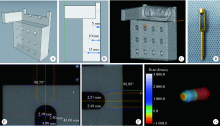
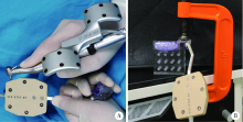
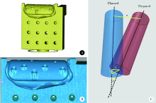
In vitro evaluation of positioning accuracy of trephine bur at different depths by dynamic navigation
LIU Si-min1,ZHAO Yi-jiao2,WANG Xiao-yan1,WANG Zu-hua1,△( )
)
- 1. Department of Conservative Dentistry and Endodontics, Peking University School and Hospital of Stomatology, Beijing 100081, China
2. Center for Digital Dentistry, Peking University School and Hospital of Stomatology & National Center of Stomatology & National Clinical Research Center for Oral Diseases & National Engineering Laboratory for Digital and Material Technology of Stomatology & Beijing Key Laboratory of Digital Stomatology & NHC Research Center of Engineering and Technology for Computerized Dentistry, Beijing 100081, China
CLC Number:
- R783.3


| [1] |
Bobek SL. Applications of navigation for orthognathic surgery[J]. Oral Maxillofac Surg Clin North Am, 2014, 26(4):587-598.
doi: 10.1016/j.coms.2014.08.003 |
| [2] | Wadley J, Dorward N, Kitchen N, et al. Pre-operative planning and intra-operative guidance in modern neurosurgery: A review of 300 cases[J]. Ann R Coll Surg Engl, 1999, 81(4):217-225. |
| [3] |
Bell RB. Computer planning and intraoperative navigation in orthognathic surgery[J]. J Oral Maxillofac Surg, 2011, 69(3):592-605.
doi: 10.1016/j.joms.2009.06.030 |
| [4] |
Emery RW, Merritt SA, Lank K, et al. Accuracy of dynamic navigation for dental implant placement-model-based evaluation[J]. J Oral Implantol, 2016, 42(5):399-405.
doi: 10.1563/aaid-joi-D-16-00025 |
| [5] |
Block MS, Emery RW, Cullum DR, et al. Implant placement is more accurate using dynamic navigation[J]. J Oral Maxillofac Surg, 2017, 75(7):1377-1386.
doi: 10.1016/j.joms.2017.02.026 |
| [6] |
Zubizarreta-Macho Á, Muñoz AP, Deglow ER, et al. Accuracy of computer-aided dynamic navigation compared to computer-aided static procedure for endodontic access cavities: An in vitro study[J]. J Clin Med, 2020, 9(1):129.
doi: 10.3390/jcm9010129 |
| [7] |
Dianat O, Nosrat A, Mostoufi B, et al. Accuracy and efficiency of guided root-end resection using a dynamic navigation system: A human cadaver study[J]. Int Endodo J, 2021, 54(5):793-801.
doi: 10.1111/iej.v54.5 |
| [8] |
Jain SD, Carrico CK, Bermanis I. 3-dimensional accuracy of dynamic navigation technology in locating calcified canals[J]. J Endod, 2020, 46(6):839-845.
doi: 10.1016/j.joen.2020.03.014 |
| [9] |
Wu D, Zhou L, Yang J, et al. Accuracy of dynamic navigation compared to static surgical guide for dental implant placement[J]. Int J Implant Dent, 2020, 6(1):78.
doi: 10.1186/s40729-020-00272-0 |
| [10] |
Wei SM, Zhu Y, Wei JX, et al. Accuracy of dynamic navigation in implant surgery: A systematic review and meta-analysis[J]. Clin Oral Implants Res, 2021, 32(4):383-393.
doi: 10.1111/clr.v32.4 |
| [11] |
Gambarini G, Galli M, Morese A, et al. Precision of dynamic navigation to perform endodontic ultraconservative access cavities: A preliminary in vitro analysis[J]. J Endod, 2020, 46(9):1286-1290.
doi: S0099-2399(20)30385-X pmid: 32553875 |
| [12] | Aydemir CA, Arısan V. Accuracy of dental implant placement via dynamic navigation or the freehand method: A split-mouth rando-mized controlled clinical trial[J]. Clin Oral Implants Res, 2020, 31(3):255-263. |
| [13] |
Mediavilla Guzmán A, Riad Deglow E, Zubizarreta-Macho Á, et al. Accuracy of computer-aided dynamic navigation compared to computer-aided static navigation for dental implant placement: An in vitro study[J]. J Clin Med, 2019, 8(12):2123.
doi: 10.3390/jcm8122123 |
| [14] |
Pellegrino G, Taraschi V, Andrea Z, et al. Dynamic navigation: A prospective clinical trial to evaluate the accuracy of implant placement[J]. Int J Comput Dent, 2019, 22(2):139-147.
pmid: 31134220 |
| [15] |
Hawkins TK, Wealleans JA, Pratt AM, et al. Targeted endodontic microsurgery and endodontic microsurgery: A surgical simulation comparison[J]. Int Endod J, 2020, 53(5):715-722.
doi: 10.1111/iej.13243 pmid: 31674678 |
| [16] |
Christofzik D, Bartols A, Faheem MK, et al. Shaping ability of four root canal instrumentation systems in simulated 3D-printed root canal models[J]. PLoS One, 2018, 13(8):e0201129.
doi: 10.1371/journal.pone.0201129 |
| [17] |
Nagy E, Fráter M, Antal M. Guided modern endodontic microsurgery by use of a trephine bur[J]. Orv Hetil, 2020, 161(30):1260-1265.
doi: 10.1556/650.2020.31778 |
| [18] |
Buniag AG, Pratt AM, Ray JJ. Targeted endodontic microsurgery: A retrospective outcomes assessment of 24 cases[J]. J Endod, 2021, 47(5):762-769.
doi: 10.1016/j.joen.2021.01.007 |
| [19] |
Collyer J. Stereotactic navigation in oral and maxillofacial surgery[J]. Br J Oral Maxillofac Surg, 2010, 48(2):79-83.
doi: 10.1016/j.bjoms.2009.04.037 pmid: 20061072 |
| [20] |
Popowicz W, Palatyńska-Ulatowska A, Kohli MR. Targeted endodontic microsurgery: Computed tomography-based guided stent approach with platelet-rich fibrin graft: A report of 2 cases[J]. J Endod, 2019, 45(12):1535-1542.
doi: S0099-2399(19)30626-0 pmid: 31606146 |
| [21] |
Rismanchian M, Bajoghli F, Gholamreza T, et al. Dental implants: Early versus standard two-stage loading (animal study)[J]. J Oral Implant, 2014, 40(1):84-93.
doi: 10.1563/AAID-JOI-D-10-00202 |
| [22] |
Gambarini G, Galli M, Stefanelli LV, et al. Endodontic microsurgery using dynamic navigation system: A case report[J]. J Endod, 2019, 45(11):1397-1402.
doi: S0099-2399(19)30544-8 pmid: 31515047 |
| [1] | Xinxin ZHAN,Lulu CAO,Dong XIANG,Hao TANG,Dandan XIA,Hong LIN. Effect of printing orientation on physical and mechanical properties of 3D printing prosthodontic base resin materials [J]. Journal of Peking University (Health Sciences), 2024, 56(2): 345-351. |
| [2] | Liang LYU,Mingjin ZHANG,Aonan WEN,Yijiao ZHAO,Yong WANG,Jing LI,Gengchen YANG,Dawei LIU. Preliminary evaluation of chin symmetry with three dimentional soft tissue spatial angle wireframe template [J]. Journal of Peking University (Health Sciences), 2024, 56(1): 106-110. |
| [3] | Bochun MAO,Yajing TIAN,Xuedong WANG,Jing LI,Yanheng ZHOU. Soft and hard tissue changes of hyperdivergent class Ⅱ patients before and after orthodontic extraction treatment [J]. Journal of Peking University (Health Sciences), 2024, 56(1): 111-119. |
| [4] | Xiaotong LING,Liuyang QU,Danni ZHENG,Jing YANG,Xuebing YAN,Denggao LIU,Yan GAO. Three-dimensional radiographic features of calcifying odontogenic cyst and calcifying epithelial odontogenic tumor [J]. Journal of Peking University (Health Sciences), 2024, 56(1): 131-137. |
| [5] | Panpan HU,Yan LI,Xiao LIU,Yanchao TANG,Zihe LI,Zhongjun LIU. Clinical outcomes of 3D-printing stand-alone artificial vertebral body in anterior cervical surgeries [J]. Journal of Peking University (Health Sciences), 2024, 56(1): 161-166. |
| [6] | Wen ZHANG,Xiao-jing LIU,Zi-li LI,Yi ZHANG. Effect of alar base cinch suture based on anatomic landmarks on the morphology of nasolabial region in patients after orthognathic surgery [J]. Journal of Peking University (Health Sciences), 2023, 55(4): 736-742. |
| [7] | Meng-en OU,Yun DING,Wei-feng TANG,Yong-sheng ZHOU. Three-dimensional finite element analysis of cement flow in abutment margin-crown platform switching [J]. Journal of Peking University (Health Sciences), 2023, 55(3): 548-552. |
| [8] | Ao-nan WEN,Wei LIU,Da-wei LIU,Yu-jia ZHU,Ning XIAO,Yong WANG,Yi-jiao ZHAO. Preliminary evaluation of the trueness of 5 chairside 3D facial scanning techniques [J]. Journal of Peking University (Health Sciences), 2023, 55(2): 343-350. |
| [9] | Shi-kai XIONG,Wei-li SHI,An-hong WANG,Xing XIE,Qin-wei GUO. Radiographic diagnosis of distal fibula avulsion fractures: Comparison of ankle X-ray and three-dimensional reconstruction of CT [J]. Journal of Peking University (Health Sciences), 2023, 55(1): 156-159. |
| [10] | Zi-xiang GAO,Yong WANG,Ao-nan WEN,Yu-jia ZHU,Qing-zhao QIN,Yun ZHANG,Jing WANG,Yi-jiao ZHAO. Automatic determination of mandibular landmarks based on three-dimensional mandibular average model [J]. Journal of Peking University (Health Sciences), 2023, 55(1): 174-180. |
| [11] | ABUDUREHEMAN Kaidierya,Rong-geng ZHANG,Hao-nan QIAN,Zhen-yang ZOU,YESITAO Danniya,Tian-yuan FAN. Preparation and in vitro evaluation of FDM 3D printed theophylline tablets with personalized dosage [J]. Journal of Peking University (Health Sciences), 2022, 54(6): 1202-1207. |
| [12] | Hai-ying XING,Yu-hui CHEN,Ke XU,Dian-dian HUANG,Qing PENG,Ran LIU,Wei SUN,Yi-ning HUANG. Evaluation of carotid atherosclerotic plaques by vascular plaque quantification (VPQ) technology of three-dimensional ultrasonography [J]. Journal of Peking University (Health Sciences), 2022, 54(5): 991-999. |
| [13] | Zhi-sheng LI,Hao-nan QIAN,Tian-yuan FAN. Preparation and in vitro evaluation of fused deposition modeling 3D printed compound tablets of captopril and hydrochlorothiazide [J]. Journal of Peking University (Health Sciences), 2022, 54(3): 572-577. |
| [14] | QIU Shu-ting,ZHU Yu-jia,WANG Shi-min,WANG Fei-long,YE Hong-qiang,ZHAO Yi-jiao,LIU Yun-song,WANG Yong,ZHOU Yong-sheng. Preliminary clinical application verification of complete digital workflow of design lips symmetry reference plane based on posed smile [J]. Journal of Peking University (Health Sciences), 2022, 54(1): 193-199. |
| [15] | SUN Yu-chun,GUO Yu-qing,CHEN Hu,DENG Ke-hui,LI Wei-wei. Independent innovation research, development and transformation of precise bionic repair technology for oral prosthesis [J]. Journal of Peking University (Health Sciences), 2022, 54(1): 7-12. |
|
||