Journal of Peking University (Health Sciences) ›› 2021, Vol. 53 ›› Issue (1): 183-187. doi: 10.19723/j.issn.1671-167X.2021.01.027
Previous Articles Next Articles
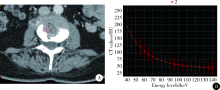
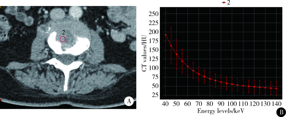
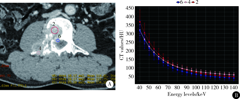
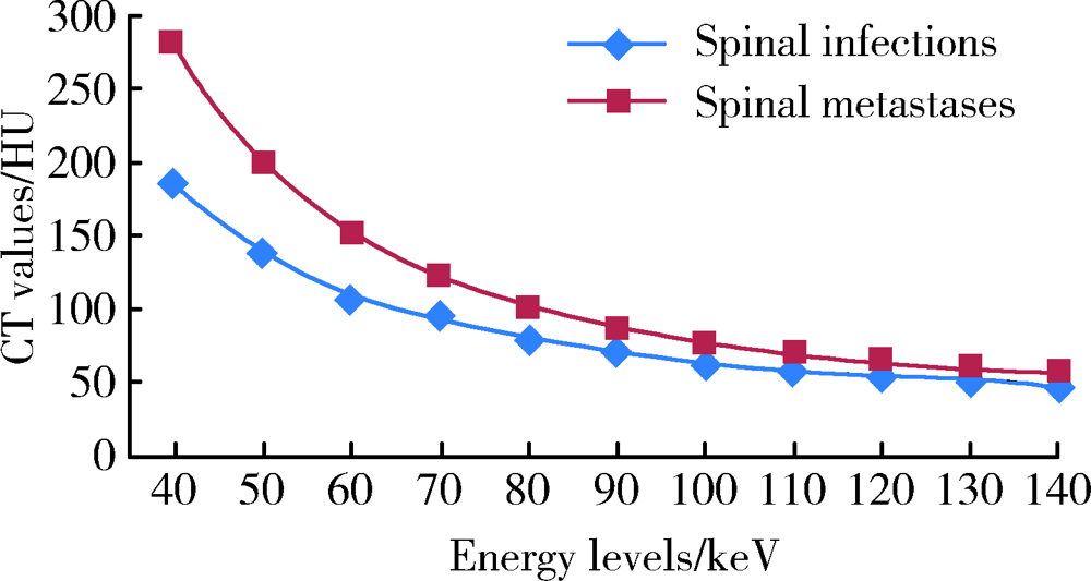
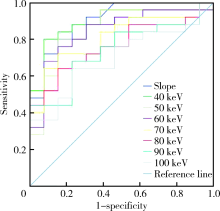
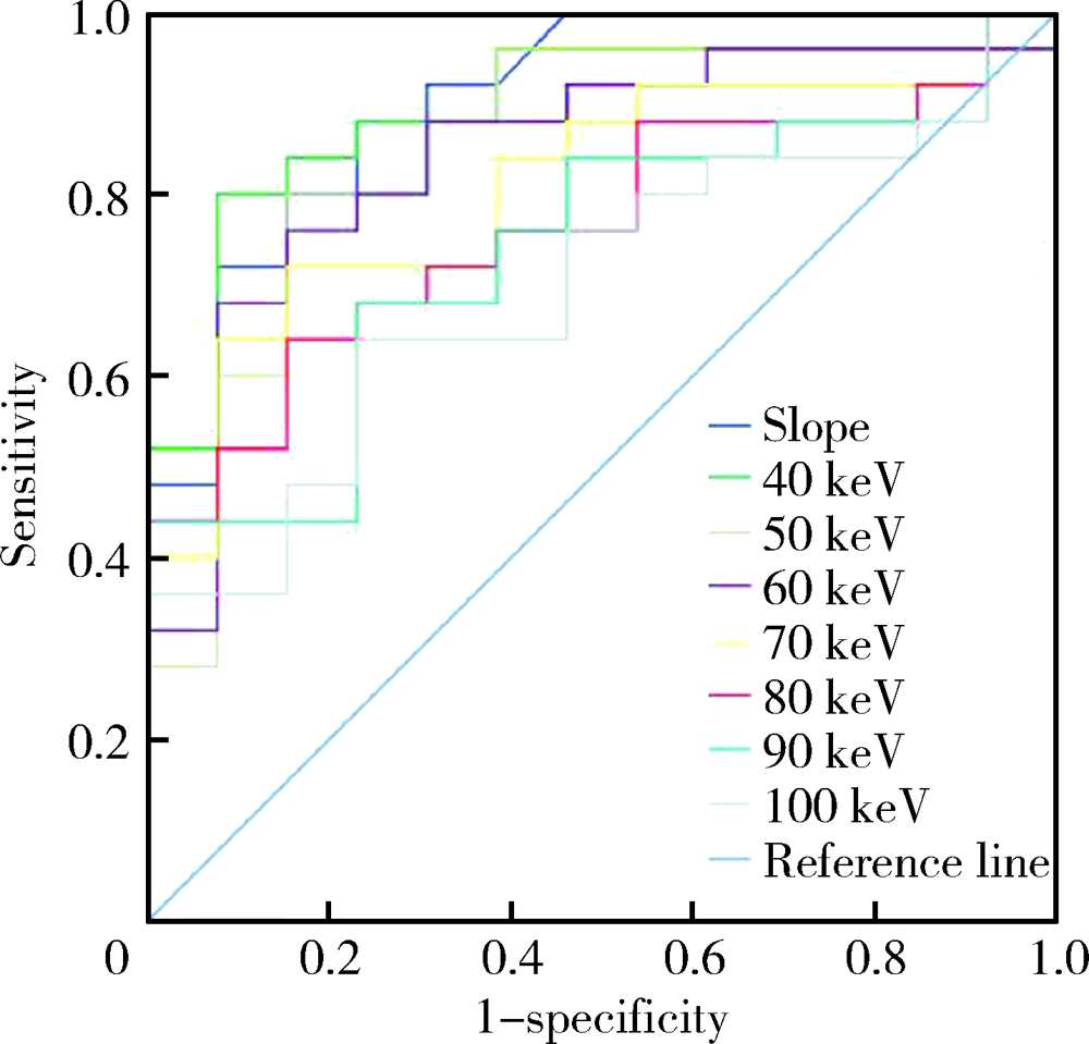
CT spectral curve in differentiating spinal tumor metastasis and infections
YUAN Yuan,LANG Ning,YUAN Hui-shu( )
)
- Department of Radiology, Peking University Third Hospital, Beijing 100191, China
CLC Number:
- R738.1


| [1] |
Lee SH, Lee JM, Kim KW, et al. Dual-energy computed tomography to assess tumor response to hepatic radiofrequency ablation: potential diagnostic value of virtual noncontrast images and iodine maps[J]. Invest Radiol, 2011,46(2):77-84.
doi: 10.1097/RLI.0b013e3181f23fcd pmid: 20856125 |
| [2] |
Lang N, Yuan H, Yu HJ, et al. Diagnosis of spinal lesions using heuristic and pharmacokinetic parameters measured by dynamic contrast-enhanced MRI[J]. Acad Radiol, 2017,24(7):867-875.
doi: 10.1016/j.acra.2016.12.014 pmid: 28162875 |
| [3] |
Babic M, Simpfendorfer CS. Infections of the Spine[J]. Infect Dis Clin North Am, 2017,31(2):279-297.
doi: 10.1016/j.idc.2017.01.003 pmid: 28366222 |
| [4] |
Dong Y, Zheng S, Machida H, et al. Differential diagnosis of osteoblastic metastases from bone islands in patients with lung cancer by single-source dual-energy CT: advantages of spectral CT imaging[J]. Eur J Radiol, 2015,84(5):901-907.
doi: 10.1016/j.ejrad.2015.01.007 pmid: 25661696 |
| [5] |
Ko JP, Brandman S, Stember J, et al. Dual-energy computed tomography: concepts, performance, and thoracic applications[J]. J Thorac Imaging, 2012,27(1):7-22.
doi: 10.1097/RTI.0b013e31823fe0e9 pmid: 22189245 |
| [6] |
Flais J, Coiffier G, Brillet E, et al. Atypical presentation of spine bone metastasis in prostate cancer mimicking Pott’s disease[J]. Clin Cases Miner Bone Metab, 2017,14(2):239-240.
doi: 10.11138/ccmbm/2017.14.1.239 pmid: 29263741 |
| [7] | 袁源, 张艳, 郎宁, 等. CT能谱曲线鉴别诊断脊柱肿瘤及肿瘤样病变[J]. 中国医学影像技术, 2015,31(4):600-603. |
| [8] |
Avrin DE, Macovski A, Zatz LE. Clinical application of Compton and photo-electric reconstruction in computed tomography: preliminary results[J]. Invest Radiol, 1978,13(3):217-222.
doi: 10.1097/00004424-197805000-00007 pmid: 711396 |
| [9] | Dilmanian FA. Computed tomography with monochromatic X rays[J]. Am J Physiol Imaging, 1992,7(3/4):175-193. |
| [10] |
Riederer SJ, Mistretta CA. Selective iodine imaging using K-edge energies in computerized X-ray tomography[J]. Med Phys, 1977,4(6):474-481.
doi: 10.1118/1.594357 pmid: 927384 |
| [11] |
Silva AC, Morse BG, Hara AK, et al. Dual-energy (spectral) CT: applications in abdominal imaging[J]. Radiographics, 2011,31(4):1031-1050.
doi: 10.1148/rg.314105159 pmid: 21768237 |
| [12] | 雷立昌, 陈建宇. 能谱CT的临床应用与研究进展[J]. 中国医学影像技术, 2013,29(1):146-149. |
| [13] | 林晓珠, 沈云, 陈克敏. CT能谱成像的基本原理与临床应用研究进展[J]. 中华放射学杂志, 2011,45(8):798-800. |
| [14] | 张靖, 周新社. 脊柱肿瘤的诊断和外科分期研究进展[J]. 中华全科医学, 2011,9(2):277-279. |
| [15] |
Go SW, Lee HY, Lim CH, et al. Atypical disseminated skeletal tuberculosis mimicking metastasis on PET-CT and MRI[J]. Intern Med, 2012,51(20):2961-2965.
doi: 10.2169/internalmedicine.51.8347 pmid: 23064577 |
| [16] |
Mittal S, Khalid M, Sabir AB, et al. Comparison of magnetic resonance imaging findings between pathologically proven cases of atypical tubercular spine and tumour metastasis: A retrospective study in 40 patients[J]. Asian Spine J, 2016,10(4):734-743.
doi: 10.4184/asj.2016.10.4.734 pmid: 27559455 |
| [17] |
Sezgin B, Atilganoglu U, Yigit O, et al. Concomitant cutaneous metastatic tuberculous abscesses and multifocal skeletal tuberculosis[J]. Indian J Dermatol, 2008,53(3):149-153.
doi: 10.4103/0019-5154.43208 pmid: 19882018 |
| [18] |
Chang DS, Rafii M, McGuinness G, et al. Primary multifocal tuberculous osteomyelitis with involvement of the ribs[J]. Skeletal Radiol, 1998,27(11):641-645.
doi: 10.1007/s002560050451 pmid: 9867183 |
| [19] |
Lang N, Su MY, Yu HJ, et al. Differentiation of tuberculosis and metastatic cancer in the spine using dynamic contrast-enhanced MRI[J]. Eur Spine J, 2015,24(8):1729-1737.
doi: 10.1007/s00586-015-3851-z pmid: 25749725 |
| [20] |
Zheng S, Dong Y, Miao Y, et al. Differentiation of osteolytic metastases and Schmorl’s nodes in cancer patients using dual-energy CT: Advantage of spectral CT imaging[J]. Eur J Radiol, 2014,83(7):1216-1221.
doi: 10.1016/j.ejrad.2014.02.003 pmid: 24820064 |
| [21] | Gupta S, Wagner-Bartak N, Jensen CT, et al. Dual-energy CT of pancreatic adenocarcinoma: Reproducibility of primary tumor measurements and assessment of tumor conspicuity and margin sharpness[J]. Abdom Radiol (NY), 2016,41(7):1317-1324. |
| [22] |
Ramon A, Bohm-Sigrand A, Pottecher P, et al. Role of dual-energy CT in the diagnosis and follow-up of gout: systematic analysis of the literature[J]. Clin Rheumatol, 2018,37(3):587-595.
doi: 10.1007/s10067-017-3976-z pmid: 29350330 |
| [1] | Doudou MA, Xiaocai MA, Tianjing CHANG, Lifang WANG, Yan DING, Lianjie SHI. Bone marrow infiltration of large B-cell lymphoma with clinical manifestations similar to systemic lupus erythematosus: A case report [J]. Journal of Peking University (Health Sciences), 2026, 58(3): 666-669. |
| [2] | Di GAN, Qiang FU, Xiaohui TANG, Chuwei LI, Zhaoping SHU. Gout of the manubriosternal joints: A case report [J]. Journal of Peking University (Health Sciences), 2026, 58(3): 670-673. |
| [3] | Ye ZHAO, Xiaoli DIAO, Yan XIONG. Application of cell transfer technology in pathological diagnosis of micro-volume cell fluid [J]. Journal of Peking University (Health Sciences), 2026, 58(1): 208-213. |
| [4] | Yue WANG, Yuhong LIANG. Florid cemento-osseous dysplasia: A case report [J]. Journal of Peking University (Health Sciences), 2026, 58(1): 220-224. |
| [5] | Yanting CHI, Hongjie JIANG, Yan CHEN, Zhixiu XU, Binbin LI. Value of direct immunofluorescence in the diagnosis of oral mucosal pemphigus vulgaris: A retrospective study based on multi-index combined analysis [J]. Journal of Peking University (Health Sciences), 2026, 58(1): 68-73. |
| [6] | Jingyan GU, Xinyi LI, Jinxia ZHAO, Rong MU. Diabetic Charcot neuroarthropathy initially misdiagnosed as rheumatoid arthritis and gout: A case report [J]. Journal of Peking University (Health Sciences), 2025, 57(6): 1193-1197. |
| [7] | Xiaodi XIAO, Youchen XIA, Jianying LIU, Peng FU. Left sided sternocleidomastoid interosseous intravascular papillary endothelial hyperplasia: A case report [J]. Journal of Peking University (Health Sciences), 2025, 57(5): 1002-1004. |
| [8] | Xiangyu SUN, Chao YUAN, Xinzhu ZHOU, Jing DIAO, Shuguo ZHENG. Application of salivary micro-ecosystem in early prevention and control of oral and systemic diseases [J]. Journal of Peking University (Health Sciences), 2025, 57(5): 859-863. |
| [9] | Jie LIU, Mingwei MA, Qing'an WANG, Ming SHI, Jinpeng YIN, Zhanping WANG, Jingtao SHEN, Xianshu GAO. Comparison of setup errors between two immobilization methods in prostate cancer radiotherapy based on cone-beam computed tomography [J]. Journal of Peking University (Health Sciences), 2025, 57(4): 692-697. |
| [10] | Yuan NING, Xiaoying ZHANG, Xue LI, Yuan LI, Jing HE, Yuebo JIN. Sjögren disease complicated by primary breast lymphoma: A case report [J]. Journal of Peking University (Health Sciences), 2025, 57(4): 808-811. |
| [11] | Peng ZHONG, Xiaodan HU, Zhenzhou WANG. Optical coherence tomography angiography and microvessel density quantification in penumbra after traumatic brain injury in rats [J]. Journal of Peking University (Health Sciences), 2025, 57(2): 262-266. |
| [12] | Jiahui YE, Shimin WANG, Zixuan WANG, Yunsong LIU, Yuchun SUN, Hongqiang YE, Yongsheng ZHOU. Comparison of two registration methods for constructing virtual craniodentofacial patients based on cone beam computed tomography images [J]. Journal of Peking University (Health Sciences), 2025, 57(2): 354-359. |
| [13] | Yuqi MA, Yuhong LIANG. In vitro study of using single cone obturation technique in artificial canals with an isthmus [J]. Journal of Peking University (Health Sciences), 2025, 57(2): 369-375. |
| [14] | Zhao CHEN, Yongkang QIU, Lei KANG. Classical Sweet syndrome with multiple organ lesions by 18F-FDG PET/CT: A case report [J]. Journal of Peking University (Health Sciences), 2025, 57(2): 403-407. |
| [15] | Guangyan YU, Xin PENG, Min GAO, Peng YE, Na GE, Mengqi JIA, Bingyu LI, Zunan TANG, Leihao HU, Wenbo ZHANG. Research progress in diagnosis and treatment of salivary gland tumors [J]. Journal of Peking University (Health Sciences), 2025, 57(1): 1-6. |
|
||