Journal of Peking University (Health Sciences) ›› 2023, Vol. 55 ›› Issue (6): 1097-1104. doi: 10.19723/j.issn.1671-167X.2023.06.022

Previous Articles     Next Articles

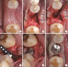
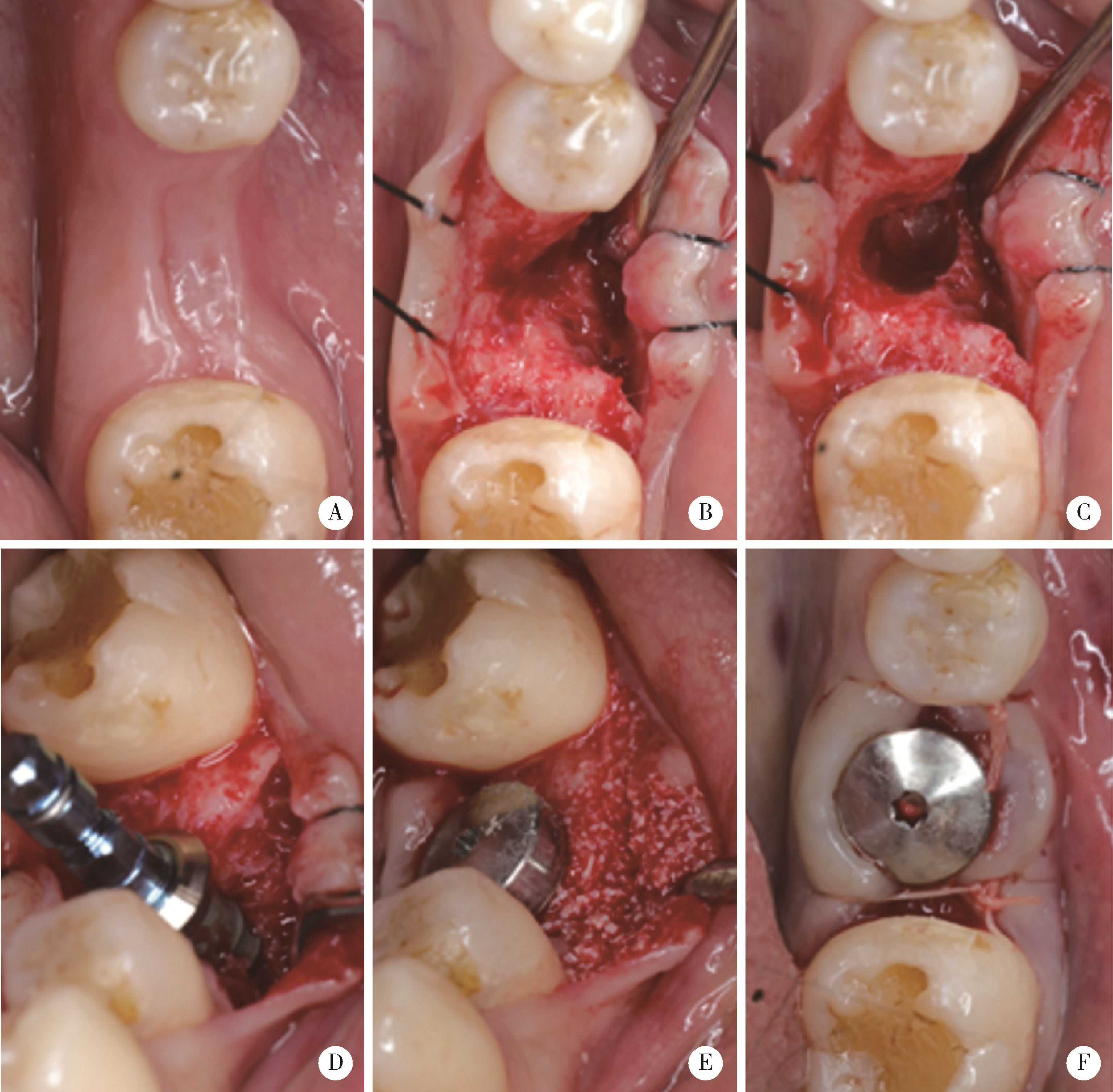
Role of collagen membrane in modified guided bone regeneration surgery using buccal punch flap approach: A retrospective and radiographical cohort study

Deng-hui DUAN1,Hom-Lay WANG2,En-bo WANG1,*()   

  1. 1. Department of Oral and Maxillofacial Surgery, Peking University School and Hospital of Stomatology & National Center for Stomatology & National Clinical Research Center for Oral Diseases & National Engineering Research Center of Oral Biomaterials and Digital Medical Devices & Beijing Key Laboratory of Digital Stomatology, Beijing 100081, China
    2. Department of Periodontics and Oral Medicine, School of Dentistry, University of Michigan, Ann Arbor, MI, 48109, USA
  • Received:2023-06-30 Online:2023-12-18 Published:2023-12-11
  • Contact: En-bo WANG E-mail:ebwang-hlg@163.com

Abstract:

Objective: To investigate whether the placement of absorbable collagen membrane increase the stability of alveolar ridge contour after guided bone regeneration (GBR) using buccal punch flap. Methods: From June 2019 to June 2023, patients who underwent GBR using buccal punch flap simultaneously with a single implant placement in posterior region (from first premolar to second molar) were divided into coverage group, in which particular bone graft was covered by collagen membrane and non-coverage group. Cone beam CT (CBCT) was taken before surgery (T0), immediately after surgery (T1), and 3-7 months after surgery (T2), and the thickness of the buccal bone plate at different levels (0, 2, 4, and 6 mm) below the smooth-rough interface of the implant (BBT-0, -2, -4, -6) was mea-sured after superimposition of CBCT models using Mimics software. Results: A total of 29 patients, including 15 patients in coverage group and 14 patients in non-coverage group, were investigated in this study. At T0, T1, and T2, there was no significant difference in BBT between the two groups (P>0.05). At T1, BBT-0 was (2.50±0.90) mm in the coverage group and (2.97±1.28) mm in the non-coverage group, with corresponding BBT-2 of (3.65±1.08) mm and (3.58±1.26) mm, respectively. At T2, BBT-0 was (1.22±0.55) mm in the coverage group and (1.70±0.97) mm in the non-coverage group, with corresponding BBT-2 of (2.32±0.94) mm and (2.57±1.26) mm, respectively. From T1 to T2, there were no statistically significant differences in the absolute values [(0.47±0.54)-(1.33±0.75) mm] and percentages [(10.04%±24.81%)-(48.43%±18.32%)] of BBT change between the two groups. The thickness of new bone formation in the buccal bone plate from T0 to T2 ranged from (1.27±1.09) mm to (2.75±2.15) mm with no statistical difference between the two groups at all levels. Conclusion: In the short term, the GBR using buccal punch flap with or without collagen membrane coverage can effectively repair the buccal implant bone defect. But collagen membrane coverage showed no additional benefit on alveolar ridge contour stability compared with non-membrane coverage.

Key words: Alveolar bone loss, Guided tissue regeneration, periodontal, Bone regeneration, Cone-beam computed tomography, Collagen membrane

CLC Number: 

  • R782.13

Figure 1

Patient enrollment procedure GBR, guided bone regeneration; CBCT, cone beam CT."

Figure 2

Guided bone regeneration with buccal punch flap and collagen membrane coverage A-C, implant placement in right mandible first molar site with buccal bone defect; D, collagen membrane inserted beneath the buccal flat; E, bone graft material grafted at implant buccal side; F, buccal flap closure with healing abutment exposure."

Figure 3

Guided bone regeneration with buccal punch flap and non-membrane coverage A-D, implant placement in left mandible first molar site with buccal bone defect; E, bone graft material grafted at implant buccal side; F, buccal flap closure with healing abutment exposure."

Figure 4

Measurement of the thickness of the buccal bone plate (green line) at different levels below the smooth-rough interface of the implant (red line) A, measurement of the thickness of the buccal bone plate before surgery (registration with the CBCT mandible and implant model immediately after surgery); B, measurement of buccal bone plate thickness immediately after surgery; C, measurement of buccal bone plate thickness 3-7 months after surgery. BBT-0, -2, -4, -6 means the thickness of buccal bone plates (BBT) at different levels of 0, 2, 4 and 6 mm."

Table 1

The thickness of buccal bone plates at different levels of 0, 2, 4 and 6 mm below the smooth-rough interface of implants"

Items Coverage group (n=15) Non-coverage group (n=14) Differences between groups P
Pre-operation (T0)
  BBT-0/mm -1.36±1.33 -1.04±1.64 -0.32 0.564
  BBT-2/mm -0.28±1.44 0.67±0.92 -0.95 0.045
  BBT-4/mm 0.96±1.52 1.16±1.03 -0.21 0.672
  BBT-6/mm 1.75±1.78 2.02±1.37 -0.26 0.661
Immediately after surgery (T1)
  BBT-0/mm 2.50±0.90 2.97±1.28 -0.47 0.262
  BBT-2/mm 3.65±1.08 3.58±1.26 0.07 0.877
  BBT-4/mm 4.40±1.39 3.82±1.22 0.58 0.247
  BBT-6/mm 4.48±1.58 3.75±1.65 0.73 0.237
3-7 months after surgery (T2)
  BBT-0/mm 1.22±0.55 1.70±0.97 -0.48 0.109
  BBT-2/mm 2.32±0.94 2.57±1.26 -0.25 0.549
  BBT-4/mm 3.45±1.29 3.25±1.52 0.20 0.706
  BBT-6/mm 3.87±1.44 3.28±1.57 0.59 0.302

Table 2

Changes in thickness of buccal bone plates at different levels of 0, 2, 4 and 6 mm under the smooth-rough interface of implants during the healing process"

Items Coverage group (n=15) Non-coverage group (n=14) Differences between groups P
Buccal bone augmentation from T0 to T1
  ABBT-0/mm 3.86±1.43 4.01±2.55 -0.15 0.848
  ABBT-2/mm 3.93±1.23 2.92±1.17 1.01 0.032
  ABBT-4/mm 3.44±1.41 2.66±0.68 0.78 0.071
  ABBT-6/mm 2.72±1.33 1.74±1.17 0.99 0.043
Buccal bone resorption from T1 to T2
  RBBT-0/mm 1.27±0.94 1.26±0.70 0.01 0.970
  RBBT-2/mm 1.33±0.75 1.02±0.62 0.32 0.227
  RBBT-4/mm 0.94±0.75 0.57±0.55 0.38 0.138
  RBBT-6/mm 0.61±0.53 0.47±0.54 0.14 0.495
  RBBT-0% 48.43%±18.32% 42.22%±16.46% 6.21 0.347
  RBBT-2% 37.01%±17.01% 29.43%±15.05% 7.58 0.216
  RBBT-4% 22.00%±15.64% 17.12%±15.55% 4.88 0.407
  RBBT-6% 13.07%±11.19% 10.04%±24.81% 3.03 0.671
New buccal bone formation from T0 to T2
  NBBT-0/mm 2.59±1.49 2.75±2.15 -0.16 0.817
  NBBT-2/mm 2.60±1.30 1.90±1.02 0.70 0.122
  NBBT-4/mm 2.50±1.25 2.09±0.84 0.41 0.315
  NBBT-6/mm 2.12±1.17 1.27±1.09 0.85 0.054
1 Hammerle CH , Jung RE , Feloutzis A . A systematic review of the survival of implants in bone sites augmented with barrier membranes (guided bone regeneration) in partially edentulous patients[J]. J Clin Periodontol, 2002, 29 (Suppl 3): 226- 231.
2 Thoma DS , Bienz SP , Figuero E , et al. Efficacy of lateral bone augmentation performed simultaneously with dental implant placement: A systematic review and meta-analysis[J]. J Clin Perio-dontol, 2019, 46 (Suppl 21): 257- 276.
3 Jung RE , Fenner N , Hämmerle CH , et al. Long-term outcome of implants placed with guided bone regeneration (GBR) using resorbable and non-resorbable membranes after 12-14 years[J]. Clin Oral Implants Res, 2013, 24 (10): 1065- 1073.
doi: 10.1111/j.1600-0501.2012.02522.x
4 Benic GI , Thoma DS , Jung RE , et al. Guided bone regeneration with particulate vs. block xenogenic bone substitutes: A pilot cone beam computed tomographic investigation[J]. Clin Oral Implants Res, 2017, 28 (11): e262- e270.
5 Fu JH , Oh TJ , Benavides E , et al. A randomized clinical trial evaluating the efficacy of the sandwich bone augmentation technique in increasing buccal bone thickness during implant placement surgery: Ⅰ. Clinical and radiographic parameters[J]. Clin Oral Implants Res, 2014, 25 (4): 458- 467.
doi: 10.1111/clr.12171
6 Ye GH , Duan DH , Wang EB . Ridge volume stability of maxillary anterior implants placed with simultaneous lateral guided bone regeneration during healing: A radiographic analysis[J]. Chin J Dent Res, 2021, 24 (4): 251- 256.
7 Wang HL , Boyapati L . "PASS" principles for predictable bone regeneration[J]. Implant Dent, 2006, 15 (1): 8- 17.
doi: 10.1097/01.id.0000204762.39826.0f
8 César Neto JB , Cavalcanti MC , Sapata VM , et al. The positive effect of tenting screws for primary horizontal guided bone regeneration: A retrospective study based on cone-beam computed tomography data[J]. Clin Oral Implants Res, 2020, 31 (9): 846- 855.
doi: 10.1111/clr.13630
9 Farias D , Caceres F , Sanz A , et al. Horizontal bone augmentation in the posterior atrophic mandible and dental implant stability using the tenting screw technique[J]. Int J Periodontics Restorative Dent, 2021, 41 (4): e147- e155.
doi: 10.11607/prd.5137
10 Duan DH , Wang HL , Xiao WC , et al. Bone regeneration using titanium plate stabilization for the treatment of peri-implant bone defects: A retrospective radiologic pilot study[J]. Clin Implant Dent Relat Res, 2022, 24 (6): 792- 800.
doi: 10.1111/cid.13139
11 Ciocca L , Lizio G , Baldissara P , et al. Prosthetically CAD-CAM-guided bone augmentation of atrophic jaws using customized tita-nium mesh: Preliminary results of an open prospective study[J]. J Oral Implantol, 2018, 44 (2): 131- 137.
doi: 10.1563/aaid-joi-D-17-00125
12 Her S , Kang T , Fien MJ . Titanium mesh as an alternative to a membrane for ridge augmentation[J]. J Oral Maxillofac Surg, 2012, 70 (4): 803- 810.
doi: 10.1016/j.joms.2011.11.017
13 Lee SR , Jang TS , Seo CS , et al. Hard tissue volume stability effect beyond the bony envelope of a three-dimensional preformed titanium mesh with two different collagen barrier membranes on peri-implant dehiscence defects in the anterior maxilla: A rando-mized clinical trial[J]. Materials (Basel), 2021, 14 (19): 5618.
doi: 10.3390/ma14195618
14 Sumida T , Otawa N , Kamata YU , et al. Custom-made titanium devices as membranes for bone augmentation in implant treatment: Clinical application and the comparison with conventional titanium mesh[J]. J Craniomaxillofac Surg, 2015, 43 (10): 2183- 2188.
doi: 10.1016/j.jcms.2015.10.020
15 Lin Z , Fateh A , Salem DM , et al. Periosteum: Biology and applications in craniofacial bone regeneration[J]. J Dent Res, 2014, 93 (2): 109- 116.
doi: 10.1177/0022034513506445
16 Duan DH , Wang HL , Wang EB . Effect of intact periosteum on alveolar ridge contour stability after horizontal guided bone regene-ration in posterior region: A retrospective and radiographical cohort study[J]. Chin J Dent Res, 2023, 26 (4): 229- 236.
17 Deng C , Yi Z , Xiong C , et al. Using the intact periosteum for horizontal bone augmentation of peri-implant defects: A retrospective cohort study[J]. Br J Oral Maxillofac Surg, 2022, 60 (10): 1325- 1331.
doi: 10.1016/j.bjoms.2022.09.012
18 Dahlin C , Linde A , Gottlow J , et al. Healing of bone defects by guided tissue regeneration[J]. Plast Reconstr Surg, 1988, 81 (5): 672- 676.
doi: 10.1097/00006534-198805000-00004
19 Dahlin C , Sennerby L , Lekholm U , et al. Generation of new bone around titanium implants using a membrane technique: An experimental study in rabbits[J]. Int J Oral Maxillofac Implants, 1989, 4 (1): 19- 25.
20 Becker W , Becker BE , Handlesman M , et al. Bone formation at dehisced dental implant sites treated with implant augmentation material: A pilot study in dogs[J]. Int J Periodontics Restorative Dent, 1990, 10 (2): 92- 101.
21 Louis PJ , Gutta R , Said-Al-Naief N , et al. Reconstruction of the maxilla and mandible with particulate bone graft and titanium mesh for implant placement[J]. J Oral Maxillofac Surg, 2008, 66 (2): 235- 245.
doi: 10.1016/j.joms.2007.08.022
22 Atef M , Tarek A , Shaheen M , et al. Horizontal ridge augmentation using native collagen membrane vs titanium mesh in atrophic maxillary ridges: Randomized clinical trial[J]. Clin Implant Dent Relat Res, 2020, 22 (2): 156- 166.
doi: 10.1111/cid.12892
23 Urban IA , Saleh MHA , Ravidà A , et al. Vertical bone augmentation utilizing a titanium-reinforced PTFE mesh: A multi-variate analysis of influencing factors[J]. Clin Oral Implants Res, 2021, 32 (7): 828- 839.
doi: 10.1111/clr.13755
24 Benic GI , Bienz SP , Song YW , et al. Randomized controlled clinical trial comparing guided bone regeneration of peri-implant defects with soft-type block versus particulate bone substitutes: Six-month results of hard-tissue changes[J]. J Clin Periodontol, 2022, 49 (5): 480- 495.
doi: 10.1111/jcpe.13606
25 Park SH , Lee KW , Oh TJ , et al. Effect of absorbable membranes on sandwich bone augmentation[J]. Clin Oral Implants Res, 2008, 19 (1): 32- 41.
doi: 10.1111/j.1600-0501.2007.01408.x
26 Spray JR , Black CG , Morris HF , et al. The influence of bone thickness on facial marginal bone response: Stage 1 placement through stage 2 uncovering[J]. Ann Periodontol, 2000, 5 (1): 119- 128.
doi: 10.1902/annals.2000.5.1.119
27 Botticelli D , Berglundh T , Lindhe J . Hard-tissue alterations following immediate implant placement in extraction sites[J]. J Clin Periodontol, 2004, 31 (10): 820- 828.
doi: 10.1111/j.1600-051X.2004.00565.x
28 Severi M , Simonelli A , Farina R , et al. Effect of lateral bone augmentation procedures in correcting peri-implant bone dehiscence and fenestration defects: A systematic review and network meta-analysis[J]. Clin Implant Dent Relat Res, 2022, 24 (2): 251- 264.
doi: 10.1111/cid.13078
29 Park JC , Kim CS , Choi SH , et al. Flap extension attained by vertical and periosteal-releasing incisions: A prospective cohort study[J]. Clin Oral Implants Res, 2012, 23 (8): 993- 998.
doi: 10.1111/j.1600-0501.2011.02244.x
30 Monje A , Pons R , Vilarrasa J , et al. Significance of barrier membrane on the reconstructive therapy of peri-implantitis: A rando-mized controlled trial[J]. J Periodontol, 2023, 94 (3): 323- 335.
doi: 10.1002/JPER.22-0511
[1] Jin-hua ZHANG,Jie PAN,Zhi-peng SUN,Xiao WANG. Effect of various intracanal materials on the diagnostic accuracy of cone-beam computed tomography in vertical root fractures [J]. Journal of Peking University (Health Sciences), 2023, 55(2): 333-338.
[2] Jia-xue YE,Yu-hong LIANG. A prevalence survey of cone-beam computed tomography use among endodontic practitioners [J]. Journal of Peking University (Health Sciences), 2023, 55(1): 114-119.
[3] Meng-qiao PAN,Jian LIU,Li XU,Xiao XU,Jian-xia HOU,Xiao-tong LI,Xiao-xia WANG. A long-term evaluation of periodontal phenotypes before and after the periodontal-orthodontic-orthognathic combined treatment of lower anterior teeth in patients with skeletal Angle class Ⅲ malocclusion [J]. Journal of Peking University (Health Sciences), 2023, 55(1): 52-61.
[4] Yu FU,Xin-nong HU,Sheng-jie CUI,Jie SHI. Decompensation effectiveness and alveolar bone remodeling analysis of mandibular anterior teeth after preoperative orthodontic treatment in high-angle patients with skeletal class Ⅱ malocclusion [J]. Journal of Peking University (Health Sciences), 2023, 55(1): 62-69.
[5] Juan GAO,Hang-miao LV,Hui-min MA,Yi-jiao ZHAO,Xiao-tong LI. Evaluation of root resorption after surgical orthodontic treatment of skeletal Class Ⅲ malocclusion by three-dimensional volumetric measurement with cone-beam CT [J]. Journal of Peking University (Health Sciences), 2022, 54(4): 719-726.
[6] LIU Wei-tao,WANG Yi-ran,WANG Xue-dong,ZHOU Yan-heng. A cone-beam computed tomography evaluation of three-dimensional changes of circummaxillary sutures following maxillary protraction with alternate rapid palatal expansions and constrictions [J]. Journal of Peking University (Health Sciences), 2022, 54(2): 346-355.
[7] Gang YANG,Wen-jie HU,Jie CAO,Deng-gao LIU. Three-dimensional morphology analysis of the supraosseous gingival profile of periodontally healthy maxillary anterior teeth [J]. Journal of Peking University (Health Sciences), 2021, 53(5): 990-994.
[8] YOU Peng-yue,LIU Yu-hua,WANG Xin-zhi,WANG Si-wen,TANG Lin. Biocompatibility and effect on bone formation of a native acellular porcine pericardium: Results of in vitro and in vivo [J]. Journal of Peking University (Health Sciences), 2021, 53(4): 776-784.
[9] WANG Si-wen,YOU Peng-yue,LIU Yu-hua,WANG Xin-zhi,TANG Lin,WANG Mei. Efficacy of two barrier membranes and deproteinized bovine bone mineral on bone regeneration in extraction sockets: A microcomputed tomographic study in dogs [J]. Journal of Peking University (Health Sciences), 2021, 53(2): 364-370.
[10] MENG Yuan,ZHANG Li-qi,ZHAO Ya-ning,LIU Deng-gao,ZHANG Zu-yan,GAO Yan. Three-dimentional radiographic features of 67 maxillary radicular cysts [J]. Journal of Peking University (Health Sciences), 2021, 53(2): 396-401.
[11] ZHOU Jing,LIU Yi. Cone-beam CT evaluation of temporomandibular joint in skeletal class Ⅱ female adolescents with different vertical patterns [J]. Journal of Peking University (Health Sciences), 2021, 53(1): 109-119.
[12] GAO Lu,GU Yan. Chinese morphological stages of midpalatal suture and its correlation with Demirjian dental age [J]. Journal of Peking University (Health Sciences), 2021, 53(1): 133-138.
[13] Wei-yi WU,Bo-wen LI,Yu-hua LIU,Xin-zhi WANG. Biodegradation properties of multi-laminated small intestinal submucosa [J]. Journal of Peking University (Health Sciences), 2020, 52(3): 564-569.
[14] Dong SHI,Jie CAO,Shi-ai DAI,Huan-xin MENG. Short-term outcome of regenerative surgery treating peri-implantitis [J]. Journal of Peking University(Health Sciences), 2020, 52(1): 58-63.
[15] WANG Yi-ran, ZHOU Yan-heng, WANG Xue-dong, WEI Song, LIU Wei-tao. Evaluation of maxillary three-dimensional changes in maxillary protraction with alternating rapid palatal expansion and constriction based on the cone-beam computed tomography [J]. Journal of Peking University(Health Sciences), 2018, 50(4): 685-693.
Viewed
Full text


Abstract

Cited

  Shared   
  Discussed   
No Suggested Reading articles found!