Journal of Peking University (Health Sciences) ›› 2021, Vol. 53 ›› Issue (5): 990-994. doi: 10.19723/j.issn.1671-167X.2021.05.030
Previous Articles Next Articles
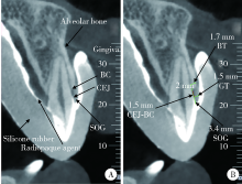
Three-dimensional morphology analysis of the supraosseous gingival profile of periodontally healthy maxillary anterior teeth
YANG Gang1,HU Wen-jie1,△( ),CAO Jie1,LIU Deng-gao2
- 1. Department of Periodontology, Beijing 100081, China
2. Department of Radiology, Peking University School and Hospital of Stomatology & National Center of Stomatology & National Clinical Research Center for Oral Diseases & National Engineering Laboratory for Digital and Material Technology of Stomatology, Beijing 100081, China
CLC Number:
- R783

| [1] |
Arora R, Narula S, Sharma R, et al. Supracrestal gingival tissue: Assessing relation with periodontal biotypes in a healthy periodon-tium [J]. Int J Periodontics Restorative Dent, 2013, 33(6):763-771.
doi: 10.11607/prd.1501 |
| [2] |
Perez JR, Smukler H, Nunn M E. Clinical evaluation of the supraosseous gingivae before and after crown lengthening [J]. J Periodontol, 2007, 78(6):1023-1030.
pmid: 17539715 |
| [3] |
Gargiulo AW, Wentz FM, Orban B. Dimensions and relations of the dentogingival junction in humans [J]. J Periodontol, 1961, 32(3):261-267.
doi: 10.1902/jop.1961.32.3.261 |
| [4] |
Kois JC. Altering gingival levels: The restorative connection part I: Biologic variables [J]. J Esthet Restor Dent, 1994, 6(1):3-7.
doi: 10.1111/j.1708-8240.1994.tb00825.x |
| [5] | Vacek JS, Gher ME, Assad DA, et al. The dimensions of the human dentogingival junction [J]. Int J Periodontics Restorative Dent, 1994, 14(2):154-165. |
| [6] |
Fischer KR, Grill E, Jockel-Schneider Y, et al. On the relationship between gingival biotypes and supracrestal gingival height, crown form and papilla height [J]. Clin Oral Implants Res, 2014, 25(8):894-898.
doi: 10.1111/clr.2014.25.issue-8 |
| [7] | 乐迪, 张豪, 胡文杰, 等. 牙周探诊法判断牙龈生物型的初步研究 [J]. 中华口腔医学杂志, 2012, 47(2):81-84. |
| [8] | 曹洁, 胡文杰, 张豪, 等. 基于锥形束计算机体层摄影术测量牙龈厚度 [J]. 北京大学学报(医学版), 2013, 45(1):135-139. |
| [9] | 张艳玲, 张豪, 胡文杰, 等. 120名汉族青年前段牙弓唇侧角化龈宽度的测量 [J]. 中华口腔医学杂志, 2010, 45(8):477-481. |
| [10] |
Zhang YL, Le D, Hu WJ, et al. Assessment of dynamic smile and gingival contour in young Chinese people [J]. Int Dent J, 2015, 65(4):182-187.
doi: 10.1111/idj.12174 |
| [11] |
Perez JR, Smukler H, Nunn ME. Clinical dimensions of the supraosseous gingivae in healthy periodontium [J]. J Periodontol, 2008, 79(12):2267-2272.
doi: 10.1902/jop.2008.080101 pmid: 19053916 |
| [12] |
Cao J, Hu WJ, Zhang H, et al. A novel technique for measurement of dentogingival tissue by cone beam computed tomography [J]. Oral Surg Oral Med Oral Pathol Oral Radiol, 2015, 119(2):e82-e87.
doi: 10.1016/j.oooo.2014.10.022 |
| [13] |
Alves PHM, Alves TCLP, Pegoraro TA, et al. Measurement pro-perties of gingival biotype evaluation methods [J]. Clin Implant Dent Relat Res, 2018, 20(3):280-284.
doi: 10.1111/cid.2018.20.issue-3 |
| [14] |
Hausmann E, Allen K, Clerehugh V. What alveolar crest level on a bite-wing radiograph represents bone loss? [J]. J Periodontol, 1991, 62(9):570-572.
pmid: 1941497 |
| [15] |
Ghassemian M, Nowzari H, Lajolo C, et al. The thickness of facial alveolar bone overlying healthy maxillary anterior teeth [J]. J Periodontol, 2012, 83(2):187-197.
doi: 10.1902/jop.2011.110172 pmid: 21692627 |
| [16] |
Nowzari H, Molayem S, Chiu CH, et al. Cone beam computed tomographic measurement of maxillary central incisors to determine prevalence of facial alveolar bone width ≥2 mm [J]. Clin Implant Dent Relat Res, 2012, 14(4):595-601.
doi: 10.1111/cid.2012.14.issue-4 |
| [17] |
Taylor R. Interpretation of the correlation coefficient: A basic review [J]. J Diagn Med Sonogr, 1990, 6(1):35-39.
doi: 10.1177/875647939000600106 |
| [18] | Kao RT, Fagan MC, Conte GJ. Thick vs thin gingival biotypes: A key determinant in treatment planning for dental implants [J]. J Calif Dent Assoc, 2008, 36(3):193-198. |
| [19] |
Müller HP, Könönen E. Variance components of gingival thickness [J]. J Periodontal Res, 2005, 40(3):239-244
pmid: 15853970 |
| [20] |
Fu JH, Yeh CY, Chan HL, et al. Tissue biotype and its relation to the underlying bone morphology [J]. J Periodontol, 2010, 81(4):569-574.
doi: 10.1902/jop.2009.090591 |
| [21] |
La Rocca AP, Alemany AS, Levi P Jr. Anterior maxillary and mandibular biotype: Relationship between gingival thickness and width with respect to underlying bone thickness [J]. Implant Dent, 2012, 21(6):507-515.
doi: 10.1097/ID.0b013e318271d487 |
| [22] | Frumkin N, Via S, Klinger A. Evaluation of the width of the alveolar bone in subjects with different gingival biotypes: A prospective cohort study using cone beam computed tomography [J]. Quintessence Int, 2017, 48(3):209-216. |
| [23] | Cook DR, Mealey BL, Verrett RG, et al. Relationship between clinical periodontal biotype and labial plate thickness: An in vivo study [J]. Int J Periodontics Restorative Dent, 2011, 31(4):345-354. |
| [24] |
Batista EL, Moreira CC, Batista FC, et al. Altered passive eruption diagnosis and treatment: A cone beam computed tomography-based reappraisal of the condition [J]. J Clin Periodontol, 2012, 39(11):1089-1096.
doi: 10.1111/j.1600-051X.2012.01940.x pmid: 22966787 |
| [1] | Yuxuan TIAN,Mingjian RUAN,Yi LIU,Derun LI,Jingyun WU,Qi SHEN,Yu FAN,Jie JIN. Predictive effect of the dual-parametric MRI modified maximum diameter of the lesions with PI-RADS 4 and 5 on the clinically significant prostate cancer [J]. Journal of Peking University (Health Sciences), 2024, 56(4): 567-574. |
| [2] | Shishi BO,Chengzhi GAO. Tooth segmentation and identification on cone-beam computed tomography with convolutional neural network based on spatial embedding information [J]. Journal of Peking University (Health Sciences), 2024, 56(4): 735-740. |
| [3] | Yuru HU,Juan LIU,Wenjing LI,Yibing ZHAO,Qiqiang LI,Ruifang LU,Huanxin MENG. Relationship between short-chain fatty acids in the gingival crevicular fluid and periodontitis of stage Ⅲ or Ⅳ [J]. Journal of Peking University (Health Sciences), 2024, 56(2): 332-337. |
| [4] | Liang LYU,Mingjin ZHANG,Aonan WEN,Yijiao ZHAO,Yong WANG,Jing LI,Gengchen YANG,Dawei LIU. Preliminary evaluation of chin symmetry with three dimentional soft tissue spatial angle wireframe template [J]. Journal of Peking University (Health Sciences), 2024, 56(1): 106-110. |
| [5] | Bochun MAO,Yajing TIAN,Xuedong WANG,Jing LI,Yanheng ZHOU. Soft and hard tissue changes of hyperdivergent class Ⅱ patients before and after orthodontic extraction treatment [J]. Journal of Peking University (Health Sciences), 2024, 56(1): 111-119. |
| [6] | Xiaotong LING,Liuyang QU,Danni ZHENG,Jing YANG,Xuebing YAN,Denggao LIU,Yan GAO. Three-dimensional radiographic features of calcifying odontogenic cyst and calcifying epithelial odontogenic tumor [J]. Journal of Peking University (Health Sciences), 2024, 56(1): 131-137. |
| [7] | Jiayun DONG,Xuefen LI,Ruifang LU,Wenjie HU,Huanxin MENG. Histopathological characteristics of peri-implant soft tissue in reconstructed jaws with vascularized bone flaps [J]. Journal of Peking University (Health Sciences), 2024, 56(1): 25-31. |
| [8] | Deng-hui DUAN,Hom-Lay WANG,En-bo WANG. Role of collagen membrane in modified guided bone regeneration surgery using buccal punch flap approach: A retrospective and radiographical cohort study [J]. Journal of Peking University (Health Sciences), 2023, 55(6): 1097-1104. |
| [9] | Yi LIU,Chang-wei YUAN,Jing-yun WU,Qi SHEN,Jiang-xi XIAO,Zheng ZHAO,Xiao-ying WANG,Xue-song LI,Zhi-song HE,Li-qun ZHOU. Diagnostic efficacy of prostate cancer using targeted biopsy with 6-core systematic biopsy for patients with PI-RADS 5 [J]. Journal of Peking University (Health Sciences), 2023, 55(5): 812-817. |
| [10] | Chang-wei YUAN,De-run LI,Zhi-hua LI,Yi LIU,Gang-zhi SHAN,Xue-song LI,Li-qun ZHOU. Application of dynamic contrast enhanced status in multiparametric magnetic resonance imaging for prostatic cancer with PI-RADS 4 lesion [J]. Journal of Peking University (Health Sciences), 2023, 55(5): 838-842. |
| [11] | Zhan-yi ZHANG,Fan ZHANG,Ye YAN,Cai-guang CAO,Chang-jian LI,Shao-hui DENG,Yue-hao SUN,Tian-liang HUANG,Yun-he GUAN,Nan LI,Min LU,Zhen-hua HU,Shu-dong ZHANG. Near-infrared targeted probe designed for intraoperative imaging of prostatic neurovascular bundles [J]. Journal of Peking University (Health Sciences), 2023, 55(5): 843-850. |
| [12] | Zhuo-hua LIN,Ru-yi CAI,Yang SUN,Rong MU,Li-gang CUI. Methodology and clinical use of superb microvascular imaging in assessing micro-circulation changes of fingertips in systemic sclerosis [J]. Journal of Peking University (Health Sciences), 2023, 55(4): 636-640. |
| [13] | Ying LIU,Ran HUO,Hui-min XU,Zheng WANG,Tao WANG,Hui-shu YUAN. Correlations between plaque characteristics and cerebral blood flow in patients with moderate to severe carotid stenosis using magnetic resonance vessel wall imaging [J]. Journal of Peking University (Health Sciences), 2023, 55(4): 646-651. |
| [14] | Qiang FU,Guan-ying GAO,Yan XU,Zhuo-hua LIN,You-jing SUN,Li-gang CUI. Comparative study of ultrasound and magnetic resonance imaging in the diagnosis of asymptomatic anterosuperior acetabular labrum tears [J]. Journal of Peking University (Health Sciences), 2023, 55(4): 665-669. |
| [15] | Xiang LIU,Hui-hui XIE,Yu-feng XU,Xiao-dong ZHANG,Xiao-feng TAO,Lin LIU,Xiao-ying WANG. Value of artificial intelligence in the improvement of diagnostic consistency of radiology residents [J]. Journal of Peking University (Health Sciences), 2023, 55(4): 670-675. |
|
||