Journal of Peking University (Health Sciences) ›› 2024, Vol. 56 ›› Issue (1): 25-31. doi: 10.19723/j.issn.1671-167X.2024.01.005

Previous Articles     Next Articles

Histopathological characteristics of peri-implant soft tissue in reconstructed jaws with vascularized bone flaps

Jiayun DONG,Xuefen LI,Ruifang LU*(),Wenjie HU,Huanxin MENG   

  1. Department of Periodontology, Peking University School and Hospital of Stomatology & National Center for Stomatology & National Clinical Research Center for Oral Diseases & National Engineering Research Center of Oral Biomaterials and Digi-tal Medical Devices, Beijing 100081, China
  • Received:2023-10-10 Online:2024-02-18 Published:2024-02-06
  • Contact: Ruifang LU E-mail:kqrflu@bjmu.edu.cn
  • Supported by:
    the National Natural Science Foundations of China(82071116);the Clinical Research Foundation of Peking University School and Hospital of Stomatology(PKUSS-2023CRF305)

RICH HTML


Abstract:

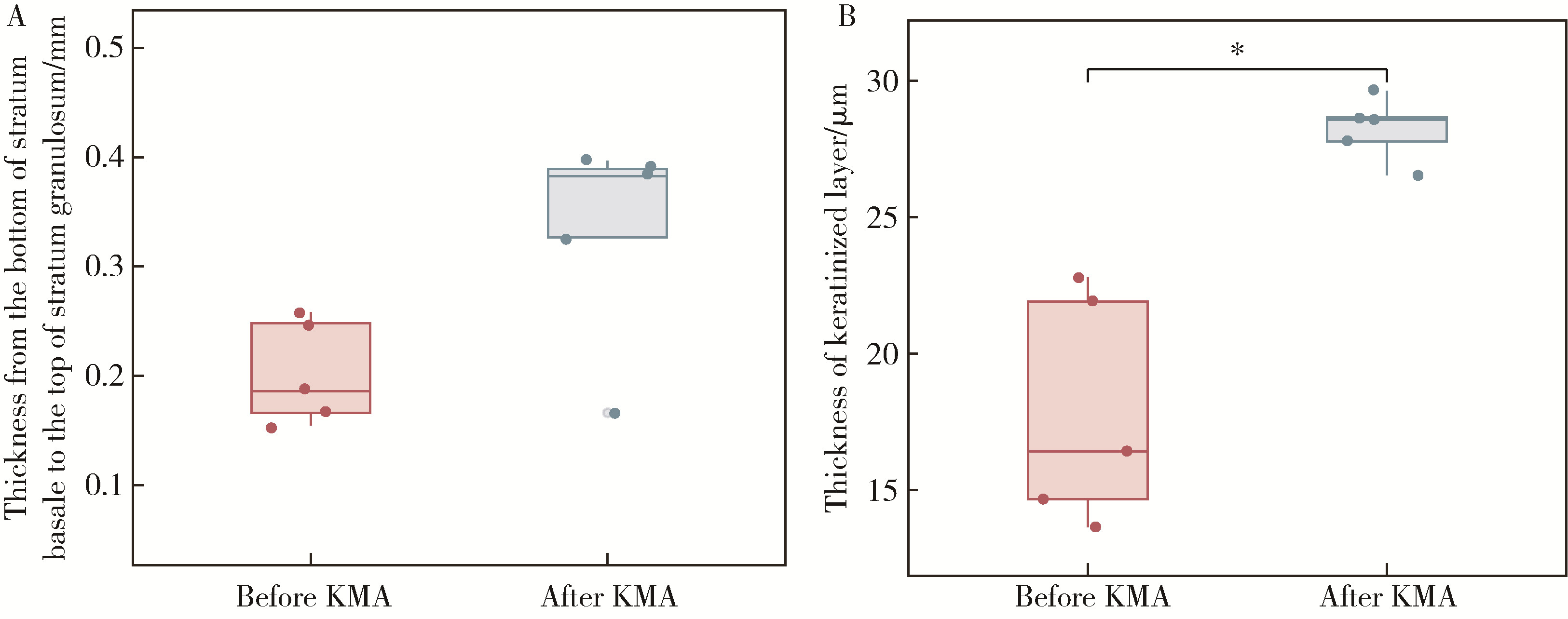
Objective: To analyze the histopathological characteristics of peri-implant soft tissue in reconstructed jaws and the changes after keratinized mucosa augmentation (KMA) with free gingival graft (FGG). Methods: Twenty patients were enrolled in this study. Five patients of them, who were periodontal and systemic healthy and referred for crown lengthening before restoration with healthy keratinized gingiva collected were enrolled as healthy controls. 15 patients of them were with fibula or iliac bone flaps jaw reconstruction (10 with fibula flap and 5 with iliac flap), who were referred to FGG and implant exposures before restoration. Soft tissue was collected before FGG in reconstructed jaws, and in 5 patients (3 with fibula flap and 2 with iliac flap) 8 weeks after FGG if a second surgery was conducted. Histological analysis with hematoxylin-eosin stain and immunological analysis to interlukin-1 (IL-1), interlukin-6 (IL-6) and tumor necrosis factor-α (TNF-α) were performed. Results: Thickness from the bottom of stratum basale to the top of stratum granulosum and thickness of keratinized layer in reconstructed jaws were significantly lower compared with that of natural healthy keratinized gingiva [0.27 (0.20, 0.30) mm vs. 0.36 (0.35, 0.47) mm, P<0.05; 16.49 (14.90, 23.37) μm vs. 26.37 (24.12, 31.53) μm, P<0.05]. In the reconstructed area, thickness from the bottom of stratum basale to the top of stratum granulosum increased after KMA with FGG [0.19 (0.16, 0.25) mm vs. 0.38 (0.25, 0.39) mm, P=0.059] and the thickness of keratinized layer significantly increased after KMA with FGG [16.42 (14.16, 22.35) μm vs. 28.57 (27.16, 29.14) μm, P<0.05], which was similar to that in the control group. Furthermore, the number of positive cells of IL-1, IL-6 and TNF-α significantly increased after KMA [0.67 (0.17, 8.93) vs. 11.00 (9.16, 18.00); 13.00 (8.50, 14.14) vs. 21.89 (15.00, 28.12); 0.22 (0.04, 0.63) vs. 2.83 (1.68, 5.00), respectively, P<0.05] as well as the average optical density value [0.15 (0.14, 0.17) vs. 0.18 (0.17, 0.21); 0.28 (0.26, 0.33) vs. 0.36 (0.33, 0.37); 0.23 (0.22, 0.29) vs. 0.30 (0.28, 0.42), respectively, P<0.05], which was similar to that in the healthy keratinized gingiva. Conclusion: The lack of rete pegs and inflammatory factors were common in soft tissue with jaw reconstruction. FGG can improve the quality of the epithelium and may improve the stability of the mucosa around implants.

Key words: Jaw reconstruction, Vascularized bone flap, Soft tissue, Free gingival graft

CLC Number: 

  • R781.4

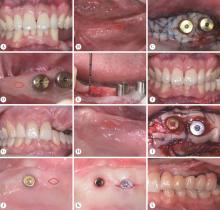
Figure 1

Soft tissue collection in reconstructed jaws A, B, soft tissue of left mandibular in reconstructed jaws with fibula bone flap before KMA, the red mark indicates collection of soft tissue before KMA, there is no keratinized tissue before FGG; C, KMA by FGG in both buccal and lingual side of implants; D, peri-implant soft tissue at 8 weeks after KMA, the red mark indicates collection of soft tissue during the second surgery; E, after removing the soft tissue; F, final restoration; G, H, soft tissue of left maxilla in reconstructed jaws with iliac bone flap before KMA, there is no keratinized tissue before FGG, the red mark indicates collection of soft tissue before KMA; I, KMA by FGG in both buccal and palatal side of implants; J, peri-implant soft tissue at 8 weeks after KMA, the red mark indicates collection of the soft tissue covering the implant during the second surgery; K, exposure of the implant after removing the soft tissue covering the implant; L, prothesis screwed into the implants. KMA, keratinized mucosa augmentation; FGG, free gingival graft."

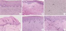
Figure 2

Histopathological characteristics of healthy keratinized gingiva and soft tissue in reconstructed jaws before and after KMA A, healthy keratinized gingiva, presenting normal structures of stratified squamous epithelium (HE ×100); B, soft tissue of a male patient with fibula bone flap before KMA, lack of normal structures of stratified squamous epithelium (HE ×40); C, soft tissue of a female patient with iliac bone flap before KMA, partial vasodilation and congestion in connective tissue(HE ×100); D, soft tissue of a male patient with fibula flap before KMA, irregular arrangement of mucosal epithelial cells (HE ×100); E, soft tissue of a female patient with fibula flap after KMA, forming rete pegs (HE ×40); F, soft tissue of a female patient with fibula flap after KMA, neovascularization and proliferation of fibrous tissue in the lamina propria (HE ×100). KMA, keratinized mucosa augmentation; HE, hematoxylin-eosin stain."

Figure 3

Comparison of the thickness of keratinized layer and thickness from the bottom of stratum basale to the top of stratum granulosum before and after KMA in reconstructed jaws A, comparison of thickness from the bottom of stratum basale to the top of stratum granulosum before and after KMA in reconstructed jaws; B, comparison of the thickness of keratinized layer before and after KMA in reconstructed jaws. * P<0.05. KMA, keratinized mucosa augmentation."

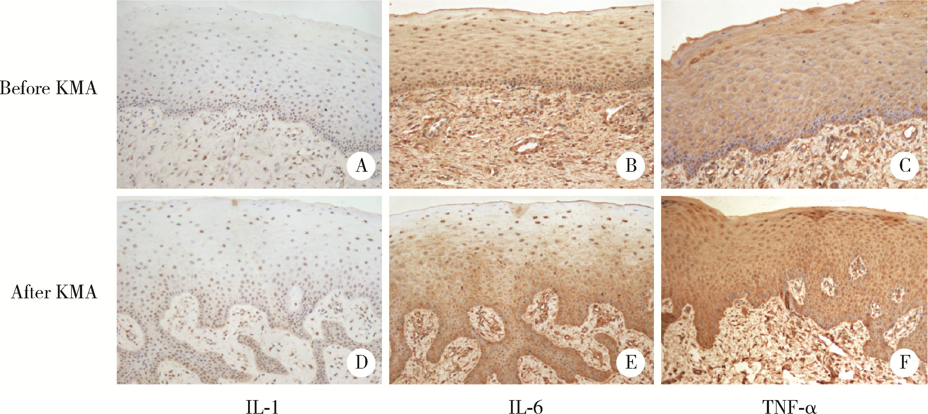
Figure 4

Immunostaining for IL-1, IL-6 and TNF-α before and after KMA of soft tissue in reconstructed jaws A, low expression of IL-1 before KMA (IHC ×100); B, positive immunostaining for IL-6 before KMA (IHC ×100); C, positive immunostaining for TNF-α before KMA (IHC ×100); D, more positive cells (brown cells) in stratum basale, indicating higher expression of IL-1 after KMA (IHC ×100); E, higher expression of IL-6 after KMA (IHC ×100); F, higher expression of TNF-α after KMA (IHC ×100). KMA, keratinized mucosa augmentation; IL-1, interlukin-1; IL-6, interlukin-6; TNF-α, tumor necrosis factor-α; IHC, immunohistochemistry."

Table 1

Comparison of the number of positive cells and AOD of healthy keratinized gingiva and peri-implant soft tissue in reconstructed jaws before and after KMA"

Variables Healthy control (n=5) Before KMA (n=5) After KMA (n=5) P2
M (P25, P75) P1 M (P25, P75) P1
Number of positive cells
  IL-1 13.50 (11.00, 14.60) 0.67 (0.17, 8.93) 0.043* 11.00 (9.16, 18.00) 0.917 0.028*
  IL-6 26.33 (20.17, 28.84) 13.00 (8.50, 14.64) 0.016* 21.89 (15.00, 28.12) 0.251 0.047*
  TNF-α 4.26 (2.75, 5.19) 0.22 (0.04, 0.63) 0.009* 2.83 (1.68, 5.00) 0.347 0.009*
AOD
  IL-1 0.20 (0.18, 0.23) 0.15 (0.14, 0.17) 0.028* 0.18 (0.17, 0.21) 0.465 0.016*
  IL-6 0.33 (0.31, 0.38) 0.28 (0.26, 0.33) 0.175 0.36 (0.33, 0.37) 0.602 0.028*
  TNF-α 0.29 (0.27, 0.35) 0.25 (0.22, 0.29) 0.016* 0.30 (0.28, 0.42) 0.917 0.047*
1 Goker F , Baj A , Bolzoni AR , et al. Dental implant-based oral rehabilitation in patients reconstructed with free fibula flaps: Clinical study with a follow-up 3 to 6 years[J]. Clin Implant Dent Relat Res, 2020, 22 (4): 514- 522.
doi: 10.1111/cid.12928
2 Pellegrino G , Tarsitano A , Ferri A , et al. Long-term results of osseointegrated implant-based dental rehabilitation in oncology patients reconstructed with a fibula free flap[J]. Clin Implant Dent Relat Res, 2018, 20 (5): 852- 859.
doi: 10.1111/cid.12658
3 Sonmez TT , Prescher A , Salama A , et al. Comparative clinico-anatomical study of ilium and fibula as two commonly used bony donor sites for maxillofacial reconstruction[J]. Br J Oral Maxillofac Surg, 2013, 51 (8): 736- 741.
doi: 10.1016/j.bjoms.2013.07.010
4 林野, 邱立新, 胡秀莲, 等. 硬腭游离黏膜移植在种植体周软组织结构重建中的应用[J]. 北京大学学报(医学版), 2007, 39 (1): 21- 25.
5 Shan X , Han D , Ge Y , et al. Clinical outcomes of keratinized mucosa augmentation in jaws reconstructed with fibula or iliac bone flaps[J]. Int J Oral Maxillofac Surg, 2021, 51 (7): 949- 956.
6 Han J , Ulevitch RJ . Limiting inflammatory responses during activation of innate immunity[J]. Nat Immunol, 2005, 6 (12): 1198- 1205.
doi: 10.1038/ni1274
7 Vignoletti F , Nunez J , de Sanctis F , et al. Healing of a xenoge-neic collagen matrix for keratinized tissue augmentation[J]. Clin Oral Implants Res, 2015, 26 (5): 545- 552.
doi: 10.1111/clr.12441
8 Wu T , Xiong X , Zhang W , et al. Morphogenesis of rete ridges in human oral mucosa: A pioneering morphological and immunohistochemical study[J]. Cells Tissues Organs, 2013, 197 (3): 239- 248.
doi: 10.1159/000342926
9 Sa GL , Liu ZK , Ren JG , et al. Keratinocyte growth factor (KGF) induces podosome formation via integrin-Erk1/2 signaling in human immortalized oral epithelial cells[J]. Cell Signal, 2019, 61, 39- 47.
doi: 10.1016/j.cellsig.2019.05.007
10 Sa GL , Xiong XP , Ren JG , et al. KGF enhances oral epithelial adhesion and rete peg elongation via integrins[J]. J Dent Res, 2017, 96 (13): 1546- 1554.
doi: 10.1177/0022034517720360
11 Squier C , Brogden KA . Human oral mucosa: Development, structure and function[M]. Chichester: John Wiley & Sons, 2011: 55- 57.
12 Lawlor K , Kaur P . Dermal contributions to human interfollicular epidermal architecture and self-renewal[J]. Int J Mol Sci, 2015, 16 (12): 28098- 28107.
doi: 10.3390/ijms161226078
13 de Luca M , Albanese E , Megna M , et al. Evidence that human oral epithelium reconstituted in vitro and transplanted onto patients with defects in the oral mucosa retains properties of the original donor site[J]. Transplantation, 1990, 50 (3): 454- 459.
doi: 10.1097/00007890-199009000-00019
14 Henry J , Toulza E , Hsu CY , et al. Update on the epidermal differentiation complex[J]. Front Biosci (Landmark Ed), 2012, 17 (4): 1517- 1532.
15 Nemes Z , Steinert PM . Bricks and mortar of the epidermal barrier[J]. Exp Mol Med, 1999, 31 (1): 5- 19.
doi: 10.1038/emm.1999.2
16 孙凯, 刘丽骏, 张倩倩, 等. Wistar大鼠自然衰老过程中口腔颊黏膜上皮厚度的改变[J]. 中华老年口腔医学杂志, 2018, 16 (6): 333- 335.
doi: 10.3969/j.issn.1672-2973.2018.06.004
17 Sabine E , Groeger JM . Epithelial barrier and oral bacterial infection[J]. Periodontology 2000, 2015, 69 (1): 46- 67.
18 Jandinski JJ . Osteoclast activating factor is now interleukin-1 beta: Historical perspective and biological implications[J]. J Oral Pathol, 1988, 17 (4): 145- 152.
19 Kondo S , Sauder DN . Tumor necrosis factor (TNF) receptor type 1 (p55) is a main mediator for TNF-alpha-induced skin inflammation[J]. Eur J Immunol, 1997, 27 (7): 1713- 1718.
[1] Ying ZHOU,Ning ZHAO,Hongyuan HUANG,Qingxiang LI,Chuanbin GUO,Yuxing GUO. Application of double-layer soft tissue suture closure technique in the surgical treatment of patients with mandible medication-related osteonecrosis of the jaw of early and medium stages [J]. Journal of Peking University (Health Sciences), 2024, 56(1): 51-56.
[2] Ran ZHAO,Yan-qing LIU,Hua TIAN. Cumulative sum control chart analysis of soft tissue balance in total knee replacement assisted by electronic pressure sensor [J]. Journal of Peking University (Health Sciences), 2023, 55(4): 658-664.
[3] Zhong ZHANG,Huan-xin MENG,Jie HAN,Li ZHANG,Dong SHI. Effect of vertical soft tissue thickness on clinical manifestation of peri-implant tissue in patients with periodontitis [J]. Journal of Peking University (Health Sciences), 2020, 52(2): 332-338.
[4] Tian-wen ZHANG,Xiao-xia WANG,Zi-li LI,Biao YI,Cheng LIANG,Xing WANG. Establishment of three-dimensional measurement methods of nasolabial soft tissue for patients with maxillary protrusion [J]. Journal of Peking University(Health Sciences), 2019, 51(5): 944-948.
[5] ZHAO Li-ping, ZHAN Ya-lin, HU Wen-jie, WANG Hao-jie, WEI Yi-ping, ZHEN Min, Xu Tao, LIU Yun-song. Dental implantation and soft tissue augmentation after ridge preservation in a molar site: a case report [J]. Journal of Peking University(Health Sciences), 2016, 48(6): 1090-1094.
Viewed
Full text


Abstract

Cited

  Shared   
  Discussed   
No Suggested Reading articles found!