Journal of Peking University(Health Sciences) ›› 2019, Vol. 51 ›› Issue (5): 944-948. doi: 10.19723/j.issn.1671-167X.2019.05.025
Previous Articles Next Articles
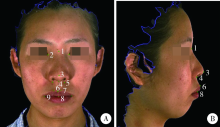
Establishment of three-dimensional measurement methods of nasolabial soft tissue for patients with maxillary protrusion
Tian-wen ZHANG,Xiao-xia WANG( ),Zi-li LI,Biao YI,Cheng LIANG,Xing WANG
- Department of Oral and Maxillofacial Surgery, Peking University School and Hospital of Stomatology & National Clinical Research Center for Oral Diseases & National Engineering Laboratory for Digital and Material Technology of Stomatology & Beijing Key Laboratory of Digital Stomatology, Beijing 100081, China
CLC Number:
- R782.2


| [1] | 李东, 白宇明, 段银钟 , 等. 双颌前突患者正颌手术及拔牙矫治的软组织变化分析[J]. 第四军医大学学报, 2004,25(18):1704-1706. |
| [2] | 张君孝, 乔鸣芳 . 60例广东省双颌前突患者头影测量分析研究[J]. 华西口腔医学杂志, 2001,19(1):32-34. |
| [3] | Ioi H, Nakata S, Nakasima A ,et a1.Effect of facial convexity on antero-posterior lip positions of the most favored Japanese facial profiles[J]. Angle Orthod, 2005,75(3):326-332. |
| [4] |
何颖, 郭传瑸, 邓旭亮 , 等. 北方正常牙合人群颅颌面三维比例测量及面部对称性分析[J]. 北京大学学报(医学版), 2015,47(4):708-713.
doi: 10.3969/j.issn.1671-167X.2015.04.031 |
| [5] | 白玉兴, 郭宏铭, 刘凤德 , 等. 面部软组织三维重建及测量系统的研制与应用[J]. 中华口腔医学杂志, 2001,36(4):61-63. |
| [6] | Ort R, Metzler P, Kruse AL , et al. The reliability of a three-Dimensional photo system- (3dMDface-) based evaluation of the face in cleft lip infants[J]. Plastic Surg Int, 2012,2012(6):1-8. |
| [7] | Lübbers H, Medinger L, Kruse A , et al. Precision and Accuracy of the 3dMD photogrammetric system in craniomaxillofacial application[J]. J Craniofac Surg, 2010,21(3):763-767. |
| [8] | Aldridge K, Boyadjiev SA, Capone GT , et al. Precision and error of three-dimensional phenotypic measures acquired from 3dMD photogrammetric images[J]. Am J Med Genet Part A, 2005,138A(3):247-253. |
| [9] | Tzou CJ, Artner NM, Pona I , et al. Comparison of three-dimensional surface-imaging systems[J]. J Plast Reconstr Aesthet Surg, 2014,67(4):489-497. |
| [10] | 袁玲君, 沈国芳, 吴勇 , 等. 骨性Ⅲ类错牙合正颌术后颊部宽度变化的三维研究[J]. 中华口腔正畸学杂志, 2012,19(2):70-74. |
| [11] | Hellak AF, Kirsten B, Schauseil M , et al. Influence of maxillary advancement surgery on skeletal and soft-tissue changes in the nose: a retrospective cone-beam computed tomography study[J]. Head Face Med, 2015,11(1):12-23. |
| [12] | Kau CH, Cronin AJ, Richmond S . A three-dimensional evaluation of postoperative swelling following orthognathic surgery at 6 months[J]. Plast Reconstr Surg, 2007,119(7):2192-2199. |
| [13] | Tian K, Li Q, Wang X , et al. Reproducibility of natural head position in normal Chinese people[J]. Am J Orthodo Dentofacial Orthop, 2015,148(3):503-510. |
| [14] | Metzger TE, Kula KS, Eckert GJ , et al. Orthodontic soft-tissue parameters: a comparison of cone-beam computed tomography and the 3dMD imaging system[J]. Am J Orthod Dentofacial Orthop, 2013,144(5):672-681. |
| [15] | Fourie Z, Damstra J, Gerrits PO , et al. Evaluation of anthropometric accuracy and reliability using different three-dimensional scanning systems[J]. Forensic Sci Int, 2011,207(1/2/3):127-134. |
| [16] | 王宗琦, 王晓霞, 李自力 , 等. 3种缝合方法控制上颌Le Fort Ⅰ型截骨术后鼻翼宽度的效果比较[J]. 北京大学学报(医学版), 2015,47(1):104-108. |
| [17] | Oh K, Seo S, Park J , et al. Post-operative soft tissue changes in patients with mandibular prognathism after bimaxillary surgery[J]. J Craniomaxillofac Surg, 2013,41(3):204-211. |
| [18] | Dantas WRM, Da Silveira MMF, Do Egito Vasconcelos BC, et al. Evaluation of the nasal shape after orthognathic surgery[J]. Braz J Otorhinolaryngol, 2015,81(1):19-23. |
| [19] | Metzler P, Geiger EJ, Chang CC , et al. Surgically assisted maxillary expansion imparts three-dimensional nasal change[J]. J Oral Maxillofac Surg, 2014,72(10):2005-2014. |
| [20] | Patel A, Islam SMS, Murray K , et al. Facial asymmetry assessment in adults using three-dimensional surface imaging[J]. Prog Orthod, 2015,16(1):36. |
| [21] | Rustemeyer J, Martin A . Soft tissue response in orthognathic surgery patients treated by bimaxillary osteotomy: cephalometry compared with 2-D photogrammetry[J]. Oral Maxillofac Surg, 2013,17(1):33-41. |
| [22] | Joss CU, Triaca A, Antonini M , et al. Soft tissue stability in segmental distraction of the anterior mandibular alveolar process. A 2-year follow-up[J]. Int J Oral Maxillofac Surg, 2012,41(5):560-565. |
| [23] | 倪媛媛, 马俊青, 李强 , 等. 江苏地区正常人群上唇三维形态相关指标的研究[J]. 口腔生物医学, 2013,4(3):125-129. |
| [1] | Xinyu XU,Ling WU,Fengqi SONG,Zili LI,Yi ZHANG,Xiaojing LIU. Mandibular condyle localization in orthognathic surgery based on mandibular movement trajectory and its preliminary accuracy verification [J]. Journal of Peking University (Health Sciences), 2024, 56(1): 57-65. |
| [2] | Congwei WANG,Min GAO,Yao YU,Wenbo ZHANG,Xin PENG. Clinical analysis of denture rehabilitation after mandibular fibula free-flap reconstruction [J]. Journal of Peking University (Health Sciences), 2024, 56(1): 66-73. |
| [3] | Andong CAI,Xiaoxia WANG,Wenjuan ZHOU,Zhonghao LIU. Comparison of the virtual surgical planning position of maxilla and condyle with the postoperative real position in patients with mandibular protrusion [J]. Journal of Peking University (Health Sciences), 2024, 56(1): 74-80. |
| [4] | Wen ZHANG,Xiao-jing LIU,Zi-li LI,Yi ZHANG. Effect of alar base cinch suture based on anatomic landmarks on the morphology of nasolabial region in patients after orthognathic surgery [J]. Journal of Peking University (Health Sciences), 2023, 55(4): 736-742. |
| [5] | Zi-xiang GAO,Yong WANG,Ao-nan WEN,Yu-jia ZHU,Qing-zhao QIN,Yun ZHANG,Jing WANG,Yi-jiao ZHAO. Automatic determination of mandibular landmarks based on three-dimensional mandibular average model [J]. Journal of Peking University (Health Sciences), 2023, 55(1): 174-180. |
| [6] | LAN Lin,HE Yang,AN Jin-gang,ZHANG Yi. Relationship between prognosis and different surgical treatments of zygomatic defects: A retrospective study [J]. Journal of Peking University (Health Sciences), 2022, 54(2): 356-362. |
| [7] | ZHU Yu-jia,ZHAO Yi-jiao,ZHENG Sheng-wen,WEN Ao-nan,FU Xiang-ling,WANG Yong. A method for constructing three-dimensional face symmetry reference plane based on weighted shape analysis algorithm [J]. Journal of Peking University (Health Sciences), 2021, 53(1): 220-226. |
| [8] | Tian-cheng QIU,Xiao-jing LIU,Zhu-lin XUE,Zi-li LI. Evaluation of the reproducibility of non-verbal facial expressions in normal persons using dynamic stereophotogrammetric system [J]. Journal of Peking University (Health Sciences), 2020, 52(6): 1107-1111. |
| [9] | Xian-tao SUN,Wei HE,Xiao-jing LIU,Zi-li LI,Xing WANG. Feasibility of Delaire cephalometric analysis to predict the ideal sagittal position of the maxilla and chin for surgery-first patients in orthognathic surgery [J]. Journal of Peking University(Health Sciences), 2020, 52(1): 90-96. |
| [10] | Shun-ji WANG,Wen-bo ZHANG,Yao YU,Xiao-yan XIE,Hong-yu YANG,Xin PENG. Application of computer-assisted design for anterolateral thigh flap in oral and maxillofacial reconstruction [J]. Journal of Peking University(Health Sciences), 2020, 52(1): 119-123. |
| [11] | Ren ZHOU,Hong-chen ZHENG,Wen-yong LI,Meng-ying WANG,Si-yue WANG,Nan LI,Jing LI,Zhi-bo ZHOU,Tao WU,Hong-ping ZHU. Exploring the association between SPRY gene family and non-syndromic oral clefts among Chinese populations using data of a next-generation sequencing study [J]. Journal of Peking University(Health Sciences), 2019, 51(3): 564-570. |
| [12] | WU Ling, LIU Xiao-jing, LI Zi-li, WANG Xing. Evaluation of accuracy of virtual occlusal definition in Angle class Ⅰ molar relationship [J]. Journal of Peking University(Health Sciences), 2018, 50(1): 154-159. |
| [13] | LIU Yu-nan, LIU Xiao-jing, YU Xiao-meng, ZHAO Fu-yun. Diagnosis accuracy of fine-needle aspiration cytology for vascular anomalies [J]. Journal of Peking University(Health Sciences), 2017, 49(3): 527-530. |
| [14] | ZHANG Wen-bo, YU Yao, WANG Yang, LIU Xiao-jing, MAO Chi, GUO Chuan-bin, YU Guang-yan, PENG Xin. Surgical reconstruction of maxillary defects using a computer-assisted techniques [J]. Journal of Peking University(Health Sciences), 2017, 49(1): 1-005. |
| [15] | WU Yu, LI Zi-li, WANG Xing, YI Biao, MA Lian. Transpalatal modified Le Fort Ⅰ osteotomy for correction of maxillary hypoplasia in cleft lip and palate patients: a preliminary clinical application [J]. Journal of Peking University(Health Sciences), 2016, 48(3): 550-554. |
|
||