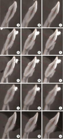
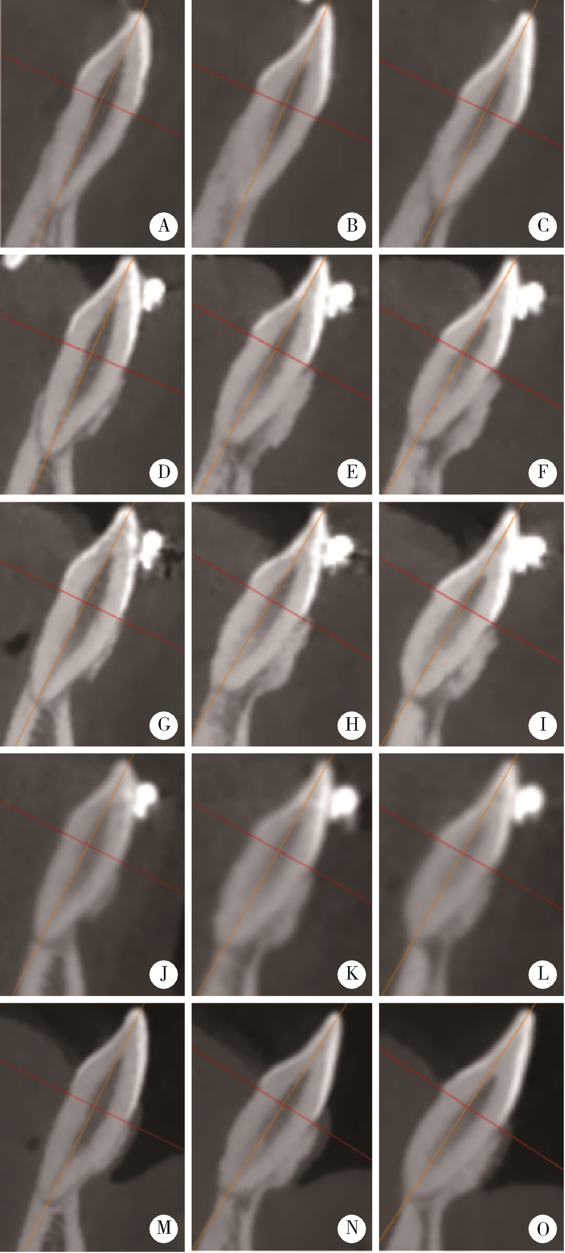
, 安氏Ⅲ类,长期观察,锥形束CT," /> , 安氏Ⅲ类,长期观察,锥形束CT,"/> A long-term evaluation of periodontal phenotypes before and after the periodontal-orthodontic-orthognathic combined treatment of lower anterior teeth in patients with skeletal Angle class Ⅲ malocclusion

Journal of Peking University (Health Sciences) ›› 2023, Vol. 55 ›› Issue (1): 52-61. doi: 10.19723/j.issn.1671-167X.2023.01.008

Previous Articles     Next Articles

A long-term evaluation of periodontal phenotypes before and after the periodontal-orthodontic-orthognathic combined treatment of lower anterior teeth in patients with skeletal Angle class Ⅲ malocclusion

Meng-qiao PAN1,Jian LIU1,Li XU1,*(),Xiao XU1,Jian-xia HOU1,Xiao-tong LI2,*(),Xiao-xia WANG3   

  1. 1. Department of Periodontology, Peking University School and Hospital of Stomatology & National Center of Stomatology & National Clinical Research Center for Oral Diseases & National Engineering Research Center of Oral Biomaterials and Digital Medical Devices & Beijing Key Laboratory of Digital Stomatology & NHC Research Center of Engineering and Technology for Computerized Dentistry & NMPA Key Laboratory for Dental Materials, Beijing 100081, China
    2. Department of Orthodontics, Peking University School and Hospital of Stomatology & National Center of Stomatology & National Clinical Research Center for Oral Diseases & National Engineering Research Center of Oral Biomaterials and Digital Medical Devices & Beijing Key Laboratory of Digital Stomatology & NHC Research Center of Engineering and Technology for Computerized Dentistry & NMPA Key Laboratory for Dental Materials, Beijing 100081, China
    3. Department of Oral and Maxillofacial Surgery, Peking University School and Hospital of Stomatology & National Center of Stomatology & National Clinical Research Center for Oral Diseases & National Engineering Research Center of Oral Biomaterials and Digital Medical Devices & Beijing Key Laboratory of Digital Stomatology & NHC Research Center of Engineering and Technology for Computerized Dentistry & NMPA Key Laboratory for Dental Materials, Beijing 100081, China
  • Received:2022-10-10 Online:2023-02-18 Published:2023-01-31
  • Contact: Li XU,Xiao-tong LI E-mail:xulihome@263.net;xiaotonglee@hotmail.com
  • Supported by:
    the Capital Characteristic Clinical Application Research Project(Z181100001718111)

Abstract:

Objective: To evaluate the changes of periodontal phenotype (width of keratinized gingiva, thickness and height of alveolar bone) of lower anterior teeth in patients with skeletal class Ⅲ malocclusion before and after the periodontal-orthodontic-orthognathic combined treatment. Methods: In the study, 20 patients with skeletal class Ⅲ malocclusion (6 males and 14 females) completed the periodontal-orthodontic-orthognathic combined treatment were included from March 2017 to June 2022, with 39 central incisors, 40 lateral incisors and 40 canines. The mean age was (25.40±4.27) years (20-34 years). The mean follow-up time was (3.70±1.05) years from the beginning of periodontal corticotomy regenerative surgery (PCRS) to the end of the combined treatment. Cone-beam computed tomography (CBCT) was used to measure the thickness, area and height of alveolar bone by the same researcher, taken before the PCRS (T0), 6 months after the PCRS (T1), 12 months after the PCRS (T2), before the orthognathic surgery (T3), and after the periodontal-orthodontic-orthognathic combined treatment (T4). The periodontal clinical parameters were used to evaluate changes in the soft tissue by another researcher, measured before the PCRS (T0) and after the combined treatment (T4). Changes of soft and hard tissue were evaluated by the periodontal phenotype. Results: The width of keratinized gingiva increased significantly (all P < 0.001) in lower anterior teeth, the central incisors, lateral incisors and canines increased by (1.82±1.57) mm, (2.03±1.48) mm and (2.05±1.27) mm, respectively. The proportion of thick periodontal biotype in the central and lateral incisors increased significantly (all P < 0.001), while the changes of periodontal biotypes in the lower canines were not obvious. The thickness of labial alveolar bone of lower anterior teeth all increased significantly after periodontal corticotomy regenerative surgery and the combined treatment (all P < 0.001). The area of labial alveolar bone of lower anterior teeth also increased significantly after the combined treatment (all P < 0.001). The whole area of labial and lingual alveolar bone of central and lateral incisors increased (P < 0.001), while the whole area of canines remained the same. All The height of the alveolar bone increased (all P < 0.001) on the labial side after the treatment. Conclusion: The periodontal phenotypes of lower anterior teeth were significantly improved after the periodontal-orthodontic-orthognathic combined treatment in patients with skeletal Angle class Ⅲ malocclusion. The improvement was long-termly stable, and the periodontal risk was reduced.

Key words: Periodontal phenotypes, Malocclusion, Angle class Ⅲ, Long-term evaluation, Cone-beam computed tomography

CLC Number: 

  • R783.5

Figure 1

Example of measuring alveolar bone thickness and area on CBCT images of the lower anterior teeth, orientated by the root long axis A, example and illustrations of morphometric measurements of the alveolar bone around the lower anterior teeth in skeletal class Ⅲ patients. B, reference points and lines: 1 and 2, cemento-enamel junction (CEJ) points; 3, midpoint of the CEJ; 4 and 5, alveolar crest points; 6, root apex; 7, root long axis, a line from points 3 to 6; 8, 9 and 10, intersecting line perpendicular to the root long axis at 4, 6 and 8 mm apical to the midpoint of the CEJ; 11, intersecting line perpendicular to the root long axis at the root apex. C, measurement variables: ABH, vertical bone level on the labial side, (distance from the CEJ to the alveolar crest parallel to the root long axis); BW-4, labial thickness at 4 mm apical to the CEJ; BW-6, labial thickness at 6 mm apical to the CEJ; BW-8, labial thickness at 8 mm apical to the CEJ; BW-a, labial thickness at the apex; BA-4, LA-4 and WA-4, labial, lingual and whole area at 4 mm apical to the CEJ; BA-6, LA-6 and WA-6, labial, lingual and whole area at 6 mm apical to the CEJ; BA-8, LA-8 and WA-8, labial, lingual and whole area at 8 mm apical to the CEJ; BA-a, LA-a and WA-a, labial, lingual and whole area at the apex."

Table 1

Comparison of periodontal clinical parameters before and after the periodontal-orthodontic-orthognathic combined treatment of lower anterior teeth in patients with skeletal Angle class Ⅲ malocclusion"

Items PLI, M(P25, P75) PD/mm, $\bar x \pm s$ BI, M(P25, P75) REC/mm, M(P25, P75) WKG/mm, $\bar x \pm s$
Central incisors
  T0 0.0(0.0, 1.0) 1.96±0.40 1.0(1.0, 2.0) 0.0(0.0, 0.0) 3.83±1.11
  T4 0.0(0.0, 1.0) 1.86±0.48 1.0(1.0, 2.0) 0.0(0.0, 0.0) 5.65±1.32
  t or Z value -1.439b 0.945a -0.762b -0.707b 7.231a
  P value 0.150 0.350 0.446 0.480 < 0.001*
Lateral incisors
  T0 1.0(0.0, 1.0) 2.18±0.47 1.0(1.0, 1.0) 0.0(0.0, 0.0) 3.97±1.10
  T4 0.0(0.0, 1.0) 1.98±0.48 1.0(1.0, 2.0) 0.0(0.0, 0.0) 6.00±1.29
  t or Z value -2.313b 1.842a -1.428b -1.380b 8.716a
  P value 0.021* 0.073 0.153 0.168 < 0.001*
Canines
  T0 1.0(0.0, 1.0) 2.31±0.46 1.0(1.0, 1.0) 0.0(0.0, 0.0) 3.41±1.11
  T4 0.0(0.0, 1.0) 2.16±0.51 1.0(1.0, 2.0) 0.0(0.0, 0.0) 5.47±1.48
  t or Z value -1.830b 1.311a -1.795b -1.000b 10.229a
  P value 0.067 0.198 0.073 0.317 < 0.001*

Table 2

Comparison of periodontal biotypes before and after the periodontal-orthodontic-orthognathic combined treatment of lower anterior teeth in patients with skeletal Angle class Ⅲ malocclusion"

Items Central incisors Lateral incisors Canines
Thick BIO
  T0 9 8 10
  T4 21 22 24
Thin BIO
  T0 30 32 30
  T4 18 18 16
  Z value -3.207 -3.742 -1.897
  P value < 0.001 < 0.001 0.058

Figure 2

Comparison of labial alveolar bone thickness before and after the periodontal-orthodontic-orthognathic combined treatment of lower anterior teeth in patients with skeletal Angle class Ⅲ malocclusion T0, before treatment; T1, 6 months after PCRS; T2, 12 months after PCRS; T3, before orthognathic surgery; T4, after the periodontal-orthodontic-orthognathic combined treatment. A, labial alveolar bone thickness at 4 mm apical to the CEJ; B, labial alveolar bone thickness at 6 mm apical to the CEJ; C, labial alveolar bone thickness at 8 mm apical to the CEJ; D, labial alveolar bone thickness from toot apex to CEJ."

Table 3

Comparison of labial alveolar bone area before and after the periodontal-orthodontic-orthognathic combined treatment of lower anterior teeth in patients with skeletal Angle class Ⅲ malocclusion"

Items Central incisors Lateral incisors Canines
BA-4
  T0 0.50±0.62 0.64±0.84 0.13±0.24
  T1 2.05±1.71 1.83±1.64 1.26±1.15
  T2 2.47±2.02 2.56±1.90 1.47±1.18
  T3 2.54±2.08 2.65±2.02 1.40±1.30
  T4 2.65±2.15 2.86±1.90 1.72±1.45
  P value < 0.001* < 0.001* < 0.001*
  Multiple comparison T0 < T1, T2, T3, T4 T0 < T1 < T2, T3, T4 T0 < T1, T2, T3, T4
BA-6
  T0 1.15±1.12 1.21±1.22 0.39±0.50
  T1 4.37±2.80 4.13±2.46 2.52±1.65
  T2 5.49±3.34 5.47±2.81 3.03±1.87
  T3 5.66±3.54 5.60±2.88 2.93±2.03
  T4 5.35±2.94 5.62±2.65 3.14±1.96
  P value < 0.001* < 0.001* < 0.001*
  Multiple comparison T0 < T1, T2, T3, T4 T0 < T1 < T2, T3, T4 T0 < T1, T2, T3, T4
BA-8
  T0 1.68±1.53 1.47±1.32 0.65±0.72
  T1 7.88±4.47 7.51±3.44 4.06±2.22
  T2 9.32±4.93 8.96±3.61 4.82±2.59
  T3 9.50±4.84 9.30±3.80 4.64±2.74
  T4 8.86±3.90 9.25±3.76 4.85±2.58
  P value < 0.001* < 0.001* < 0.001*
  Multiple comparison T0 < T1, T2, T3, T4 T0 < T1 < T2, T3, T4 T0 < T1, T2, T3, T4
BA-a
  T0 4.04±3.18 4.48±2.43 6.10±4.66
  T1 14.97±6.85 16.88±6.19 11.86±6.73
  T2 16.59±8.03 18.62±6.83 14.08±6.62
  T3 16.68±7.57 18.82±7.11 15.14±7.29
  T4 15.80±7.35 19.50±10.34 14.98±7.75
  P value < 0.001* < 0.001* < 0.001*
  Multiple comparison T0 < T1, T2, T3, T4 T0 < T1, T2, T3, T4 T0 < T1, T2, T3, T4

Table 4

Comparison of the whole alveolar bone area before and after the periodontal-orthodontic-orthognathic combined treatment of lower anterior teeth in patients with skeletal Angle class Ⅲ malocclusion"

Items Central incisors Lateral incisors Canines
WA-4
  T0 0.87±0.93 1.21±1.14 1.57±2.20
  T1 2.37±1.95 2.25±2.01 2.88±2.83
  T2 2.67±2.17 2.83±2.05 2.30±1.93
  T3 2.78±2.50 2.91±2.24 2.31±2.44
  T4 2.85±2.37 3.20±2.09 3.28±3.18
  P value < 0.001* < 0.001* 0.038*
  Multiple comparison T0 < T1, T2, T3, T4 T0 < T1, T2, T3, T4 T0 < T1, T4
WA-6
  T0 2.37±2.05 2.72±2.34 4.19±5.12
  T1 5.08±3.16 5.18±3.15 6.10±3.91
  T2 5.91±3.55 6.04±3.04 5.29±3.64
  T3 6.02±3.93 6.19±3.19 5.22±4.34
  T4 5.79±3.25 6.48±3.13 6.55±5.18
  P value < 0.001* < 0.001* 0.174
  Multiple comparison T0 < T1, T2, T3, T4 T0 < T1, T2, T3, T4 T0=T1, T2, T3, T4
WA-8
  T0 4.35±3.39 4.30±3.72 7.34±6.91
  T1 9.32±4.88 9.31±4.49 10.55±4.97
  T2 10.03±5.14 9.90±3.95 9.11±4.90
  T3 10.12±5.18 10.21±4.34 8.88±6.04
  T4 9.66±4.35 10.70±4.47 10.47±7.12
  P value < 0.001* < 0.001* 0.124
  Multiple comparison T0 < T1, T2, T3, T4 T0 < T1, T2, T3, T4 T0=T1, T2, T3, T4
WA-a
  T0 13.71±8.30 15.53±8.66 29.53±15.91
  T1 19.51±8.24 22.32±7.85 32.50±14.43
  T2 18.58±8.58 21.37±7.85 29.51±14.40
  T3 18.46±8.07 21.38±8.10 29.87±14.66
  T4 18.01±8.91 22.93±11.59 30.81±17.84
  P value 0.028* 0.002* 0.911
  Multiple comparison T0 < T1, T2, T3, T4 T0 < T1, T2, T3, T4 T0=T1, T2, T3, T4

Figure 3

Comparison of alveolar bone heights before and after the periodontal-orthodontic-orthognathic combined treatment of lower anterior teeth in patients with skeletal Angle class Ⅲ malocclusion T0, before treatment; T1, 6 months after PCRS; T2, 12 months after PCRS; T3, before orthognathic surgery; T4, after the periodontal-orthodontic-orthognathic combined treatment. *P < 0.05, a significant difference."

Figure 4

Example of soft tissue changes of lower anterior teeth in a patient with skeletal Angle class Ⅲ malocclusion during the periodontal-orthodontic-orthognathic combined treatment A, before treatment; B, 6 months after PCRS; C, 12 months after PCRS; D, before orthognathic surgery; E, after the periodontal-orthodontic-orthognathic combined treatment."

Figure 5

Example of alveolar bone changes of lower anterior teeth in a patient with skeletal Angle class Ⅲ malocclusion during the periodontal-orthodontic-orthognathic combined treatment A, canine before treatment; B, lateral incisor before treatment; C, central incisor before treatment; D, canine 6 months after PCRS; E, lateral incisor 6 months after PCRS; F, central incisor 6 months after PCRS; G, canine 12 months after PCRS; H, lateral incisor 12 months after PCRS; I, central incisor 12 months after PCRS; J, canine before orthognathic surgery; K, lateral incisor before orthognathic surgery; L, central incisor before orthognathic surgery; M, canine after the periodontal-orthodontic-orthognathic combined treatment; N, lateral incisor after the periodontal-orthodontic-orthognathic combined treatment; O, central incisor after the periodontal-orthodontic-orthognathic combined treatment."

1 傅民魁, 林久祥. 口腔正畸学[M]. 北京: 北京大学医学出版社, 2014: 270- 271.
2 Kaya Y , Alkan Ö , Keskin S . An evaluation of the gingival biotype and the width of keratinized gingiva in the mandibular anterior region of individuals with different dental malocclusion groups and levels of crowding[J]. Korean J Orthod, 2017, 47 (3): 176- 185.
doi: 10.4041/kjod.2017.47.3.176
3 毛铭馨, 徐莉, 靖无迪, 等. 骨性安氏Ⅲ类错畸形患者前牙唇侧牙槽嵴顶位置及相关因素分析[J]. 北京大学学报(医学版), 2020, 52 (1): 77- 82.
4 Guo R , Zhang L , Hu M , et al. Alveolar bone changes in maxillary and mandibular anterior teeth during orthodontic treatment: A systematic review and meta-analysis[J]. Orthod Craniofac Res, 2021, 24 (2): 165- 179.
doi: 10.1111/ocr.12421
5 Lee KM , Kim YI , Park SB , et al. Alveolar bone loss around lower incisors during surgical orthodontic treatment in mandibular prognathism[J]. Angle Orthod, 2012, 82 (4): 637- 644.
doi: 10.2319/081711-526.1
6 Ma H , Li W , Xu L , et al. Morphometric evaluation of the alveolar bone around central incisors during surgical orthodontic treatment of high-angle skeletal class Ⅲ malocclusion[J]. Orthod Craniofac Res, 2021, 24 (1): 87- 95.
doi: 10.1111/ocr.12408
7 Jing WD , Jiao J , Xu L , et al. Periodontal soft- and hard-tissue changes after augmented corticotomy in Chinese adult patients with skeletal Angle class Ⅲ malocclusion: A non-randomized controlled trial[J]. J Periodontol, 2020, 91 (11): 1419- 1428.
doi: 10.1002/JPER.19-0522
8 Xu X , Wu JQ , Jiang JH , et al. Periodontal effect of periodontally accelerated osteogenic orthodontics in skeletal Angle class Ⅲ: A nonrandomized, controlled trial[J]. Int J Periodontics Restorative Dent, 2020, 40 (4): e169- e177.
doi: 10.11607/prd.4545
9 Wang B , Shen G , Fang B , et al. Augmented corticotomy-assisted surgical orthodontics decompensates lower incisors in Class Ⅲ malocclusion patients[J]. J Oral Maxillofac Surg, 2014, 72 (3): 596- 602.
doi: 10.1016/j.joms.2013.08.021
10 Brugnami F , Meuli S , Caiazzo A , et al. Three-dimensional digital planning of class Ⅲ decompensation with clear aligners: Hard and soft tissue augmentation with concomitant corticotomy to stretch the limits of safe orthodontic treatment[J]. J Oral Biol Craniofac Res, 2021, 11 (2): 297- 302.
doi: 10.1016/j.jobcr.2021.02.011
11 Malpartida-Carrillo V , Tinedo-Lopez PL , Guerrero ME , et al. Periodontal phenotype: A review of historical and current classifications evaluating different methods and characteristics[J]. J Esthet Restor Dent, 2021, 33 (3): 432- 445.
doi: 10.1111/jerd.12661
12 Chapple ILC , Mealey BL , van Dyke TE , et al. Periodontal health and gingival diseases and conditions on an intact and a reduced periodontium: Consensus report of workgroup 1 of the 2017 World Workshop on the Classification of Periodontal and Peri-Implant Diseases and Conditions[J]. J Clin Periodontol, 2018, 45 (Suppl 20): S68- S77.
13 徐筱, 靖无迪, 侯建霞, 等. 牙周组织再生结合骨皮质切开术辅助骨性Ⅲ类错正畸-正颌治疗一例[J]. 中华口腔医学杂志, 2019, 54 (10): 686- 690.
14 韩烨, 苗莉莉, 靖无迪, 等. 牙周组织再生结合骨皮质切开术对骨性Ⅲ类错牙龈厚度影响的数字化评估[J]. 中华口腔医学杂志, 2020, (2): 73- 79.
doi: 10.3760/cma.j.issn.1002-0098.2020.02.001
15 徐筱, 徐莉, 江久汇, 等. 改良骨皮质切开术对牙周组织影响的临床观察[J]. 中华口腔医学杂志, 2014, 49 (6): 343- 346.
doi: 10.3760/cma.j.issn.1002-0098.2014.06.006
16 马慧敏, 张婕, 徐莉, 等. 骨性Ⅲ类错畸形患者正畸正颌联合治疗前后前牙区牙槽骨厚度的变化[J]. 中华口腔正畸学杂志, 2018, 25 (3): 121- 124.
17 Choi YJ , Chung CJ , Kim KH . Periodontal consequences of mandibular incisor proclination during presurgical orthodontic treatment in class Ⅲ malocclusion patients[J]. Angle Orthod, 2015, 85 (3): 427- 433.
doi: 10.2319/021414-110.1
18 Sun L , Yuan L , Wang B , et al. Changes of alveolar bone dehiscence and fenestration after augmented corticotomy-assisted orthodontic treatment: A CBCT evaluation[J]. Prog Orthod, 2019, 20 (1): 7.
doi: 10.1186/s40510-019-0259-z
19 Wang CW , Yu SH , Mandelaris GA , et al. Is periodontal phenotype modification therapy beneficial for patients receiving ortho-dontic treatment? An American Academy of Periodontology best evidence review[J]. J Periodontol, 2020, 91 (3): 299- 310.
20 Zweers J , Thomas RZ , Slot DE , et al. Characteristics of periodontal biotype, its dimensions, associations and prevalence: A systematic review[J]. J Clin Periodontol, 2014, 41 (10): 958- 971.
21 王高南, 焦剑, 周彦恒, 等. 正畸牙齿位置的移动对角化龈宽度的影响[J]. 北京大学学报(医学版), 2019, 51 (5): 931- 936.
22 Vlachodimou E , Fragkioudakis I , Vouros I . Is there an association between the gingival phenotype and the width of keratinized gingiva? A systematic review[J]. Dent J (Basel), 2021, 9 (3): 34.
[1] Jin-hua ZHANG,Jie PAN,Zhi-peng SUN,Xiao WANG. Effect of various intracanal materials on the diagnostic accuracy of cone-beam computed tomography in vertical root fractures [J]. Journal of Peking University (Health Sciences), 2023, 55(2): 333-338.
[2] Jia-xue YE,Yu-hong LIANG. A prevalence survey of cone-beam computed tomography use among endodontic practitioners [J]. Journal of Peking University (Health Sciences), 2023, 55(1): 114-119.
[3] Yu FU,Xin-nong HU,Sheng-jie CUI,Jie SHI. Decompensation effectiveness and alveolar bone remodeling analysis of mandibular anterior teeth after preoperative orthodontic treatment in high-angle patients with skeletal class Ⅱ malocclusion [J]. Journal of Peking University (Health Sciences), 2023, 55(1): 62-69.
[4] Juan GAO,Hang-miao LV,Hui-min MA,Yi-jiao ZHAO,Xiao-tong LI. Evaluation of root resorption after surgical orthodontic treatment of skeletal Class Ⅲ malocclusion by three-dimensional volumetric measurement with cone-beam CT [J]. Journal of Peking University (Health Sciences), 2022, 54(4): 719-726.
[5] LIU Wei-tao,WANG Yi-ran,WANG Xue-dong,ZHOU Yan-heng. A cone-beam computed tomography evaluation of three-dimensional changes of circummaxillary sutures following maxillary protraction with alternate rapid palatal expansions and constrictions [J]. Journal of Peking University (Health Sciences), 2022, 54(2): 346-355.
[6] Gang YANG,Wen-jie HU,Jie CAO,Deng-gao LIU. Three-dimensional morphology analysis of the supraosseous gingival profile of periodontally healthy maxillary anterior teeth [J]. Journal of Peking University (Health Sciences), 2021, 53(5): 990-994.
[7] MENG Yuan,ZHANG Li-qi,ZHAO Ya-ning,LIU Deng-gao,ZHANG Zu-yan,GAO Yan. Three-dimentional radiographic features of 67 maxillary radicular cysts [J]. Journal of Peking University (Health Sciences), 2021, 53(2): 396-401.
[8] ZHOU Jing,LIU Yi. Cone-beam CT evaluation of temporomandibular joint in skeletal class Ⅱ female adolescents with different vertical patterns [J]. Journal of Peking University (Health Sciences), 2021, 53(1): 109-119.
[9] GAO Lu,GU Yan. Chinese morphological stages of midpalatal suture and its correlation with Demirjian dental age [J]. Journal of Peking University (Health Sciences), 2021, 53(1): 133-138.
[10] WANG Yi-ran, ZHOU Yan-heng, WANG Xue-dong, WEI Song, LIU Wei-tao. Evaluation of maxillary three-dimensional changes in maxillary protraction with alternating rapid palatal expansion and constriction based on the cone-beam computed tomography [J]. Journal of Peking University(Health Sciences), 2018, 50(4): 685-693.
[11] JIA Peng-chen, YANG Gang, HU Wen-jie, ZHAO Yi-jiao, LIU Mu-qing. Preliminary study on the accuracy of infrabony root surface area of single-root teeth by periapical films [J]. Journal of Peking University(Health Sciences), 2018, 50(1): 91-97.
[12] MA Jing, JIANG Jiu-hui. Morphological analysis of alveolar bone of anterior mandible in high-angle skeletal class Ⅱ and class Ⅲ malocclusions assessed with cone-beam computed tomography [J]. Journal of Peking University(Health Sciences), 2018, 50(1): 98-103.
[13] XU Xiao, XU Li, JIANG Jiu-hui, WU Jia-qi, LI Xiao-tong, JING Wu-di. Accuracy analysis of alveolar dehiscence and fenestration of maxillary anterior teeth of Angle class Ⅲ by cone-beam CT [J]. Journal of Peking University(Health Sciences), 2018, 50(1): 104-109.
[14] CAO Jie, MENG Huan-xin. Evaluation of using cone beam computed tomography as a regular test before and after periodontal regenerative surgery#br# [J]. Journal of Peking University(Health Sciences), 2018, 50(1): 110-116.
[15] CHANG Da-tong, ZHOU Yan-heng, LIU Wei-tao. Evaluation of cone-beam computed tomography on upper airway changes after alternating rapid palatal expansion and constriction [J]. Journal of Peking University(Health Sciences), 2017, 49(4): 685-690.
Viewed
Full text


Abstract

Cited

  Shared   
  Discussed   
[1] . [J]. Journal of Peking University(Health Sciences), 2001, 33(1): 50 -53 .
[2] . [J]. Journal of Peking University(Health Sciences), 2002, 34(2): 140 -143 .
[3] . [J]. Journal of Peking University(Health Sciences), 2010, 42(4): 476 -479 .
[4] . [J]. Journal of Peking University(Health Sciences), 2008, 40(2): 208 -210 .
[5] . [J]. Journal of Peking University(Health Sciences), 2008, 40(5): 459 -464 .
[6] . [J]. Journal of Peking University(Health Sciences), 2007, 39(4): 434 -436 .
[7] . [J]. Journal of Peking University(Health Sciences), 2001, 33(3): 288 -289 .
[8] . [J]. Journal of Peking University(Health Sciences), 2002, 34(2): 97 -98 .
[9] . [J]. Journal of Peking University(Health Sciences), 2002, 34(2): 112 -116 .
[10] . [J]. Journal of Peking University(Health Sciences), 2011, 43(1): 29 -33 .