Journal of Peking University (Health Sciences) ›› 2022, Vol. 54 ›› Issue (5): 896-906. doi: 10.19723/j.issn.1671-167X.2022.05.017

Previous Articles     Next Articles

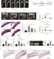
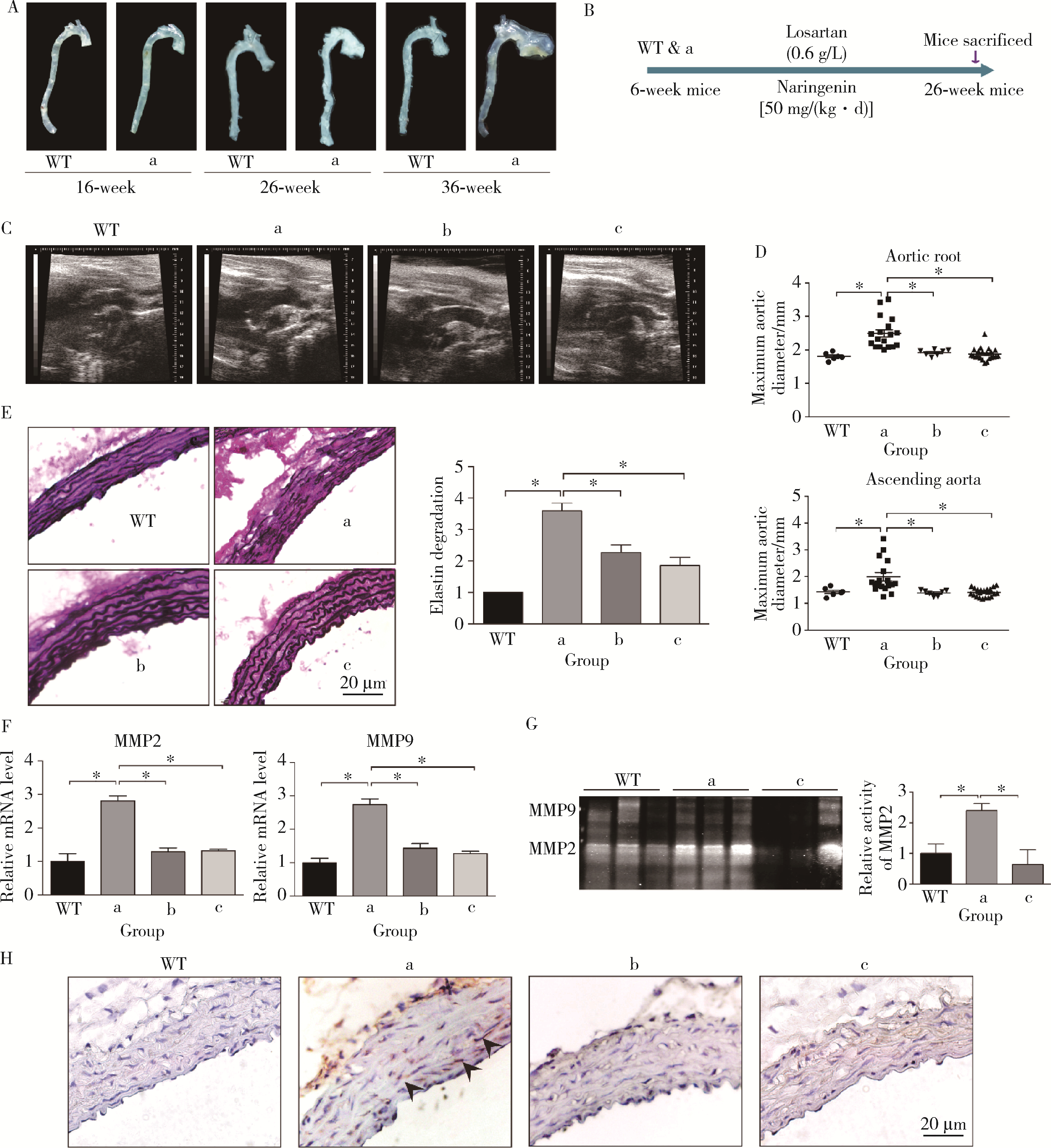
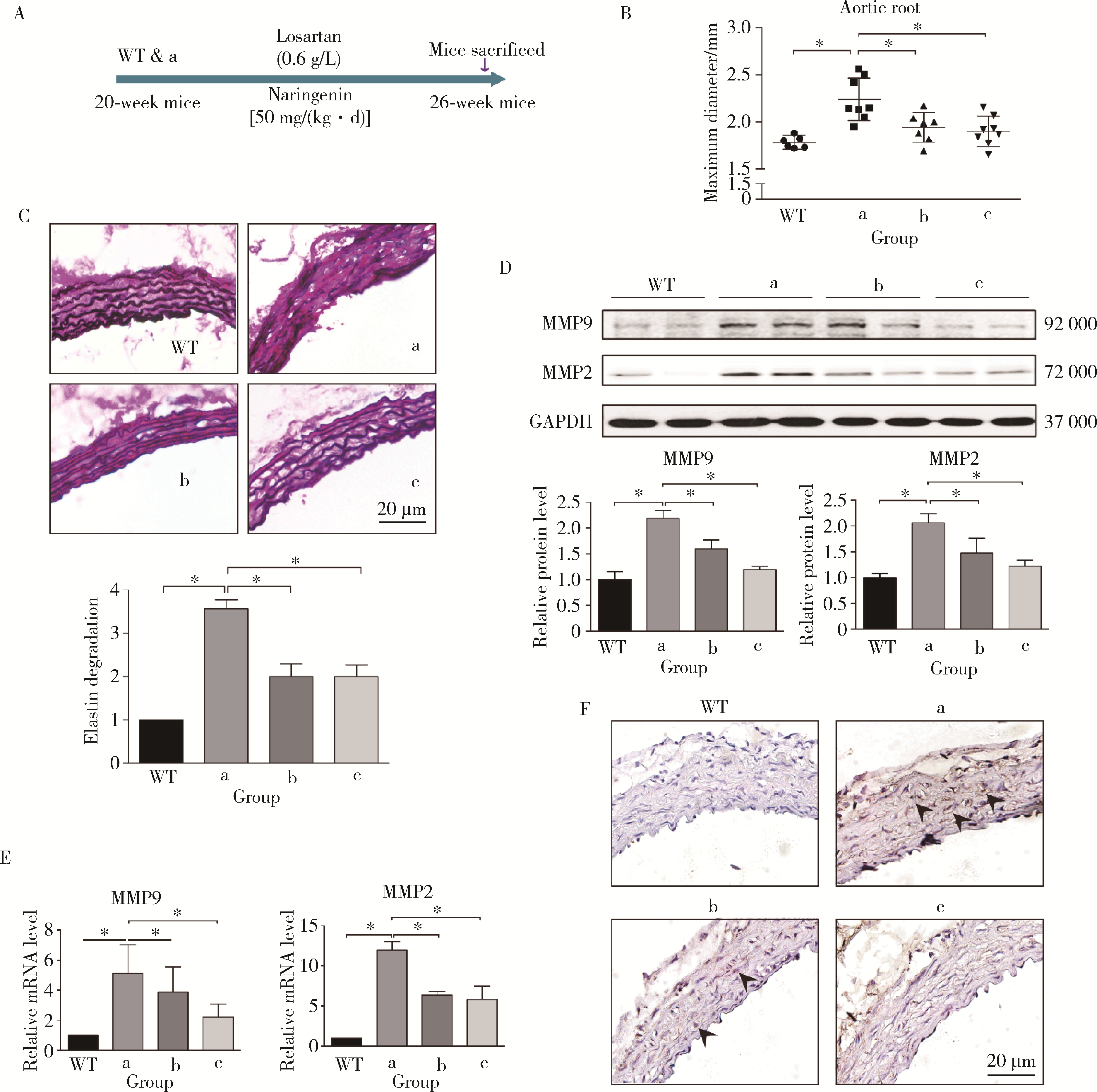
Naringenin inhibits thoracic aortic aneurysm formation in mice with Marfan syndrome

Zhi-qing LI1,Bing YU1,Ze-yu CAI1,Ying-bao WANG1,Xu ZHANG1,Biao ZHOU2,Xiao-hong FANG3,Fang YU1,Yi FU1,Jin-peng SUN1,Wei LI4,Wei KONG1,*()   

  1. 1. Department of Physiology and Pathophysiology, Peking University School of Basic Medical Sciences, Beijing 100191, China
    2. Department of General Surgery, China-Japan Friendship Hospital, Beijing 100029, China
    3. Institute of Chemistry, Chinese Academy of Sciences, Beijing 100190, China
    4. Department of Vascular Surgery, Peking University People's Hospital, Beijing 100044, China
  • Received:2022-06-03 Online:2022-10-18 Published:2022-10-14
  • Contact: Wei KONG E-mail:kongw@bjmu.edu.cn
  • Supported by:
    the National Natural Science Foundation of China(81730010);the National Natural Science Foundation of China(82100480);the National Natural Science Foundation of China(31930056);the National Natural Science Foundation of China(81921001);China Postdoctoral Science Foundation(2020M680257)

RICH HTML


Abstract:

Objective: To identify whether naringenin plays a protective role during thoracic aneurysm formation in Marfan syndrome. Methods: To validate the effect of naringenin, Fbn1C1039G/+ mice, the mouse model of Marfan syndrome, were fed with naringenin, and the disease progress was evaluated. The molecular mechanism of naringenin was further investigated via in vitro studies, such as bioluminescence resonance energy transfer (BRET), atomic force microscope and radioligand receptor binding assay. Results: Six-week-old Fbn1C1039G/+ mice were fed with naringenin for 20 weeks. Compared with the control group, naringenin significantly suppressed the aortic expansion [Fbn1C1039G/+ vs. Fbn1C1039G/++naringenin: (2.49±0.47) mm, n=18 vs. (1.87±0.19) mm, n=22, P < 0.05], the degradation of elastin, and the expression and activity of matrix metalloproteinase 2 (MMP2) and MMP9 in the ascending aorta of Fbn1C1039G/+ mice. Besides, treatment with naringenin for 6 weeks also attenuated the disease progress among the 20-week-old Fbn1C1039G/+ mice with established thoracic aortic aneurysms [Fbn1C1039G/+ vs. Fbn1C1039G/++naringenin: (2.24±0.23) mm, n=8 vs. (1.90±0.17) mm, n=8, P < 0.05]. To understand the underlying molecular mechanisms, we examined the effects of naringenin on angiotensin Ⅱ type 1 receptor (AT1) signaling and transforming growth factor-β (TGF-β) signaling respectively, which were the dominant signaling pathways contributing to aortopathy in Marfan syndrome as previously reported. The results showed that naringenin decreased angiotensin Ⅱ (Ang Ⅱ)-induced phosphorylation of protein kinase C (PKC) and extracellular regulating kinase 1/2 (ERK1/2) in HEK293A cell overexpressing AT1 receptor. Moreover, naringenin inhibited Ang Ⅱ-induced calcium mobilization and uclear factor of activated T-cells (NFAT) signaling. The internalization of AT1 receptor and its binding to β-arrestin-2 with Ang Ⅱ induction were also suppressed by naringenin. As evidenced by atomic force microscope and radioligand receptor binding assay, naringenin inhibited Ang Ⅱ binding to AT1 receptor. In terms of TGF-β signaling, we found that feeding the mice with naringenin decreased the phosphorylation of Smad2 and ERK1/2 as well as the expression of TGF-β downstream genes. Besides, the serum level of TGF-β was also decreased by naringenin in the Fbn1C1039G/+ mice. Furthermore, we detected the effect of naringenin on platelet, a rich source of TGF-β, both in vivo and in vitro. And we found that naringenin markedly decreased the TGF-β level by inhibiting the activation of platelet. Conclusion: Our study showed that naringenin has a protective effect on thoracic aortic aneurysm formation in Marfan syndrome by suppressing both AT1 and TGF-β signaling.

Key words: Naringenin, Marfan syndrome, Thoracic aortic aneurysm, Angiotensin Ⅱ type 1 receptor (AT1), Transforming growth factor-β (TGF-β)

CLC Number: 

  • R33

Figure 1

Naringenin inhibits the formation of thoracic aortic aneurysm in Fbn1C1039G/+ mice A, representative ex vivo morphology of aortic root and ascending aorta in wild-type (WT) and Fbn1C1039G/+ mice of different ages; B, schematic diagram for long-term naringenin feeding animal experiment, and six-week-old wild type and Fbn1C1039G/+ mice were treated with losartan (0.6 g/L) or naringenin [NGN, 50 mg/(kg·d)] until 26 weeks old; C and D, representative ultrasound images and quantification of thoracic aorta in naringenin or losartan-fed wild type and Fbn1C1039G/+ mice, n=6-23; E, representative images of elastic Van Gieson staining and quantification of elastin degradation of thoracic aorta, n=6-23, scale bar=20 μm; F, real-time PCR analysis of MMP2 and MMP9 expression in thoracic aorta of naringenin and losartan-fed wild type and Fbn1C1039G/+ mice, n=6; G, representative images of gelatin zymogram to detect the activity and expression of MMP2 and MMP9 in aortic root of wild-type and Fbn1C1039G/+ mice fed with naringenin and losartan, n=6; H, representative images of p-Smad2 immunohistochemical staining of ascending aorta in wild-type mice and Fbn1C1039G/+ mice fed with naringenin and losartan, n=6, scale bar=20 μm. *P < 0.05. a, Fbn1C1039G/+; b, Fbn1C1039G/++losartan; c, Fbn1C1039G/++NGN."

Figure 2

Naringenin (NGN) attenuates the progression of established aneurysms in Fbn1C1039G/+ mice A, schematic diagram for short-term naringenin feeding animal experiment; B, quantification of the maximal thoracic aortic diameters of mice, n=6-23; C, representative images of elastica van gieson (EVG) staining of aortic root and ascending aorta in naringenin and losartan-fed wild-type mice and Fbn1C1039G/+ mice, n=6-23, scale bar=20 μm; D, Western blot and quantification of MMP2 and MMP9 expression in thoracic aorta of mice, n=6; E, real-time PCR analysis of MMP2 and MMP9 expression in thoracic aorta of mice, n=6; F, representative images of p-Smad2 immunohistochemical staining of ascending aorta plate in wild-type mice and Fbn1C1039G/+ mice fed with naringenin and losartan, n=6, scale bar=20 μm. *P < 0.05. WT, wide-type; a, Fbn1C1039G/+; b, Fbn1C1039G/++losartan; c, Fbn1C1039G/++NGN."

Figure 3

Naringenin(NGN) inhibits angiotensin Ⅱ-AT1 signaling A and B, representative Western blot and quantification of phosphorylated (p-) and total (t-) PKC and ERK1/2 in A7r5 cells (A) and AT1-overexpressing HEK293A cells (B), n=6; C, calcium signals in HEK293A cells, transfected with the AT1 plasmid and stimulated continuously by Ang Ⅱ (1 μmol/L) with or without naringenin (NGN, 100 μmol/L) pretreatment, were detected using Fluo 3-AM and quantified by the Leica confocal microscope system, n=6; D and E, HEK293A cells were transfected with AT1, NFAT and TK plasmids and cultured for 40 hours, and cells were pretreated with naringenin (100, 200 μmol/L) for 30 minutes and then stimulated with Ang Ⅱ (1 μmol/L in D, or different concentrations as indicated in E) for 8 hours to detect the luciferase fluorescence intensity, n=6; F, COS7 cells were transfected by pEGFP-AT1 plasmid, cultured for 48 hours and starved for 24 hours, pretreated with naringenin (100 μmol/L) for 30 minutes, stimulated with Ang Ⅱ (1 μmol/L) for 15 minutes, and examined the distribution of AT1 receptors by confocal microscope, n=6; G, the BRET signal of binding of AT1 and β-arrestin-2, n=6. *P < 0.05 vs. control group or the indicated group. # P < 0.05 vs. Ang Ⅱ group at the same time point in B. CTL, control; d, Ang Ⅱ; e, Ang Ⅱ+NGN 100 μmol/L; f, Ang Ⅱ+NGN 200 μmol/L; g, NGN 200 μmol/L."

Figure 4

Naringenin antagonizes the binding of angiotensin Ⅱ to AT1 A and B, HeLa cells were transfected with 0.2 μg of AT1 plasmid and cultured for 8 h, and then changed to DMEM medium without phenol red to detect fluorescence, and cells were pretreated with naringenin (NGN, 100 μmol/L) or ethanol (control, CTL) for 1 hour and then treated with angiotensin Ⅱ (Ang Ⅱ, 1 μmol/L) for 15 minutes to detect the binding probability (A) and binding force (B) of receptor-ligated receptors under an atomic force microscope system. n=6, *P < 0.05; C, the competition binding curve of [125I]-Ang Ⅱ with Ang Ⅱ, naringenin and valsartan, n=6;D, the saturation binding curve of [125I]-Ang Ⅱ and the AT1 receptor in the presence and absence of naringenin; n=6."

Figure 5

Naringenin inhibits TGF-β signaling both in vivo and in vitro A and B, representative western blots and quantification of phosphorylated (p-) and total (t-) Smad2 and ERK1/2 in ascending aorta of wild-type mice and Fbn1C1039G/+ mice after long-term (A) or short-term (B) feeding with naringenin (NGN) or losartan, n=6; C, real-time PCR analysis of TGF-β target genes in thoracic aorta of wild-type and Fbn1C1039G/+ mice after long-term feeding with naringenin or losartan, n=6; D, representative Western blot and quantification of phosphorylated (p-) and total (t-) Smad2. HEK293A cells transfected with AT1 plasmid were pretreated with naringenin (100 μmol/L) for 30 min, and stimulated with Ang Ⅱ (1 μmol/L) for different times (0-60 min), n=6. *P < 0.05 vs. control group or the indicated group. # P < 0.05 vs. Ang Ⅱ group at the same time point in D. WT, wide-type; a, Fbn1C1039G/+; b, Fbn1C1039G/++losartan; c, Fbn1C1039G/++NGN."

Figure 6

Naringenin decreases the level of platelet-derived TGF-β A and B, ELISA analysis of free TGF-β and total TGF-β levels in wild-type mice and Fbn1C1039G/+ mice after long-term (A) or short-term (B) feeding with naringenin (NGN) or losartan, n=6-15; C, blood clotting time of 8-week-old wild-type mice fed with naringenin for a week, n=6; D, the right carotid artery complete occlusion time of mice following FeCl3 injury; E, platelets of 8-week-old wild-type mice were extracted, pretreated with naringenin (100 μmol/L) for 30 minutes, stimulated with thrombin (0.1 U/mL) for 30 minutes, and the content of released TGF-β was detected by ELISA, n=6-15; F, the platelets of wild type mice and Fbn1C1039G/+ mice after long-term feeding with naringenin and losartan were extracted, and the proportion of CD62P-positive platelets was measured by flow cytometry, n=6-7. *P < 0.05. WT, wide-type; CTL, control; a, Fbn1C1039G/+; b, Fbn1C1039G/++losartan; c, Fbn1C1039G/++NGN; h, NGN 100 μmol/L; i, thrombin; j, NGN 1 μmol/L+thrombin; k, NGN 100 μmol/L+thrombin."

1 Milewicz DM , Braverman AC , De Backer J , et al. Marfan syndrome[J]. Nat Rev Dis Primers, 2021, 7 (1): 64.
doi: 10.1038/s41572-021-00298-7
2 Holm TM , Habashi JP , Doyle JJ , et al. Noncanonical TGFβ signaling contributes to aortic aneurysm progression in Marfan syndrome mice[J]. Science, 2011, 332 (6027): 358- 361.
doi: 10.1126/science.1192149
3 Habashi JP , Judge DP , Holm TM , et al. Losartan, an AT1 antagonist, prevents aortic aneurysm in a mouse model of Marfan syndrome[J]. Science, 2006, 312 (5770): 117- 121.
doi: 10.1126/science.1124287
4 Cook JR , Clayton NP , Carta L , et al. Dimorphic effects of transforming growth factor-beta signaling during aortic aneurysm progression in mice suggest a combinatorial therapy for Marfan syndrome[J]. Arterioscler Thromb Vasc Biol, 2015, 35 (4): 911- 917.
doi: 10.1161/ATVBAHA.114.305150
5 Jia Y , Zhang L , Liu Z , et al. Targeting macrophage TFEB-14-3-3 epsilon Interface by naringenin inhibits abdominal aortic aneurysm[J]. Cell Discov, 2022, 8 (1): 21.
doi: 10.1038/s41421-021-00363-1
6 Rodríguez-Vita J , Sánchez-López E , Esteban V , et al. Angiotensin Ⅱ activates the Smad pathway in vascular smooth muscle cells by a transforming growth factor-beta-independent mechanism[J]. Circulation, 2005, 111 (19): 2509- 2517.
doi: 10.1161/01.CIR.0000165133.84978.E2
7 Touat Z , Lepage L , Ollivier V , et al. Dilation-dependent activation of platelets and prothrombin in human thoracic ascending aortic aneurysm[J]. Arterioscler Thromb Vasc Biol, 2008, 28 (5): 940- 946.
doi: 10.1161/ATVBAHA.107.158576
8 Metelli A , Salem M , Wallace CH , et al. Immunoregulatory functions and the therapeutic implications of GARP-TGF-β in inflammation and cancer[J]. J Hematol Oncol, 2018, 11 (1): 24.
doi: 10.1186/s13045-018-0570-z
9 Fusi F , Trezza A , Tramaglino M , et al. The beneficial health effects of flavonoids on the cardiovascular system: Focus on K(+) channels[J]. Pharmacol Res, 2020, 152, 104625.
doi: 10.1016/j.phrs.2019.104625
10 Burke AC , Sutherland BG , Telford DE , et al. Naringenin enhances the regression of atherosclerosis induced by a chow diet in Ldlr (-/-) mice[J]. Atherosclerosis, 2019, 286, 60- 70.
doi: 10.1016/j.atherosclerosis.2019.05.009
11 Wisler JW , Harris EM , Raisch M , et al. The role of beta-arrestin2-dependent signaling in thoracic aortic aneurysm formation in a murine model of Marfan syndrome[J]. Am J Physiol Heart Circ Physiol, 2015, 309 (9): H1516- 1527.
doi: 10.1152/ajpheart.00291.2015
12 Teixido-Tura G , Forteza A , Rodríguez-Palomares J , et al. Losartan versus atenolol for prevention of aortic dilation in patients with marfan syndrome[J]. J Am Coll Cardiol, 2018, 72 (14): 1613- 1618.
doi: 10.1016/j.jacc.2018.07.052
13 van Andel MM , Indrakusuma R , Jalalzadeh H , et al. Long-term clinical outcomes of losartan in patients with Marfan syndrome: follow-up of the multicentre randomized controlled COMPARE trial[J]. Eur Heart J, 2020, 41 (43): 4181- 4187.
doi: 10.1093/eurheartj/ehaa377
14 Rifkin DB , Rifkin WJ , Zilberberg L . LTBPs in biology and medicine: LTBP diseases[J]. Matrix Biol, 2018, 71/72, 90- 99.
doi: 10.1016/j.matbio.2017.11.014
15 Li W , Li Q , Jiao Y , et al. Tgfbr2 disruption in postnatal smooth muscle impairs aortic wall homeostasis[J]. J Clin Invest, 2014, 124 (2): 755- 767.
doi: 10.1172/JCI69942
16 Wei H , Hu JH , Angelov SN , et al. Aortopathy in a mouse model of marfan syndrome is not mediated by altered transforming growth factor beta signaling[J]. J Am Heart Assoc, 2017, 6 (1): e004968.
doi: 10.1161/JAHA.116.004968
17 Hernandez-Aquino E , Zarco N , Casas-Grajales S , et al. Naringenin prevents experimental liver fibrosis by blocking TGFbeta-Smad3 and JNK-Smad3 pathways[J]. World J Gastroenterol, 2017, 23 (24): 4354- 4368.
doi: 10.3748/wjg.v23.i24.4354
18 Lim W , Song G . Naringenin-induced migration of embrynoic trophectoderm cells is mediated via PI3K/AKT and ERK1/2 MAPK signaling cascades[J]. Mol Cell Endocrinol, 2016, 428, 28- 37.
doi: 10.1016/j.mce.2016.03.018
19 Yang Y , Xu Y , Xia T , et al. A single-molecule study of the inhibition effect of Naringenin on transforming growth factor-beta ligand-receptor binding[J]. Chem Commun (Camb), 2011, 47 (19): 5440- 5442.
doi: 10.1039/C1CC10778J
20 Lopez JJ , El Haouari M , Jardin I , et al. Flavonoids and platelet-derived thrombotic disorders[J]. Curr Med Chem, 2019, 26 (39): 7035- 7047.
doi: 10.2174/0929867325666180417170218
[1] Jia-hui DENG,Xiao-lin HUANG,Xiao-xing LIU,Jie SUN,Lin LU. The past, present and future of sleep medicine in China [J]. Journal of Peking University (Health Sciences), 2023, 55(3): 567-封三.
[2] Yang HUO,Bing ZHOU,Hong-yan HE,Long ZHAO,Xue-li ZHANG,Jing LI,Yu-hua ZUO,Yu ZHENG,Zheng-hong REN,Fang HAN,Jun ZHANG. Comparison and correlation analysis of sleep parameters between watch-type sleep monitor (Actiwatch) and polysomnography [J]. Journal of Peking University (Health Sciences), 2021, 53(5): 942-945.
[3] Qian REN,Jian ZHOU,Ming-gang WANG,Ke-ming CHEN. Pulsed electromagnetic fields stimulating osteogenic differentiation and maturation involves primary cilia-PI3K/AKT pathway [J]. Journal of Peking University(Health Sciences), 2019, 51(2): 245-251.
[4] LI Man, LI Yuan, SUN Lin, SONG Jun-lai, LV Cong. High mobility group box 1 promotes apoptosis of astrocytes after oxygen glucose deprivation/reoxygenation by regulating the expression of Bcl-2 and Bax [J]. Journal of Peking University(Health Sciences), 2018, 50(5): 785-791.
[5] FENG Yong-liang, FAN Jing-hui, LIN Xian-juan, YANG Ji-chun, CUI Qing-hua, TANG Xin-jing, XU Guo-heng, GENG Bin. Facilitating the measurement of circulatory hydrogen sulfide with fluorescence probe-coated microplates [J]. Journal of Peking University(Health Sciences), 2017, 49(6): 1060-1065.
[6] ZHANG Xiao-wei, LAN Ke, YANG Wen-bo, LI Qing, ZHAO Yong-ping, YIN Hua-qi, Kite Brandes, BAI Wen-jun, XU Tao. Expression and localization of transmembrane protein CMTM2 in human testis and sperm [J]. Journal of Peking University(Health Sciences), 2017, 49(4): 575-579.
[7] WANG Yu-jie, GUO Xiang-yang, WANG Jun. Influences of repeated propofol anesthesia on hippocampal apoptosis and long-term learning and memory abilities of neonatal rats [J]. Journal of Peking University(Health Sciences), 2017, 49(2): 310-314.
[8] XIAO Yang, DU Yao-yao, GAO Cheng, KONG Wei. Dynamic alteration of microRNA in high phosphorus induced calcification of vascular smooth muscle cell [J]. Journal of Peking University(Health Sciences), 2016, 48(5): 756-765.
[9] WANG Qi, HU Hao, LIANG Chen, WANG Jia, XU Ke-xin. Effect of the night shift work on micturition patterns of nurses [J]. Journal of Peking University(Health Sciences), 2016, 48(4): 659-662.
[10] WANG Ying, Obada BARRY, Gerhard WAHL, CHEN Bo, LIN Ye. Pilot study of laser-doppler flowmetry measurement of oral mucosa blood flow [J]. Journal of Peking University(Health Sciences), 2016, 48(4): 697-701.
[11] QIN Xue-yan, ZHAO Hua-xiang, ZHANG Qian, CHEN Feng, LIN Jiu-xiang. NELL-1: a novel highly efficient and specific growth factor [J]. Journal of Peking University(Health Sciences), 2016, 48(2): 380-383.
[12] YANG Guang-Ju, CAO Ye, ZHANG Lei, QIN Xue-Ying, XIE Qiu-Fei. Data of the quantitative orofacial somatosensory functions of healthy subjects and its influence factors analysis [J]. Journal of Peking University(Health Sciences), 2015, 47(3): 521-528.
[13] ZHENG Yang, LI Wei-Shi, LIU Zhong-Jun. [J]. Journal of Peking University(Health Sciences), 2015, 47(2): 203-206.
Viewed
Full text


Abstract

Cited

  Shared   
  Discussed   
No Suggested Reading articles found!