Journal of Peking University (Health Sciences) ›› 2023, Vol. 55 ›› Issue (4): 729-735. doi: 10.19723/j.issn.1671-167X.2023.04.026

Previous Articles     Next Articles

Retrospective evaluation of treatment outcomes in immature teeth treated with regenerative endodontic procedures with an over-36-month review

Yun-fei DAI,He LIU,Chu-fang PENG,Xi-jun JIANG*()   

  1. Department of Paediatric Dentistry, Peking University School and Hospital of Stomatology & National Center for Stomatology & National Clinical Research Center for Oral Diseases & National Engineering Research Center of Oral Biomaterials and Digital Medical Devices, Beijing 100081, China
  • Received:2020-11-11 Online:2023-08-18 Published:2023-08-03
  • Contact: Xi-jun JIANG E-mail:tsyxyjxj@163.com

RICH HTML


Abstract:

Objective: To evaluate the clinical outcomes of immature teeth treated with regenerative endodontic procedures with an over-36-month review, to identify potential contributing factors of root deve-lopment, and to provide new reference for long-time prognosis of regenerative endodontic procedures. Methods: We recruited teeth that had undergone regenerative endodontic procedures at the Department of Pediatric Dentistry in Peking University School and Hospital of Stomatology from January 2013 to June 2017 and had a follow-up period of more than 36 months.Clinical and radiographic records were collected.We evaluated the treatment outcomes and summarized different patterns of root development.Changes in root length, root canal wall thickness were compared between preoperative and recall radiographs.A statistical analysis was performed using software SPSS 22.0 to identify potential contributing factors of root development. Results: In this study, 84 teeth were recruited and the mean follow-up period was (44.7±19.3) months.The longest follow-up period was 81 months.Sixty-eight teeth (81.0%) were clinical success with bony healing, and 55 teeth (80.9%) gained the continued root development.Forty teeth completed root development with apical closure.The rate of the apical closure reached 58.8%.Twenty-four teeth gained normal root morphology with the increasing of root length and canal wall thickness and apical closure.The rate of continued root development was 92.5% in teeth with broken central cusp and 58.3% in teeth with trauma, which was statistically significant (P < 0.05).There was a statistically significant difference (P < 0.05) between the root development rates of teeth with different induced bleeding heights (orifice/middle/tip of the root)(92.9%/81.0%/63.2%). Conclusion: Most of the teeth treated with regenerative endodontic procedures achieved continued root development with an over 36-month follow-up.However, the patterns of root development were different.The morphology of some teeth were close to the physiological state.Etiology and the height of induced bleeding are two factors significantly associated with the rate of the continued development root.

Key words: Regenerative endodontics, Tooth root, Tooth calcification, Immature teeth

CLC Number: 

  • R788.2

Figure 1

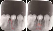
Measurement of root dimensions The straight line from A (cementoenamel junction) to B (midpoint of the radiographic apex) indicates the root length. The straight line from C to D indicates the root width at the level of two thirds of the length of the preoperative root canal. The straight line from E to F is the pulp space width at the same level. Measurements of root canal wall thickness were made by subtracting EF from CD. The straight line from G (distal apical end of the root) to H (mesial apical end of the root) indicates the apical foramen diameter."

Table 1

Demographics and clinical characteristics of the teeth (n=84)"

Variables Data
Age/years, $\bar x \pm s$ 10.25±2.13
Gender, n(%)
   Male 54 (64.3)
  Female 30 (35.7)
Tooth type, n(%)
  Anterior 37 (44.0)
  Premolar 47 (55.9)
Etiology, n(%)
  Broken central cusp 45 (53.6)
  Trauma 35 (41.7)
  Caries 4 (4.8)
Diagnosis, n(%)
  Pulp necrosis 5 (6.0)
  Acute apical abscess 15 (17.9)
  Asymptomatic apical periodontitis 5 (6.0)
  Chronic apical abscess 31 (36.9)
  Symptomatic apical periodontitis 28 (33.3)
Follow-up period/months, $\bar x \pm s$ 44.7±19.3

Table 2

Basic information of clinical failure teeth (n=16)"

Variables Data
Age/years, $\bar x \pm s$ 9.69±2.41
Gender, n(%)
  Male 10 (62.5)
  Female 6 (37.5)
Tooth type, n(%)
  Anterior 11 (68.8)
  Premolar 5 (31.2)
Etiology, n(%)
  Broken central cusp 5 (68.8)
  Trauma 11 (31.2)
Diagnosis, n(%)
  Acute apical abscess 1 (6.3)
  Asymptomatic apical periodontitis 1 (6.3)
  Chronic apical abscess 8 (50.0)
  Symptomatic apical periodontitis 6 (37.5)

Figure 2

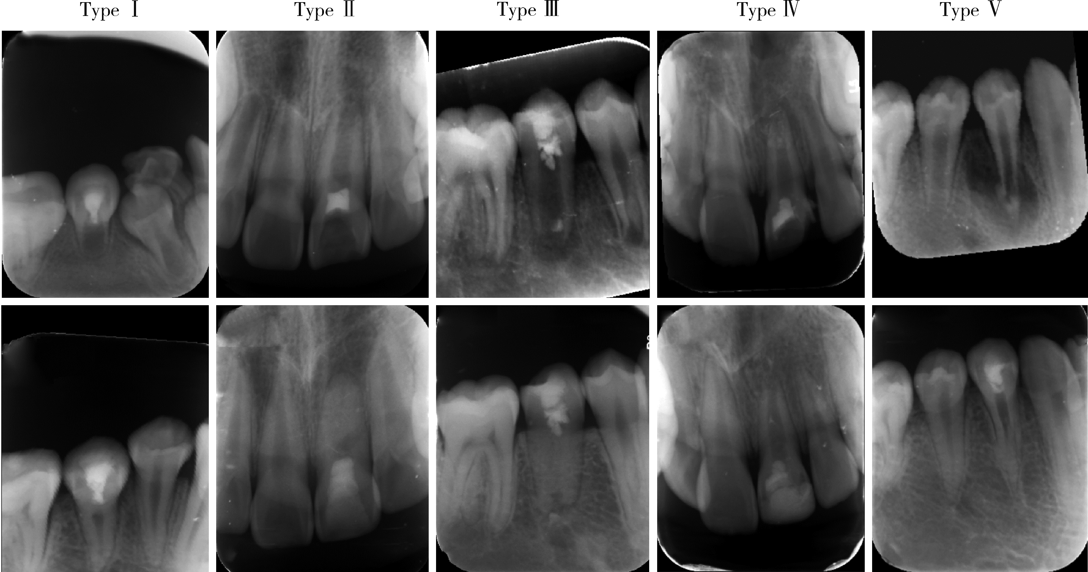
Five types of continued root development Type Ⅰ: Periapical radiograph after induced bleeding showing a wide apex of #29 (Upper); Periapical radiograph at 63-month follow-up showing a closed apical foramen and increased root length and root canal wall thickness (Lower). Type Ⅱ: Periapical radiograph after induced bleeding showing a wide apex of #9 (Upper); Periapical radiograph at 60-month follow-up showing a blunt and closed apical foramen and increased root canal wall thickeness (Lower). Type Ⅲ: Periapical radiograph after induced bleeding showing a wide apex of #29 (Upper); Periapical radiograph at 69-month follow-up showing a reduced apical foramen diameter and continued development of root length and/or root canal wall thickness (Lower). Type Ⅳ: Periapical radiograph after induced bleeding showing a wide apex of #9 (Upper); Periapical radiograph at 72-month follow-up showing no significant change in apical foramen diameter, root length or root canal wall thickness (Lower). Type Ⅴ: Preoperative periapical radiograph showing a wide apex of #28 (Upper); At 69-month follow-up, there was a segmented root development, with the apical portion of the root continuing to develop away from the main root (Lower)."

Table 3

Dimensions of different morphology root"

Dimensions of different morphology rootTypes of root morphologyP
Ⅰ (n=24) Ⅱ (n=16) Ⅲ (n=11) Ⅳ (n=13)
Increase of root length/% 32.51 5.35 16.78 5.94 <0.01
Increase of root canal wall thickness/% 53.53 39.16 43.19 12.29 <0.01

Table 4

Statistical analysis of potential contributing factors of root development"

Variables Failed group (n=13) Success group (n=55) P
Age/years, $\bar x \pm s$ 9.69±2.56 10.56±1.90 0.170
Etiology, n 0.003
  Broken central cusp 3 37
  Trauma 10 14
Develop stage, $\bar x \pm s$ 8.00±0.41 7.91±0.62 0.523
Induced bleeding, n 0.014
  Orifice of the root 2 26
  Middle of the root 4 17
  Tip of the root 7 12
Canal sealing material, n 0.322
  Bioceramic materials 11 51
  Glass ionomer cement 2 4
Period of canal sealing/d, $\bar x \pm s$ 42.00±30.37 36.83±23.26 0.501
1 Cehreli ZC , Isbitiren B , Sara S , et al. Regenerative endodontic treatment (revascularization) of immature necrotic molars medicated with calcium hydroxide: A case series[J]. J Endod, 2011, 37 (9): 1327- 1330.
doi: 10.1016/j.joen.2011.05.033
2 Iwaya SI , Ikawa M , Kubota M . Revascularization of an immature permanent tooth with apical periodontitis and sinus tract[J]. Dent Traumatol, 2001, 17 (4): 185- 187.
doi: 10.1034/j.1600-9657.2001.017004185.x
3 Almutairi W , Yassen GH , Aminoshariae A , et al. Regenerative endodontics: A systematic analysis of the failed cases[J]. J Endod, 2019, 45 (5): 567- 577.
doi: 10.1016/j.joen.2019.02.004
4 Saoud TM , Zaazou A , Nabil A , et al. Clinical and radiographic outcomes of traumatized immature permanent necrotic teeth after revascularization/revitalization therapy[J]. J Endod, 2014, 40 (12): 1946- 1952.
doi: 10.1016/j.joen.2014.08.023
5 Torabinejad M , Nosrat A , Verma P , et al. Regenerative endodontic treatment or mineral trioxide aggregate apical plug in teeth with necrotic pulps and open apices: A systematic review and meta-analysis[J]. J Endod, 2017, 43 (11): 1806- 1820.
doi: 10.1016/j.joen.2017.06.029
6 Shamszadeh S , Asgary S , Nosrat A . Regenerative endodontics: A scientometric and bibliometric analysis[J]. J Endod, 2019, 45 (3): 272- 280.
doi: 10.1016/j.joen.2018.11.010
7 Dohan DM , Choukroun J , Diss A , et al. Platelet-rich fibrin (PRF): A second-generation platelet concentrate. Part Ⅰ: Technological concepts and evolution[J]. Oral Surg Oral Med Oral pathol Oral Radiol Endod, 2006, 101 (3): e37- e44.
doi: 10.1016/j.tripleo.2005.07.008
8 Chan EKM , Desmeules M , Cielecki M , et al. Longitudinal cohort study of regenerative endodontic treatment for immature necrotic permanent teeth[J]. J Endod, 2017, 43 (3): 395- 400.
doi: 10.1016/j.joen.2016.10.035
9 Bose R , Nummikoski P , Hargreaves K . A retrospective evaluation of radiographic outcomes in immature teeth with necrotic root canal systems treated with regenerative endodontic procedures[J]. J Endod, 2009, 35 (10): 1343- 1349.
doi: 10.1016/j.joen.2009.06.021
10 Song M , Cao Y , Shin SJ , et al. Revascularization-associated intracanal calcification: Assessment of prevalence and contributing factors[J]. J Endod, 2017, 43 (12): 2025- 2033.
doi: 10.1016/j.joen.2017.06.018
11 Lin J , Zeng Q , Wei X , et al. Regenerative endodontics versus apexification in immature permanent teeth with apical periodontitis: A prospective randomized controlled study[J]. J Endod, 2017, 43 (11): 1821- 1827.
doi: 10.1016/j.joen.2017.06.023
12 Chen MYH , Chen KL , Chen CA , et al. Responses of immature permanent teeth with infected necrotic pulp tissue and apical periodontitis/abscess to revascularization procedures[J]. Int Endod J, 2012, 45 (3): 294- 305.
doi: 10.1111/j.1365-2591.2011.01978.x
13 Nolla CM . The development of the permanent teeth[J]. J Dent Child, 1960, 27, 254- 266.
14 Jung IY , Kim ES , Lee CY , et al. Continued development of the root separated from the main root[J]. J Endod, 2011, 37 (5): 711- 714.
doi: 10.1016/j.joen.2011.01.015
15 Banchs F , Trope M . Revascularization of immature permanent teeth with apical periodontitis: New treatment protocol?[J]. J Endod, 2004, 30 (4): 196- 200.
doi: 10.1097/00004770-200404000-00003
16 Civinini R , Macera A , Redl B , et al. The use of autologous blood-derived growth factors in bone regeneration[J]. Clin Cases Miner Bone Metab, 2011, 8 (1): 25- 31.
17 Yassen GH , Sabrah AHA , Eckert GJ , et al. Effect of different endodontic regeneration protocols on wettability, roughness, and chemical composition of surface dentin[J]. J Endod, 2015, 41 (6): 956- 960.
doi: 10.1016/j.joen.2015.02.023
18 Shah N , Logani A , Bhaskar U , et al. Efficacy of revascularization to induce apexification/apexogensis in infected, nonvital, immature teeth: A pilot clinical study[J]. J Endod, 2008, 34 (8): 919- 925.
doi: 10.1016/j.joen.2008.05.001
[1] Juan GAO,Hang-miao LV,Hui-min MA,Yi-jiao ZHAO,Xiao-tong LI. Evaluation of root resorption after surgical orthodontic treatment of skeletal Class Ⅲ malocclusion by three-dimensional volumetric measurement with cone-beam CT [J]. Journal of Peking University (Health Sciences), 2022, 54(4): 719-726.
[2] LU Jin-hui, QIAN Jun, LIU He, ZHU Jun-xia. Clinical study on autologus platelet-rich fibrin aided revascularization of immature permanent teeth [J]. Journal of Peking University(Health Sciences), 2018, 50(4): 672-679.
[3] ZHAO Yi-jiao, LIU Yi, SUN Yu-chun, WANG Yong. Three-dimensional data fusion method for tooth crown and root based on curvature continuity algorithm#br# [J]. Journal of Peking University(Health Sciences), 2017, 49(4): 719-723.
[4] ZHAO Yi-jiao, WANG Si-wei, LIU Yi, WANG Yong. A method for rapid extracting three-dimensional root model of vivo tooth from cone beam computed tomography data based on the anatomical characteristics of periodontal ligament [J]. Journal of Peking University(Health Sciences), 2017, 49(1): 54-059.
[5] WEN Fu-jia,CHEN Gui,LIU Yi. Morphological analysis of roots and alveolar bone changes after upper anterior #br# retraction with maximum anchorage based on cone-beam computed tomography [J]. Journal of Peking University(Health Sciences), 2016, 48(4): 702-708.
[6] ZHEN Min, HU Wen-jie, RONG Qi-guo. Finite element analysis of the maxillary central incisor with crown lengthening surgery and post-core restoration in management of crown-root fracture [J]. Journal of Peking University(Health Sciences), 2015, 47(6): 1015-1021.
Viewed
Full text


Abstract

Cited

  Shared   
  Discussed   
No Suggested Reading articles found!