Journal of Peking University (Health Sciences) ›› 2025, Vol. 57 ›› Issue (5): 903-910. doi: 10.19723/j.issn.1671-167X.2025.05.014

Previous Articles     Next Articles

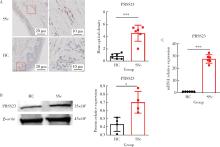
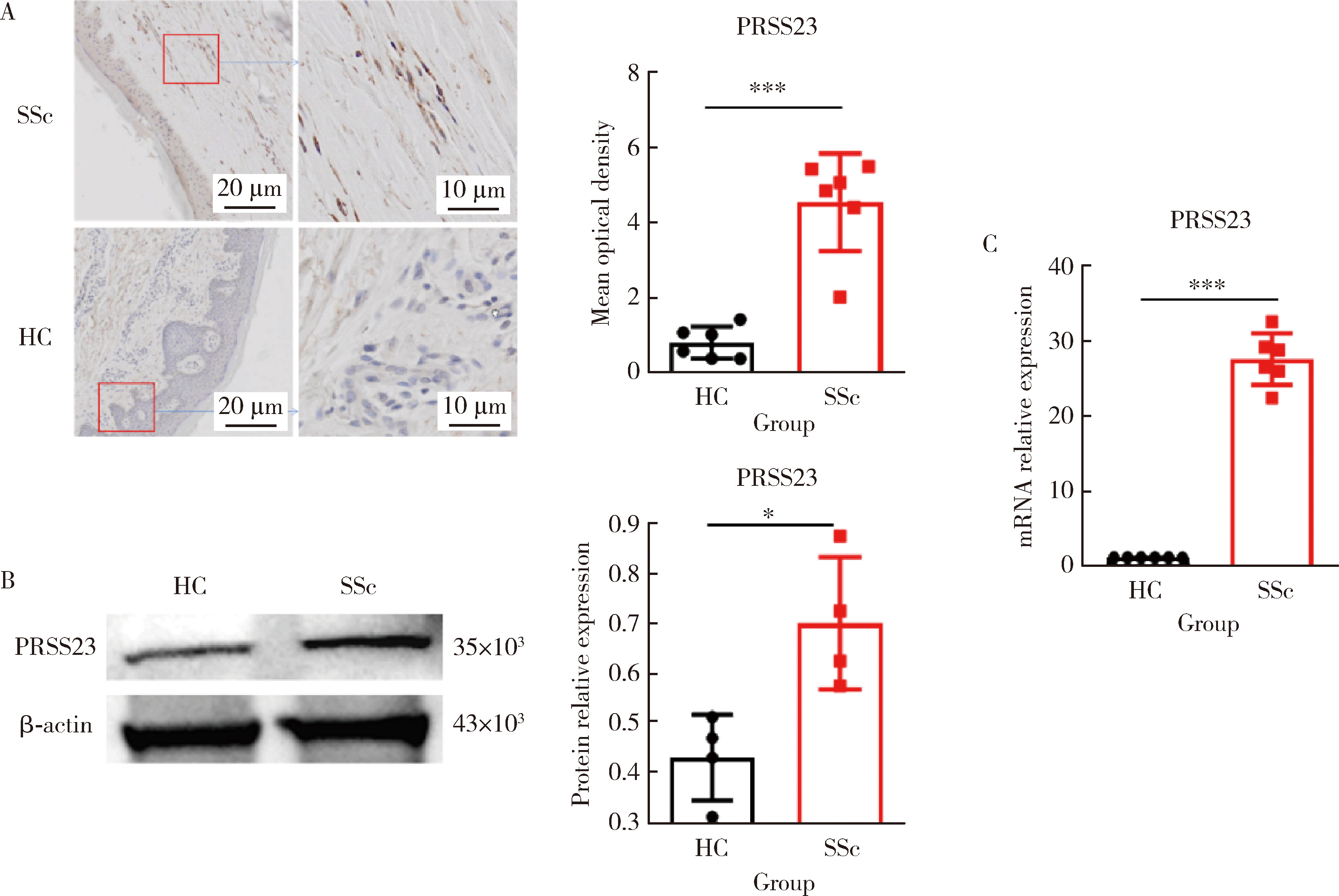
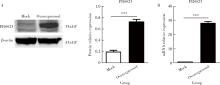
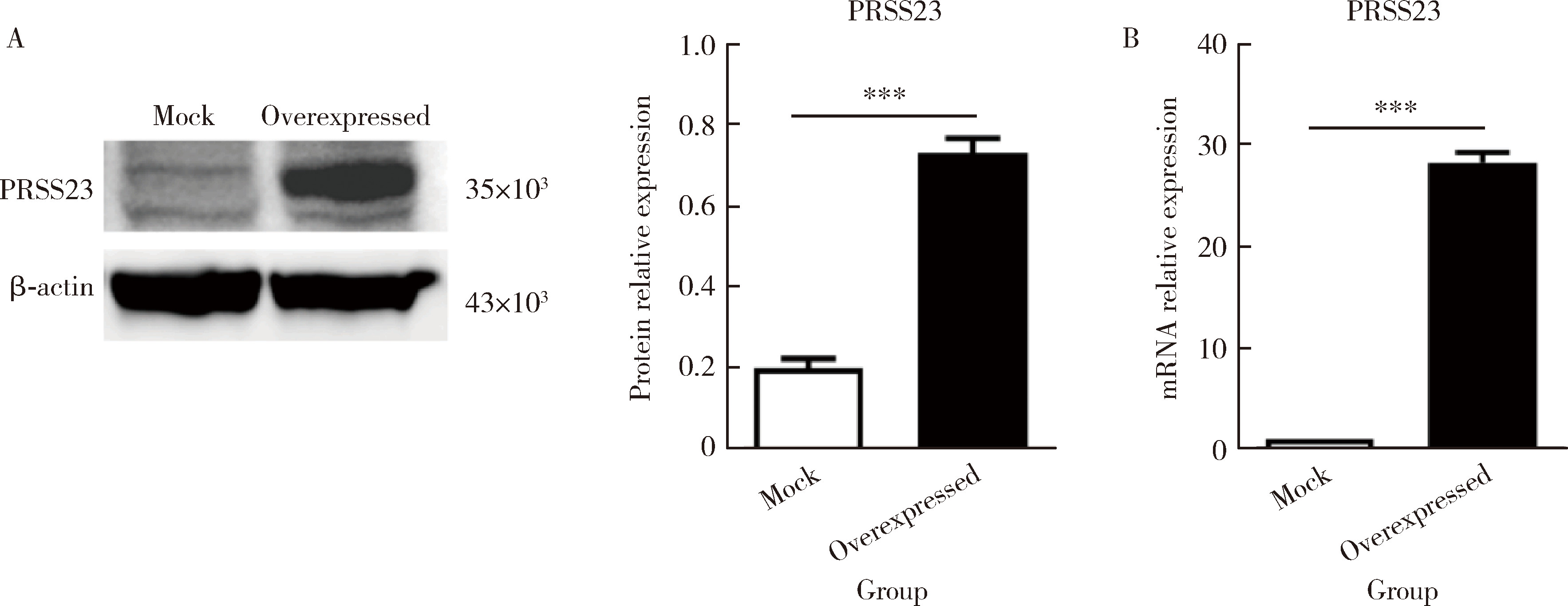
Pathogenesis and mechanism of serine protease 23 in skin fibrosis of systemic sclerosis

Xiandun YUAN, Zhaohua LI, Dan XU, Ting LI, Dan FANG, Rong MU*()   

  1. Department of Rheumatology and Immunology, Peking University Third Hospital, Beijing 100191, China
  • Received:2023-02-28 Online:2025-10-18 Published:2024-04-10
  • Contact: Rong MU
  • Supported by:
    the National Natural Science Foundation of China(81771706)

RICH HTML


Abstract: Objective: It has been reported that the mRNA expression of serine protease 23 (PRSS23) was increased in skin fibroblasts from systemic sclerosis patients (SSc). The purpose of this study is to explore the pathogenetic effect and mechanism of PRSS23 in skin fibrosis of SSc. Methods: The expression of PRSS23 in skin tissues from the SSc patients and healthy controls was detected by immunohisto-chemistry. Fibroblasts isolated from fresh skin tissue were used to detect the expression of PRSS23 by real-time quantitative PCR (RT-qPCR) and Western blot. Overexprssion of PRSS23 in BJ, the fibroblasts cell line of skin, was constructed by lentivirus. After stimulation with 400 μmol/L hydrogen peroxide for 12 h, Annexin V/7-AAD staining was used to detect apoptosis of fibroblasts; flow cytometry and Western blot were used to detect the expression of apoptosis-related protein cleaved Caspase-3. The expression of interleukin 6 (IL-6) and tumor necrosis factor alpha (TNF-α) in fibroblasts was detected by RT-qPCR and enzyme linked immunosorbent assay (ELISA). Results: Compared with the healthy controls, the expression of PRSS23 in skin tissues of the SSc patients was significantly increased [4.952 (3.806-5.439) vs. 0.806 (0.395-1.173), P < 0.001], and fibroblast was the main cell that expressed PRSS23. The mRNA [27.59 (25.02-30.00) vs. 1.00, P < 0.001] and protein [0.675 (0.587-0.837) vs. 0.451 (0.342-0.502), P=0.029] of PRSS23 in skin fibroblasts isolated from the SSc patients were significantly up-regulated. Compared with the control group, the anti-apoptotic ability of skin fibroblasts overexpressing PRSS23 was enhanced, and the proportion of apoptotic cells was significantly reduced after hydrogen peroxide induction [(5.043±1.097)% vs. (17.480±3.212)%, P=0.022], the expression of apoptosis-related protein cleaved Caspase-3 was also markedly reduced [(0.718±0.022) vs. (1.422±0.105), P=0.003]. In addition, the mRNA [(99.780±1.796) vs. (1.000±0.004), P < 0.001] and protein [(211.600±2.431) ng/L vs. (65.930±1.768) ng/L, P < 0.001] of IL-6 in the fibroblasts overexpressing PRSS23 were significantly up-regulated; the mRNA[(3.555±0.555) vs. (1.000±0.004), P < 0.001] and protein levels [(41.190±0.949) ng/L vs. (31.150±0.360) ng/L, P < 0.001] of TNF-α in the fibroblasts overexpressing PRSS23 were also significantly up-regulated. Conclusion: The expression of PRSS23 is increased in skin fibroblasts of SSc patients. PRSS23 can inhibit cell apoptosis, promote the secretion of inflammatory factors such as IL-6 and TNF-α, and regulate the process that skin fibroblasts transform into pro-inflammatory type. So, PRSS23 is associated with the development of skin fibrosis.

Key words: Systemic sclerosis, Fibroblasts, Serine proteases, Apoptosis

CLC Number: 

  • R593.2

Table 1

Primer sequences for IL-6, TNF-α and PRSS23"

Gene GenBank ID Primer sequences (5′ to 3′) Length/bp
IL-6 NM_000600 Forward: 5′-ACTCACCTCAGAACGAATTG-3′
Reversed: 5′-CCATCTTGGAAGGTTCAGGTTG-3′
149
TNF-α NM_000594 Forward: 5′-CCTCTAATCAGCCTCTG-3′
Reversed: 5′-GAGGACCTGGAGTAGAG-3′
220
PRSS23 NM_009104 Forward: 5′-TGTGCTGGGCAAGTGAG-3′
Reversed: 5′-AGTTCCCTTATGACTGGGG-3′
174

Figure 1

Expression of PRSS23 in fibroblasts from SSc patients' skin tissue A, immunohistochemical staining was performed to compare the expression of PRSS23 in skin tissue slices (the magnification is 20× for the left image and 40× for the right image, and the right image shows an enlarged view of the red box in the left image). Compared with the healthy controls, PRSS23 positive expression in skin tissue slices from SSc patients was significantly increased (n=6, P < 0.001). B, protein immunoblotting was used to quantify the protein expression of PRSS23 in human skin fibroblasts. Compared with the healthy controls, PRSS23 protein expression in skin tissue from SSc patients was significantly increased (n=6, P=0.029). C, RT-qPCR was used to quantify the mRNA expression of PRSS23 in human skin fibroblasts. Compared with the healthy controls, PRSS23 mRNA expression in skin tissue from SSc patients was significantly increased (n=6, P < 0.001), and the statistical method used was the Mann-Whitney U test. * P < 0.05, *** P < 0.001. HC, healthy control; SSc, systemic sclerosis; PRSS23, serine protease 23; RT-qPCR, quantitative real-time PCR."

Table 2

Expression of PRSS23 in fibroblasts of skin tissue of SSc patients"

Items Sample size (n) Value, M (P25-P75)
Mean optical density
  HC 6 0.806 (0.395-1.173)
  SSc 6 4.952 (3.806-5.439)***
Protein relative expression
  HC 4 0.451 (0.342-0.502)
  SSc 4 0.675 (0.587-0.837)*
mRNA relative expression
  HC 6 1.000
  SSc 6 27.590 (25.020-30.000)***

Figure 2

Overexpression of PRSS23 in skin fibroblasts transfected with PRSS23 plasmid A, quantification of PRSS23 protein expression levels in fibroblasts transfected with PRSS23 plasmid or empty vector by Western blot. The PRSS23 protein expression was significantly increased in the overexpression group compared to the empty vector control group (n=4, P < 0.001). B, quantification of PRSS23 mRNA expression levels in two types of cells by RT-qPCR. The mRNA expression of PRSS23 was significantly higher in the overexpression group than in the empty vector control group (n=4, P < 0.001), indicating a successful transfection. The statistical method used was unpaired t test. *** P < 0.001. Mock, empty vector control group; Overexpressed, PRSS23 overexpression group; PRSS23, serine protease 23."

Table 3

Expression of PRSS23 in skin fibroblasts transfected with PRSS23 plasmid"

Items Sample size (n) Value, ${\bar x}$±SE
Protein relative expression (PRSS23)
  Mock 3 0.199±0.023
  Overexpressed 3 0.730±0.037***
mRNA relative expression
  Mock 4 1.000
  Overexpressed 4 28.160±0.507***

Figure 3

Overexpression of PRSS23 inhibits apoptosis of skin fibroblasts A, BJ cells transfected with PRSS23 plasmid or empty vector were treated with 400 μmol/L hydrogen peroxide for 12 h to induce apoptosis. After staining with the Annexin V-FITC/7-AAD apoptosis detection kit, cells were analyzed by flow cytometry. Left panel shows the flow cytometry results, and the right panel shows the statistical analysis of apoptosis rate. Compared to the control group, the overexpression of PRSS23 decreased the apoptosis rate (n=4, P=0.022). B, after hydrogen peroxide treatment, cells were stained with anti-cleaved Caspase-3 antibody and analyzed by flow cytometry. Left panel shows the flow cytometry results, and the right panel shows the statistical analysis of cleaved Caspase-3 positive cells. Compared to the control group, the overexpression of PRSS23 reduced the ratio of cleaved Caspase-3 positive cells (n=4, P=0.026). C, the expression level of cleaved Caspase-3 in the two cell groups was quantified by Western blot. Top panel shows the Western blotting results, and the bottom panel shows the relative expression levels of cleaved Caspase-3 (n=4, P=0.003). The statistical analysis was performed using the unpaired t test. * P < 0.05, ** P < 0.01. Mock, empty vector control group; Overexpressed, PRSS23 overexpression group; PRSS23, serine protease 23; FITC, fluorescein isothiocyanate."

Table 4

Determination of the inhibition of apoptosis rate and related apoptotic proteins by PRSS23 in skin fibroblasts"

Items Sample size (n) Value, ${\bar x}$±SE
Apoptosis ratio
  Mock 3 (17.480±3.212)%
  Overexpressed 3 (5.043±1.097)%*
Positive cell ratio
  Mock 4 (12.510±1.472)%
  Overexpressed 4 (7.460±0.866)%*
Protein relative expression (cleaved Caspase 3)
  Mock 4 1.422±0.105
  Overexpressed 4 0.718±0.022**

Figure 4

Overexpression of PRSS23 promotes the secretion of pro-inflammatory cytokines IL-6 and TNF-α in skin fibroblasts A, quantification of IL-6 mRNA expression in cells using RT-qPCR. Compared to the control group with empty vector, the PRSS23 overexpression group showed a significant increase in IL-6 mRNA expression (n=4, P < 0.001). B, quantification of TNF-α mRNA expression in cells using RT-qPCR. Compared to the control group with empty vector, the PRSS23 overexpression group showed a significant increase in TNF-α mRNA expression (n=4, P=0.004). C, quantification of IL-6 protein concentration in cell supernatants using ELISA. Compared to the control group with empty vector, the PRSS23 overexpression group showed a significant increase in IL-6 protein concentration (n=4, P < 0.001). D, quantification of TNF-α protein concentration in cell supernatants using ELISA. Compared to the control group with empty vector, the PRSS23 overexpression group showed a significant increase in TNF-α protein concentration (n=4, P < 0.001). Statistical analysis was performed using unpaired t test. ** P < 0.01, *** P < 0.001. Mock, empty vector control group; Overexpressed, PRSS23 overexpression group; PRSS23, serine protease 23; IL-6, interleukin-6; TNF-α, tumor necrosis factor-alpha; RT-qPCR, quantitative real-time PCR; ELISA, enzyme linked immunosorbent assay."

Table 5

Effect of PRSS23 on the secretion of inflammatory cytokines in skin fibroblasts"

Items Sample size (n) Value, ${\bar x}$±SE
IL-6 mRNA relative expression
  Mock 3 1.000
  Overexpressed 3 99.780±1.796***
TNF-α mRNA relative expression
  Mock 4 1.000
  Overexpressed 4 3.555±0.555**
IL-6 protein content (cell supernatant)/(ng/L)
  Mock 4 65.930±1.768
  Overexpressed 4 211.600±2.431***
TNF-α protein content (cell supernatant)/(ng/L)
  Mock 4 31.150±0.360
  Overexpressed 4 41.190±0.949***
1
Domsic RT, Rodriguez-Reyna T, Lucas M, et al. Skin thickness progression rate: A predictor of mortality and early internal organ involvement in diffuse scleroderma[J]. Ann Rheum Dis, 2011, 70(1): 104- 109.

doi: 10.1136/ard.2009.127621
2
Hinz B, Lagares D. Evasion of apoptosis by myofibroblasts: A hallmark of fibrotic diseases[J]. Nat Rev Rheumatol, 2020, 16(1): 11- 31.

doi: 10.1038/s41584-019-0324-5
3
Dodi AE, Ajayi IO, Chang C, et al. Regulation of fibroblast Fas expression by soluble and mechanical pro-fibrotic stimuli[J]. Respir Res, 2018, 19(1): 91.

doi: 10.1186/s12931-018-0801-4
4
Xu C, Meng LB, Duan YC, et al. Screening and identification of biomarkers for systemic sclerosis via microarray technology[J]. Int J Mol Med, 2019, 44(5): 1753- 1770.
5
Tabib T, Huang M, Morse N, et al. Myofibroblast transcriptome indicates SFRP2(hi) fibroblast progenitors in systemic sclerosis skin[J]. Nat Commun, 2021, 12(1): 4384.

doi: 10.1038/s41467-021-24607-6
6
Kanno Y. The role of fibrinolytic regulators in vascular dysfunction of systemic sclerosis[J]. Int J Mol Sci, 2019, 20(3): 619.

doi: 10.3390/ijms20030619
7
Menou A, Duitman J, Crestani B. The impaired proteases and anti-proteases balance in idiopathic pulmonary fibrosis[J]. Matrix Biol, 2018(68/69): 382- 403.
8
Page MJ, Di Cera E. Serine peptidases: Classification, structure and function[J]. Cell Mol Life Sci, 2008, 65(7/8): 1220- 1236.
9
Han B, Yang Y, Chen J, et al. PRSS23 knockdown inhibits gastric tumorigenesis through EIF2 signaling[J]. Pharmacol Res, 2019, 142, 50- 57.

doi: 10.1016/j.phrs.2019.02.008
10
Rawlings ND, Barrett AJ, Bateman A. MEROPS: The peptidase database[J]. Nucleic Acids Res, 2010, 38(Database issue): D227- D233.
11
Deng CC, Hu YF, Zhu DH, et al. Single-cell RNA-seq reveals fibroblast heterogeneity and increased mesenchymal fibroblasts in human fibrotic skin diseases[J]. Nat Commun, 2021, 12(1): 3709.

doi: 10.1038/s41467-021-24110-y
12
Xiong J, Li Y, Tan X, et al. Targeting PRSS23 with tipranavir induces gastric cancer stem cell apoptosis and inhibits growth of gastric cancer via the MKK3/p38 MAPK-IL24 pathway[J]. Acta Pharmacol Sin, 2024, 45(2): 405- 421.

doi: 10.1038/s41401-023-01165-9
13
Fan L, Wang Q, Liu R, et al. Citrullinated fibronectin inhibits apoptosis and promotes the secretion of pro-inflammatory cytokines in fibroblast-like synoviocytes in rheumatoid arthritis[J]. Arthritis Res Ther, 2012, 14(6): R266.

doi: 10.1186/ar4112
14
Chaudhuri V, Zhou L, Karasek M. Inflammatory cytokines induce the transformation of human dermal microvascular endothelial cells into myofibroblasts: A potential role in skin fibrogenesis[J]. J Cutan Pathol, 2007, 34(2): 146- 153.

doi: 10.1111/j.1600-0560.2006.00584.x
15
Johnson BZ, Stevenson AW, Prêle CM, et al. The role of IL-6 in skin fibrosis and cutaneous wound healing[J]. Biomedicines, 2020, 8(5): 101.

doi: 10.3390/biomedicines8050101
16
Borthwick LA, Wynn TA, Fisher AJ. Cytokine mediated tissue fibrosis[J]. Biochim Biophys Acta, 2013, 1832(7): 1049- 1060.

doi: 10.1016/j.bbadis.2012.09.014
17
Li Y, Zhao J, Yin Y, et al. The role of IL-6 in fibrotic diseases: Molecular and cellular mechanisms[J]. Int J Biol Sci, 2022, 18(14): 5405- 5414.

doi: 10.7150/ijbs.75876
18
Khan K, Xu S, Nihtyanova S, et al. Clinical and pathological significance of interleukin 6 overexpression in systemic sclerosis[J]. Ann Rheum Dis, 2012, 71(7): 1235- 1242.

doi: 10.1136/annrheumdis-2011-200955
19
Khanna D, Denton CP, Jahreis A, et al. Safety and efficacy of subcutaneous tocilizumab in adults with systemic sclerosis (faSScinate): A phase 2, randomised, controlled trial[J]. Lancet, 2016, 387(10038): 2630- 2640.

doi: 10.1016/S0140-6736(16)00232-4
20
Bahcecioglu IH, Koca SS, Poyrazoglu OK, et al. Hepatoprotective effect of infliximab, an anti-TNF-alpha agent, on carbon tetrachloride-induced hepatic fibrosis[J]. Inflammation, 2008, 31(4): 215- 221.

doi: 10.1007/s10753-008-9067-1
21
Messadi DV, Doung HS, Zhang Q, et al. Activation of NFkappaB signal pathways in keloid fibroblasts[J]. Arch Dermatol Res, 2004, 296(3): 125- 133.
22
Zhu G, Cai J, Zhang J, et al. Abnormal nuclear factor (NF)-kappaB signal pathway and aspirin inhibits tumor necrosis factor alpha-induced NF-kappaB activation in keloid fibroblasts[J]. Dermatol Surg, 2007, 33(6): 697- 708.
23
Li X, Sun Z, Peng G, et al. Single-cell RNA sequencing reveals a pro-invasive cancer-associated fibroblast subgroup associated with poor clinical outcomes in patients with gastric cancer[J]. Theranostics, 2022, 12(2): 620- 638.

doi: 10.7150/thno.60540
24
Solé-Boldo L, Raddatz G, Schütz S, et al. Single-cell transcriptomes of the human skin reveal age-related loss of fibroblast priming[J]. Commun Biol, 2020, 3(1): 188.

doi: 10.1038/s42003-020-0922-4
25
Griffin MF, desJardins-Park HE, Mascharak S, et al. Understanding the impact of fibroblast heterogeneity on skin fibrosis[J]. Dis Model Mech, 2020, 13(6): dmm044164.

doi: 10.1242/dmm.044164
26
Sato Y, Yanagita M. Resident fibroblasts in the kidney: A major driver of fibrosis and inflammation[J]. Inflamm Regen, 2017, 37, 17.

doi: 10.1186/s41232-017-0048-3
[1] Wei PAN, Yun LI, Junjia LUO, Chun LI, Hua YE, Xue LI, Yuan JIA. COVID-19 vaccines efficacy and infection features in patients with systemic sclerosis: A single-center cohort study [J]. Journal of Peking University (Health Sciences), 2024, 56(6): 1041-1046.
[2] Kelin ZHAO, Xue XIA, Naixu SHI, Han ZHOU, Jingwen GAI, Ping LI. Expression and significance of ferroptosis marker 4-HNE in in vitro model of systemic sclerosis [J]. Journal of Peking University (Health Sciences), 2024, 56(6): 950-955.
[3] Shan HE,Xin CHEN,Qi CHENG,Lingjiang ZHU,Peiyu ZHANG,Shuting TONG,Jing XUE,Yan DU. Tofacitinib inhibits the transformation of lung fibroblasts into myofibroblasts through JAK/STAT3 pathway [J]. Journal of Peking University (Health Sciences), 2024, 56(3): 505-511.
[4] Wen-gen LI,Xiao-dong GU,Rui-qiang WENG,Su-dong LIU,Chao CHEN. Expression and clinical significance of plasma exosomal miR-34-5p and miR-142-3p in systemic sclerosis [J]. Journal of Peking University (Health Sciences), 2023, 55(6): 1022-1027.
[5] Yun-chong LIU,Zong-long WU,Li-yuan GE,Tan DU,Ya-qian WU,Yi-meng SONG,Cheng LIU,Lu-lin MA. Mechanism of nuclear protein 1 in the resistance to axitinib in clear cell renal cell carcinoma [J]. Journal of Peking University (Health Sciences), 2023, 55(5): 781-792.
[6] Zhuo-hua LIN,Ru-yi CAI,Yang SUN,Rong MU,Li-gang CUI. Methodology and clinical use of superb microvascular imaging in assessing micro-circulation changes of fingertips in systemic sclerosis [J]. Journal of Peking University (Health Sciences), 2023, 55(4): 636-640.
[7] LOU Xue,LIAO Li,LI Xing-jun,WANG Nan,LIU Shuang,CUI Ruo-mei,XU Jian. Methylation status and expression of TWEAK gene promoter region in peripheral blood of patients with rheumatoid arthritis [J]. Journal of Peking University (Health Sciences), 2021, 53(6): 1020-1025.
[8] GAO Ya-dong,ZHU An,LI Lu-di,ZHANG Tao,WANG Shuo,SHAN Dan-ping,LI Ying-zi,WANG Qi. Cytotoxicity and underlying mechanism of evodiamine in HepG2 cells [J]. Journal of Peking University (Health Sciences), 2021, 53(6): 1107-1114.
[9] MA Xiang-bo,ZHANG Xue-wu,JIA Ru-lin,GAO Ying,LIU Hong-jiang,LIU Yu-fang,LI Ying-ni. Application of lymphocytes test in peripheral blood of patients with systemic sclerosis during the treatment [J]. Journal of Peking University (Health Sciences), 2021, 53(4): 721-727.
[10] GAO Hong-yu,MENG Huan-xin,HOU Jian-xia,HUANG Bao-xin,LI Wei. Expression and distribution of calprotectin in healthy and inflamed periodontal tissues [J]. Journal of Peking University (Health Sciences), 2021, 53(4): 744-749.
[11] Jing ZHAO,Feng SUN,Yun LI,Xiao-zhen ZHAO,Dan XU,Ying-ni LI,Yu-hui LI,Xiao-lin SUN. Significance of anti-tubulin-α-1C autoantibody in systemic sclerosis [J]. Journal of Peking University (Health Sciences), 2020, 52(6): 1009-1013.
[12] Lei-zhen SU,Jie CHEN,Xian LI,Ping JI. Effects of salinomycin on proliferation and apoptosis of oral squamous cell carcinoma [J]. Journal of Peking University (Health Sciences), 2020, 52(5): 902-906.
[13] Liang GENG,Jing LV,Jing FAN. Effect of Fei-Liu-Ping ointment combined with cyclophosphamide on lung cancer cell proliferation and acidic microenvironment [J]. Journal of Peking University (Health Sciences), 2020, 52(2): 247-253.
[14] Bing-qing SHI,Xiao-jing YUAN,Yu-ming ZHAO. Effects of mineral trioxide aggregate and ethanolic extracts of Shandong propolis on the biological properties of human dental pulp fibroblasts [J]. Journal of Peking University(Health Sciences), 2019, 51(6): 1108-1114.
[15] Hong-lin ZHU,Qian DU,Wei-lin CHEN,Xiao-xia ZUO,Quan-zhen LI,Si-jia LIU. Altered serum cytokine expression profile in systemic sclerosis and its regulatory mechanisms [J]. Journal of Peking University(Health Sciences), 2019, 51(4): 716-722.
Viewed
Full text


Abstract

Cited

  Shared   
  Discussed   
No Suggested Reading articles found!