Journal of Peking University (Health Sciences) ›› 2026, Vol. 58 ›› Issue (3): 641-649. doi: 10.19723/j.issn.1671-167X.2026.03.026

Previous Articles     Next Articles

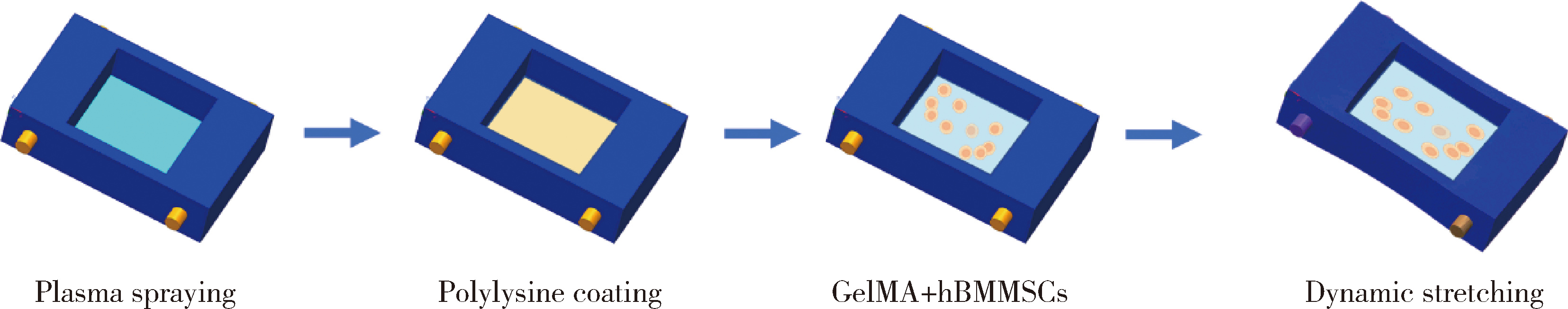
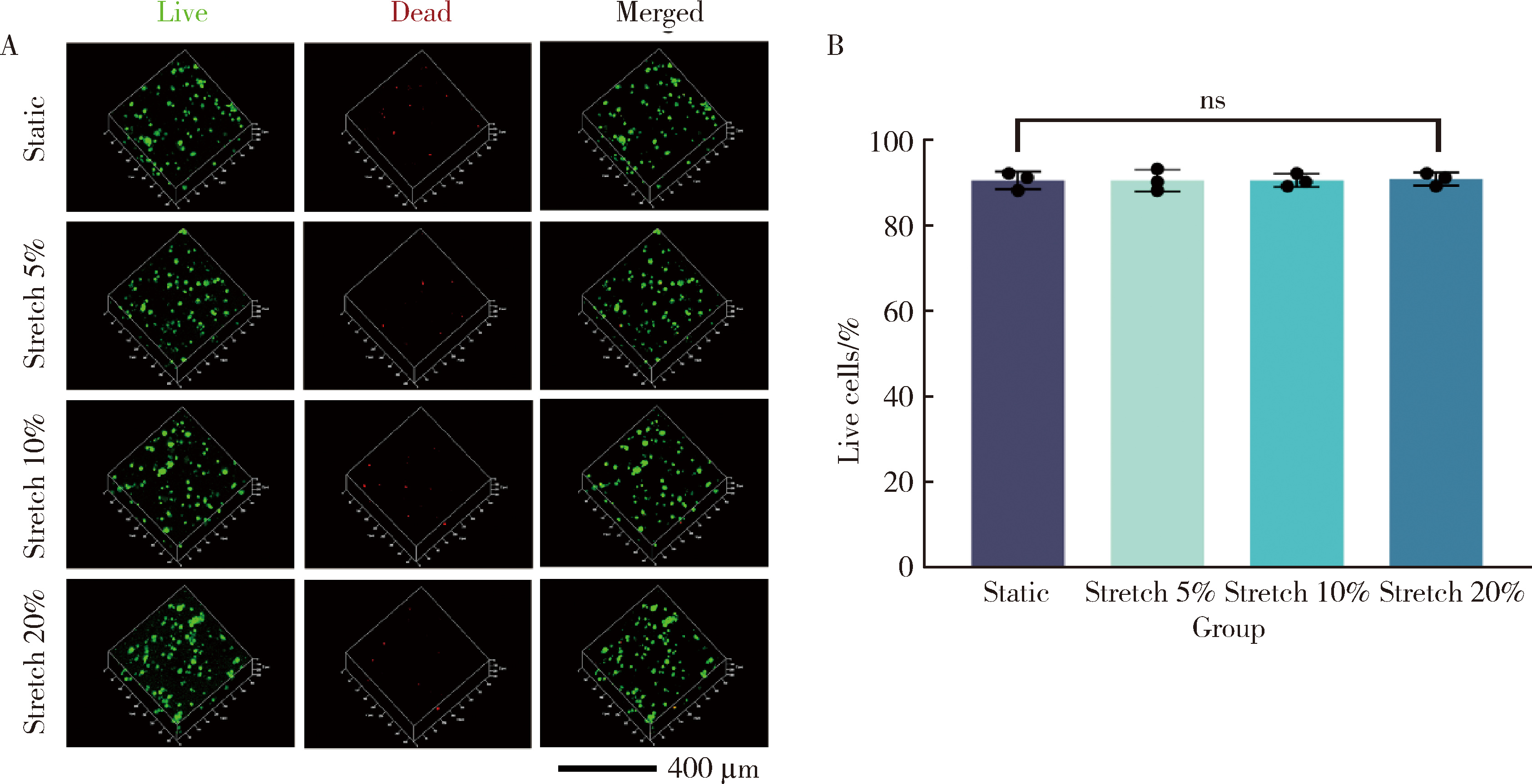
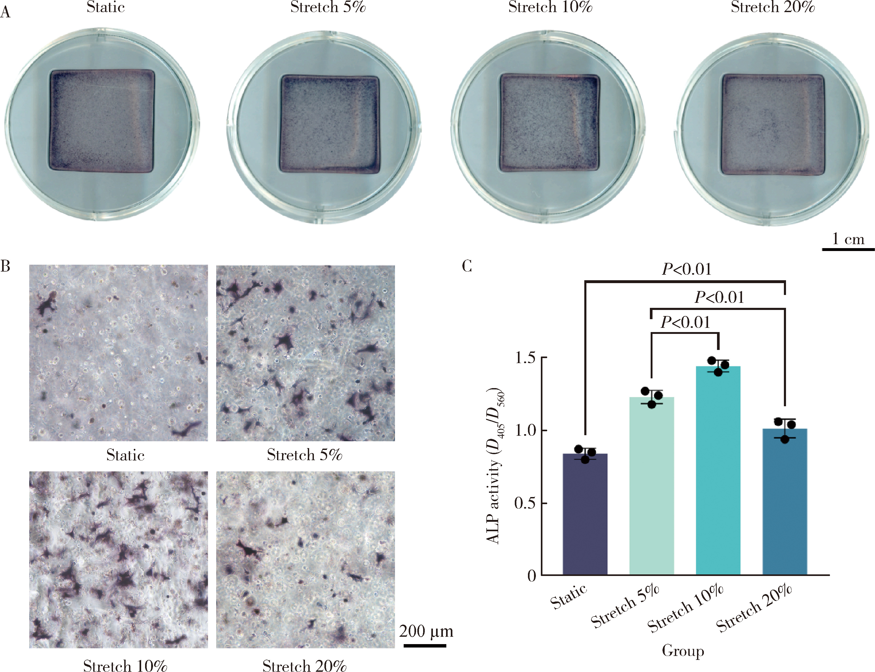
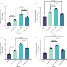
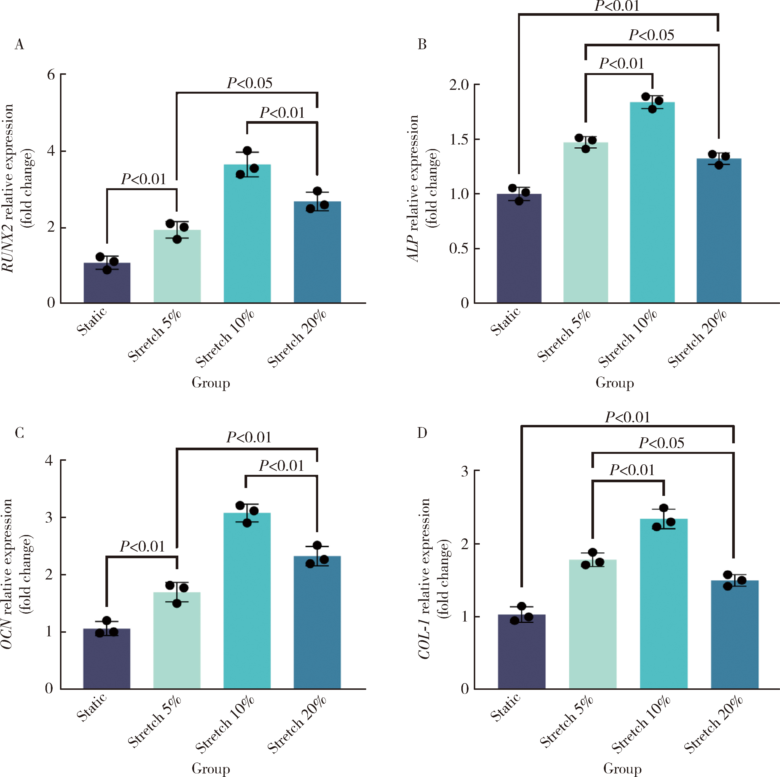
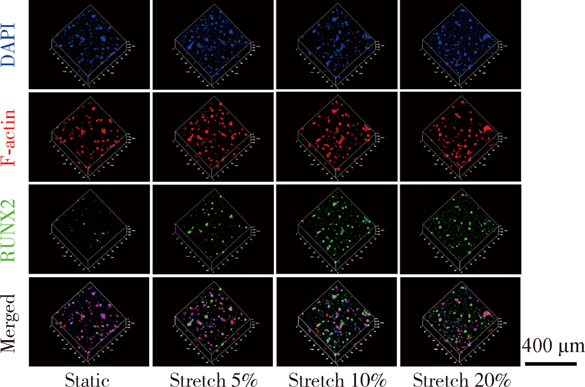
Dynamic stretching promotes osteogenic differentiation of human bone marrow mesenchymal stem cells in three-dimensional culture

Xiaoqiang BAI1,2, Zhiruo YUAN2, Yongsheng ZHOU1,2,*(), Longwei LV2,3,*()   

  1. 1. Institute of Medical Technology, Peking University Health Science Center, Beijing 100191, China
    2. Department of Prosthodontics, Peking University School and Hospital of Stomatology & National Center for Stomatology & National Clinical Research Center for Oral Diseases & National Engineering Research Center of Oral Biomaterials and Digital Medical Devices & Beijing Key Laboratory for Intelligent Biomanufacturing and Regeneration of Craniofacial Tissues & NHC Key Laboratory of Digital Stomatology, Beijing 100081, China
    3. Peking University Hospital of Stomatology Sanya Division(Sanya Stomatology Center), Sanya 572013, Hainan, China
  • Received:2024-01-16 Online:2026-06-18 Published:2026-04-09
  • Contact: Yongsheng ZHOU, Longwei LV
  • Supported by:
    the National Natural Science Foundation of China(82271032); the National Natural Science Foundation of China(82470943); the Hainan Provincial Natural Science Foundation of China(825YXQN602); the Peking University Clinical Scientist Training Program(BMU2025PYJH011)

RICH HTML


Abstract:

Objective: To explore the promotion of dynamic distraction on osteogenic differentiation of human bone marrow mesenchymal stem cells (hBMMSCs) in three-dimensional culture. Methods: Dynamic stretching in a three-dimensional culture for hBMMSCs was achieved with proportions set at 5%, 10%, and 20%, a frequency of 0.5 Hz, and a dynamic stretching duration of 2 hours per day. Static culture was used as the control group. Deformation of hBMMSCs induced by dynamic stretching was observed through cytoplasmic fluorescence staining. After 7 days of dynamic stretching culture, the impact of dynamic stretching on the viability of hBMMSCs was observed through cell live/dead staining. The effect of dynamic stretching on the osteogenic differentiation of hBMMSCs was observed through alkaline phosphatase (ALP) staining and the expression of osteogenic related genes and proteins after 7 days of dynamic stretching culture. Results: Dynamic stretching in a three-dimensional culture for hBMMSCs was successfully constructed, which could achieve different durations, frequencies, and ratios of dynamic stretching. Dynamic stretching led to deformation of hBMMSCs, and the greater the stretching ratio, the more pronounced the cell deformation, transitioning from a circular to a flat oval shape. After 7 days of dynamic stretching culture, hBMMSCs in the static control group and dynamic stretching groups were mostly green stained live cells, with only a few red stained dead cells. The difference in the proportion of live cells between the groups was not statistically significant (P>0.05). The ALP staining in the dynamic stretching group was deeper than that in the static control group, and the number of ALP staining positive cells observed under the microscope was higher. The expression of osteogenic related genes and proteins increased after 7 days of dynamic stretching culture, and the difference was statistically significant (P < 0.05). Among them, the dynamic stretching group with 10% had the deepest ALP staining, the highest number of positive cells, and the most significant increase in the expression of osteogenic related genes and proteins compared with the static control group. Conclusion: Dynamic stretching caused deformation of hBMMSCs without a significant impact on cell viability, and it could effectively promote the osteogenic differentiation of hBMMSCs.

Key words: Dynamic stretching, Three-dimensional culture, Human bone marrow mesenchymal stem cells, Osteogenic differentiation

CLC Number: 

  • R78

Table 1

Prime sequences used for quantitative real-time PCR"

Gene Forward primer(5′to 3′) Reverse prime(5′to 3′)
GAPDH GAAGGTGAAGGTCGGAGTC GAAGATGGTGATGGGATTTC
RUNX2 GAACCACAAGTGCGGTGCAA ACTGCTTGCAGCCTTAAATGACT
ALP ATGGGATGGGTGTCTCCACA CCACGAAGGGGAACTTGTC
OCN CATGAGAGCCCTCACACTCCTC CCTGCTTGGACACAAAGGCTGC
COL-1 ACAGGGCTCTAATGATGTTGA AGGCGTGATGGCTTATTTGT

Figure 1

Construction of dynamic stretching three-dimensional culture hBMMSCs, human bone marrow mesenchymal stem cells; GelMA, gelatin methacryloyl."

Figure 2

Deformation of hBMMSCs under dynamic stretching at different proportions"

Figure 3

Live/dead staining (A) and the proportion of live cells (B) of hBMMSCs under dynamic stretching at different proportions hBMMSCs, human bone marrow mesenchymal stem cells; ns, not significant."

Figure 4

ALP staining' s general view (A), microscope view (B) and relative quantification of activity (C) of hBMMSCs under dynamic stretching at different proportions hBMMSCs, human bone marrow mesenchymal stem cells; ALP, alkaline phosphatase."

Figure 5

Expression of osteogenic related genes of hBMMSCs under dynamic stretching at different proportions hBMMSCs, human bone marrow mesenchymal stem cells; RUNX2, runt-related transcription factor 2 (A); ALP, alkaline phosphatase (B); OCN, osteocalcin (C); COL-1, collagen-1 (D)."

Figure 6

Expression of osteogenic related proteins RUNX2 underdynamic stretching at different proportions hBMMSCs, human bone marrow mesenchymal stem cells; RUNX2, runt-related transcription factor 2. DAPI, 4′, 6-diamidino-2-phenylindole."

Figure 7

Expression of osteogenic related proteins OCN under dynamic stretching at different proportions hBMMSCs, human bone marrow mesenchymal stem cells; OCN, osteocalcin. DAPI, 4′, 6-diamidino-2-phenylindole."

Figure 8

Expression of osteogenic related proteins COL-1 under dynamic stretching at different proportions hBMMSCs, human bone marrow mesenchymal stem cells; COL-1, collagen-1. DAPI, 4′, 6-diamidino-2-phenylindole."

Figure 9

Semi quantitative analysis of the expression of osteogenic related proteins RUNX2(A), OCN(B), and COL-1(C) of hBMMSCs under dynamic stretching at different proportions hBMMSCs, human bone marrow mesenchymal stem cells; RUNX2, runt-related transcription factor 2; OCN, osteocalcin; COL-1, collagen-1."

1
Hartmann A , Hildebrandt H , Younan Z , et al. Long-term results in three-dimensional, complex bone augmentation procedures with customized titanium meshes[J]. Clin Oral Implants Res, 2022, 33 (11): 1171- 1181.

doi: 10.1111/clr.14000
2
Khachatryan L , Khachatryan G , Hakobyan G . The treatment of lower jaw defects using vascularized fibula graft and dental implants[J]. J Craniofacial Surg, 2018, 29 (8): 2214- 2217.

doi: 10.1097/SCS.0000000000005015
3
Fernandez de Grado G , Keller L , Idoux-Gillet Y , et al. Bone substitutes: A review of their characteristics, clinical use, and perspectives for large bone defects management[J]. J Tissue Eng, 2018, 9, 2041731418776819.

doi: 10.1177/2041731418776819
4
Tang XY , Wu S , Wang D , et al. Human organoids in basic research and clinical applications[J]. Sig Transduct Target Ther, 2022, 7, 168.

doi: 10.1038/s41392-022-01024-9
5
Kratochvil MJ , Seymour AJ , Li TL , et al. Engineered materials for organoid systems[J]. Nat Rev Mater, 2019, 4 (9): 606- 622.

doi: 10.1038/s41578-019-0129-9
6
Zhang J , Griesbach J , Ganeyev M , et al. Long-term mechanical loading is required for the formation of 3D bioprinted functional osteocyte bone organoids[J]. Biofabrication, 2022, 14 (3): 035018.

doi: 10.1088/1758-5090/ac73b9
7
Boccafoschi F , Mosca C , Ramella M , et al. The effect of mechanical strain on soft (cardiovascular) and hard (bone) tissues: Common pathways for different biological outcomes[J]. Cell Adhes Migr, 2013, 7 (2): 165- 173.

doi: 10.4161/cam.23020
8
Chen X , Yan J , He F , et al. Mechanical stretch induces antioxidant responses and osteogenic differentiation in human mesenchymal stem cells through activation of the AMPK-SIRT1 signaling pathway[J]. Free Radic Biol Med, 2018, 126, 187- 201.

doi: 10.1016/j.freeradbiomed.2018.08.001
9
Wang L , You X , Lotinun S , et al. Mechanical sensing protein PIEZO1 regulates bone homeostasis via osteoblast-osteoclast crosstalk[J]. Nat Commun, 2020, 11, 282.

doi: 10.1038/s41467-019-14146-6
10
Li W , Zhao J , Sun W , et al. Osteocytes promote osteoclastogenesis via autophagy-mediated RANKL secretion under mechanical compressive force[J]. Arch Biochem Biophys, 2020, 694, 108594.

doi: 10.1016/j.abb.2020.108594
11
Xu H , Xia M , Sun L , et al. Osteocytes enhance osteogenesis by autophagy-mediated FGF23 secretion under mechanical tension[J]. Front Cell Dev Biol, 2022, 9, 782736.

doi: 10.3389/fcell.2021.782736
12
Xie C , Liang R , Ye J , et al. High-efficient engineering of osteo-callus organoids for rapid bone regeneration within one month[J]. Biomaterials, 2022, 288, 121741.

doi: 10.1016/j.biomaterials.2022.121741
13
Sun J , Chan YT , Ho KWK , et al. "Slow walk" mimetic tensile loading maintains human meniscus tissue resident progenitor cells homeostasis in photocrosslinked gelatin hydrogel[J]. Bioact Mater, 2023, 25, 256- 272.
14
Wu S , Wu X , Wang X , et al. Hydrogels for bone organoid construction: From a materiobiological perspective[J]. J Mater Sci Technol, 2023, 136, 21- 31.
15
Skerry TM . The response of bone to mechanical loading and disuse: Fundamental principles and influences on osteoblast/osteocyte homeostasis[J]. Arch Biochem Biophys, 2008, 473 (2): 117- 123.

doi: 10.1016/j.abb.2008.02.028
16
Williams D , Kuipers A , Mukai C , et al. Acclimation during space flight: Effects on human physiology[J]. Can Med Assoc J, 2009, 180 (13): 1317- 1323.

doi: 10.1503/cmaj.090628
17
Man J , Graham T , Squires-Donelly G , et al. The effects of microgravity on bone structure and function[J]. NPJ Microgravity, 2022, 8, 9.

doi: 10.1038/s41526-022-00194-8
18
Cooper AN , Cunningham CB , Morris JS , et al. Musculoskeletal mass and shape are correlated with competitive ability in male house mice (Mus musculus)[J]. J Exp Biol, 2020, 223 (Pt 3): jeb213389.
19
Fahy N , Alini M , Stoddart MJ . Mechanical stimulation of mesenchymal stem cells: Implications for cartilage tissue engineering[J]. J Orthop Res, 2018, 36 (1): 52- 63.

doi: 10.1002/jor.23670
20
Zhang Y , Habibovic P . Delivering mechanical stimulation to cells: State of the art in materials and devices design[J]. Adv Mater, 2022, 34 (32): 2110267.

doi: 10.1002/adma.202110267
21
Chen S , Chen X , Geng Z , et al. The horizon of bone organoid: A perspective on construction and application[J]. Bioact Mater, 2022, 18, 15- 25.
22
Weivoda MM , Bradley EW . Macrophages and bone remodeling[J]. J Bone & Mineral Res, 2023, 38 (3): 359- 369.
23
Yang C , Liu Y , Wang Z , et al. Controlled mechanical loading improves bone regeneration by regulating type H vessels in aS1Pr1-dependent manner[J]. FASEB J, 2022, 36 (10): e22530.

doi: 10.1096/fj.202200339RRR
24
Wu T , Yin F , Wang N , et al. Involvement of mechanosensitive ion channels in the effects of mechanical stretch induces osteogenic differentiation in mouse bone marrow mesenchymal stem cells[J]. J Cell Physiol, 2021, 236 (1): 284- 293.

doi: 10.1002/jcp.29841
25
Wang D , Cai J , Zeng Z , et al. The interactions between mTOR and NF-κB: A novel mechanism mediating mechanical stretch-stimulated osteoblast differentiation[J]. J Cell Physiol, 2021, 236 (6): 4592- 4603.

doi: 10.1002/jcp.30184
26
Wang H , Yu H , Huang T , et al. Hippo-YAP/TAZ signaling in osteogenesis and macrophage polarization: Therapeutic implications in bone defect repair[J]. Genes Dis, 2023, 10 (6): 2528- 2539.
27
Qin L , He T , Chen S , et al. Roles of mechanosensitive channel Piezo1/2 proteins in skeleton and other tissues[J]. Bone Res, 2021, 9, 44.
28
Fischer C , Milting H , Fein E , et al. Long-term functional and structural preservation of precision-cut human myocardium under continuous electromechanical stimulation in vitro[J]. Nat Commun, 2019, 10, 117.
29
Mihic A , Li J , Miyagi Y , et al. The effect of cyclic stretch on maturation and 3D tissue formation of human embryonic stem cell-derived cardiomyocytes[J]. Biomaterials, 2014, 35 (9): 2798- 2808.
30
Abdel Fattah AR , Daza B , Rustandi G , et al. Actuation enhances patterning in human neural tube organoids[J]. Nat Commun, 2021, 12, 3192.
31
Qian X , Nguyen HN , Song MM , et al. Brain-region-specific organoids using mini-bioreactors for modeling ZIKV exposure[J]. Cell, 2016, 165 (5): 1238- 1254.
[1] Liting ZENG, Kaiyuan CHENG, Zhongning LIU, Jian LI, Jingwen YANG, Ting JIANG. miR-488-5p promotes osteogenic and neurogenic differentiation of rat bone marrow mesenchymal stem cells and enhances neuralized bone regeneration [J]. Journal of Peking University (Health Sciences), 2026, 58(1): 10-21.
[2] Chunhui SHENG, Xiao ZHANG, Longwei LV, Yongsheng ZHOU. Exosome derived from human adipose-derived mesenchymal stem cells prevented bone loss induced by estrogen deficiency [J]. Journal of Peking University (Health Sciences), 2025, 57(2): 217-226.
[3] Yibo HU, Weijia LYU, Wei XIA, Yihong LIU. Hydrodynamic finite element analysis of biological scaffolds with different pore sizes for cell growth and osteogenic differentiation [J]. Journal of Peking University (Health Sciences), 2025, 57(1): 97-105.
[4] Ting SHUAI, Yanyan GUO, Chunping LIN, Xiaomei HOU, Chanyuan JIN. Knockdown of NPTX1 promotes osteogenic differentiation of human bone marrow mesenchymal stem cells [J]. Journal of Peking University (Health Sciences), 2025, 57(1): 7-12.
[5] Xia LIU,Ying ni LI,Xiao li SUN,Qing lin PENG,Xin LU,Guo chun WANG. Effects of integrin metalloproteinases on osteogenic differentiation [J]. Journal of Peking University(Health Sciences), 2018, 50(6): 962-967.
Viewed
Full text


Abstract

Cited

  Shared   
  Discussed   
No Suggested Reading articles found!