Journal of Peking University (Health Sciences) ›› 2026, Vol. 58 ›› Issue (1): 10-21. doi: 10.19723/j.issn.1671-167X.2026.01.002

Previous Articles     Next Articles

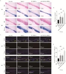
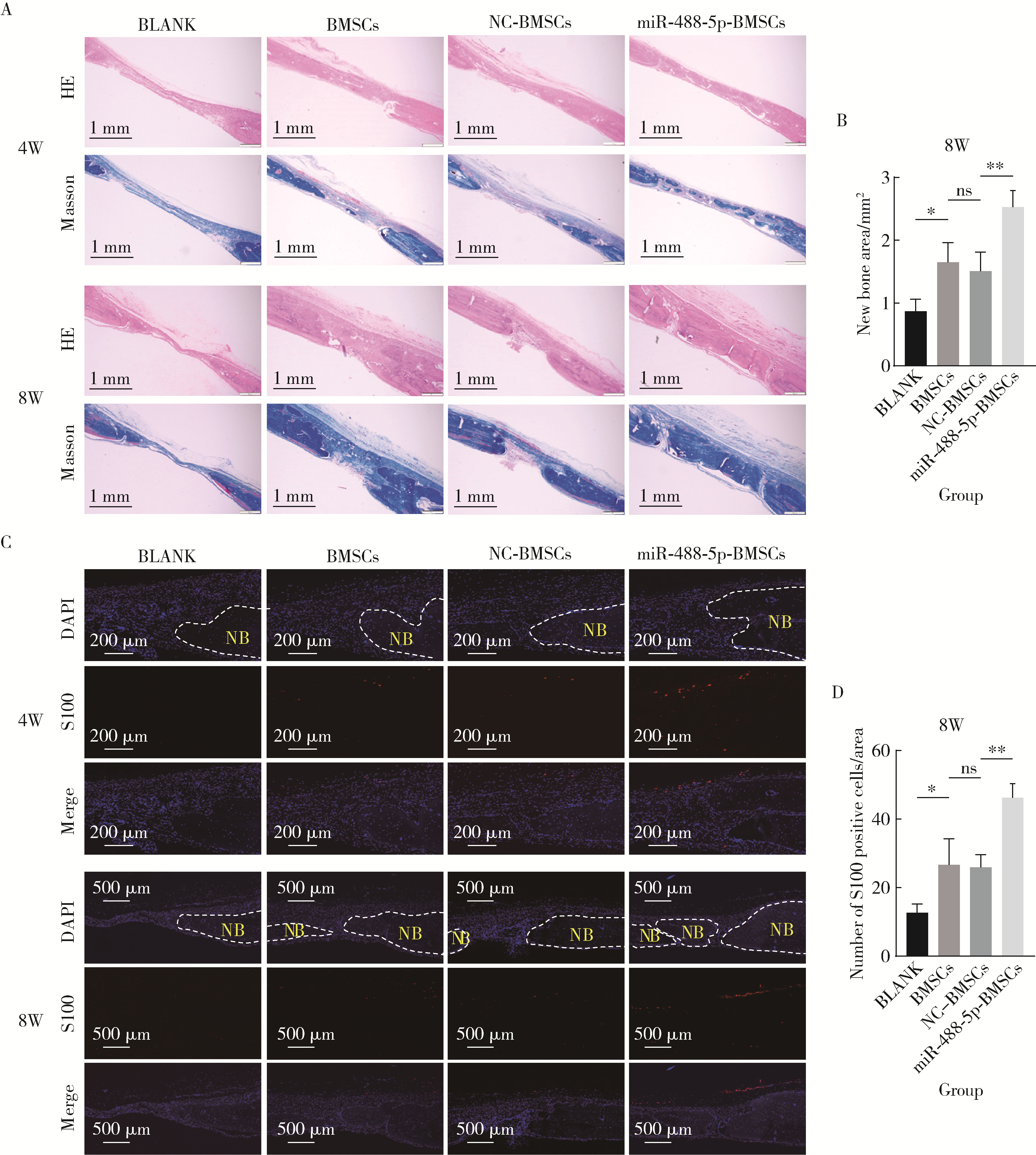
miR-488-5p promotes osteogenic and neurogenic differentiation of rat bone marrow mesenchymal stem cells and enhances neuralized bone regeneration

Liting ZENG1, Kaiyuan CHENG1,2, Zhongning LIU1, Jian LI1, Jingwen YANG1,*(), Ting JIANG1,*()   

  1. 1. Department of Prosthodontics, Peking University School and Hospital of Stomatology & National Center for Stomatology & National Clinical Research Center for Oral Diseases & National Engineering Research Center of Oral Biomaterials and Digital Medical Devices, Beijing 100081, China
    2. Department Ⅰ of Prosthodontics, Tianjin Stomatological Hospital, School of Medicine, Nankai University, Tianjin Key Laboratory of Oral and Maxillofacial Function Reconstruction, Tianjin 300041, China
  • Received:2025-10-10 Online:2026-02-18 Published:2025-12-10
  • Contact: Jingwen YANG, Ting JIANG
  • Supported by:
    the National Natural Science Foundation of China(82170928); the National Natural Science Foundation of China(82201022)

RICH HTML


Abstract:

Objective: To investigate the role of microRNA miR-488-5p, which showed increased expression after the disconnection of the inferior alveolar nerve, in promoting the osteogenic and neurogenic differentiation of rat bone marrow mesenchymal stem cells (rBMSCs), as well as its effect on promoting the neuralized tissue engineered bone regeneration. Methods: rBMSCs were subjected to in vitro neural or osteogenic differentiation induction cultures. The expression levels of miR-488-5p at different time points (days 0, 2, 4, and 7) were detected by quantitative real-time polymerase chain reaction (qRT-PCR). miR-488-5p overexpression or low expression in rBMSCs was achieved by transfection with miR-488-5p mimics or inhibitors. Four groups, the miR-488-5p mimics, the miR-488-5p inhibitor, and their respective negative controls (NC), were established to investigate the effects of miR-488-5p on the neural differentiation and osteogenic differentiation of rBMSCs.A 5 mm diameter, full-thickness circular critical bone defect was created in the rat calvaria. The rats were treated with light-cured gelatin methacryloyl (GelMA) seeded with rBMSCs. The rats were divided into four groups: ①BLANK group: GelMA; ②BMSCs group: GelMA + rBMSCs; ③NC-BMSCs group: GelMA + rBMSCs transfected with miR-488-5p mimics NC; and ④miR-488-5p-BMSCs group: GelMA + rBMSCs transfected with miR-488-5p mimics. Specimens were obtained 4 and 8 weeks after surgery, and micro-CT was performed to measure and analyze bone mineral density (BMD), bone volume/total volume (BV/TV), bone surface area/total volume (BS/TV) and trabecular number (Tb.N). The effects of neuralized tissue engineering bone formation in the defect area were assessed using Hematoxylin-Eosin (HE) staining, Masson staining, and tissue immunofluorescence staining of the nerve-specific protein soluble protein-100 (S100). Results: As rBMSCs progressed toward neural or osteogenic differentiation, miR-488-5p expression increased significantly from day 0 to day 7. Regarding neural differentiation, the mimics group showed increased expression of neural-related genes and proteins compared with the mimics NC group, while the opposite result was observed in the inhibitor group. As for osteogenic differentiation, the mimics group showed increased expression of osteogenic genes and proteins, more intense alkaline phosphatase (ALP) and alizarin red staining (ARS) staining, and enhanced ALP activity compared with the mimics NC group, while the opposite result was observed in the inhibitor group. 4 and 8 weeks after critical calvarial defect construction in rats, the BLANK group had the least amount of new bone formation, while the BMSCs group and the NC-BMSCs group had similar and intermediate amounts of new bone formation. The miR-488-5p-BMSCs group had the most new bone formation. At 4 weeks, the BMD [(0.63±0.05) g/cm3 vs. (0.51±0.03) g/cm3], BV/TV (33.17%±6.43% vs. 18.11%±1.52%), BS/TV [(3.43±0.69) /mm vs. (2.46±0.20) /mm], and Tb.N [(0.92±0.21) /mm vs.(0.59±0.07) /mm] in the miR-488-5p-BMSCs group were significantly higher than those in the NC-BMSCs group. At 8 weeks, the BMD [(0.80±0.04) g/cm3 vs. (0.68±0.04) g/cm3], BV/TV (56.69%±6.22% vs. 42.36%±3.86%), and the number of S100-labeled nerve cells around the new bone (46.33±4.04 vs. 26.00±3.61) in the miR-488-5p-BMSCs group was also significantly higher than that in the NC-BMSCs group. Conclusion: miR-488-5p promoted the osteogenic and neurogenic differentiation of rBMSCs and promoted the formation of neuralized tissue-engineered bone in rat calvarial defects.

Key words: miR-488-5p, Bone marrow mesenchymal stem cells, Osteogenic differentiation, Neurogenic differentiation, Neuro-bone tissue engineering

CLC Number: 

  • R68

Table 1

Primer sequences used for qRT-PCR"

Gene Forward primer (5′ to 3′) Reverse primer (5′ to 3′)
U6 CGCTTCGGCAGCACATATAC CGAATTTGCGTGTCATCCTT
miR-488-5p GGCACCCAGATAATGGCAC AGTGCAGGGTCCGAGGTATT
Gapdh AGGTCGGTGTGAACGGATTT GAACTTGCCGTGGGTAGAGT
*Map2 ACAGCAACAAGTGGTGAATCAG GGAGGATGGAGGAAGGTCTTG
*Ng2 TTCTCACACAGAGGAGCCC CACTCAAGCTCTGGCTGCT
*Tubb3 CAGATGCTGGCCATTCAGAGTAAG TGTTGCCGATGAAGGTGGAC
*Ngf TGCCAAGGACGCAGCTTTC TGAAGTTTAGTCCAGTGGGCTTCAG
*Nes TGAACAAGAGACCCAACAAACAC TTCCAAGAGGCTTCGGTAACT
*S100 CTGTCAAGAACCTGCTCCGA AGTGGGCATGGAACACATTGA
#Runx2 GCCTTCAAGGTTGTAGCCCT TGAACCTGGCCACTTGGTTT
#Alp CATGGTGAGTGACACGGACA CCATGACGTGGGGGATGTAG
#Ocn CCGTTTAGGGCATGTGTTGC TTTCGAGGCAGAGAGAGGGA
#Bmp2 CGGGAACAAATGCAGGAAGC AAGGACATTCCCCATGGCAG
#Col1a1 CCCCAGCCGCAAAGAGTCTA CAGCTGACTTCAGGGATGTCTTC
#Opn CCAGCCAAGGACCAACTACA AGTGTTTGCTGTAATGCGCC

Figure 1

miR-488-5p promotes neurogenic differentiation of rBMSCs A, mRNA expression levels of neurogenic-related genes Nes, S100, Map2, Ng2, Tubb3, Ngf on day 12 of neurogenic differentiation in the NM and PM groups as detected by qRT-PCR; B, immunofluorescence detection of Tubb3 in the NM and PM groups; C, miR-488-5p expression levels on days 0, 2, 4 and 7 of neurogenic differentiation as detected by qRT-PCR; D, mRNA expression levels of neurogenic-related genes Nes, S100, Map2, Ng2, Tubb3, Ngf on day 12 of neurogenic differentiation as detected by qRT-PCR; E, protein expression levels of Tubb3 and Gap43 as detected by Western blot; F, immunofluorescence detection of Tubb3 and quantification of Tubb3 fluorescence intensity. ns, not significant. * P < 0.05, * * P < 0.01, * * * P < 0.001. PM, proliferation medium; NM, neurogenic medium; Mimics NC, negative control for miR-488-5p mimics; Inhibitor NC, negative control for miR-488-5p inhibitor; Nes, nestin; S100, soluble protein-100; Map2, microtubule-associated protein 2; Ng2, neural/glial antigen 2; Tubb3, class Ⅲ β-tubulin; Ngf, nerve growth factor; Gap43, growth associated protein 43;DAPI, 4', 6-diamidino-2-phenylindole; rBMSCs, rat bone marrow mesenchymal stem cells; qRT-PCR, quantitative real-time polymerase chain reaction."

Figure 2

miR-488-5p promotes osteogenic differentiation of rBMSCs A, mRNA expression levels of osteogenic-related genes Runx2, Alp, Col1a1, Opn, Bmp2, Ocn on day 7 of osteogenic differentiation in the OM and PM groups as detected by qRT-PCR; B, ALP staining on day 7 of osteogenic differentiation in the OM and PM groups; C, miR-488-5p expression levels on days 0, 2, 4 and 7 of osteogenic differentiation as detected by qRT-PCR; D, mRNA expression levels of osteogenic-related genes Runx2, Alp, Col1a1, Opn, Bmp2, Ocn on day 7 of osteogenic differentiation as detected by qRT-PCR; E, protein expression levels of Runx2 and Osx as detected by Western blot; F, ALP staining on day 7 and ARS staining on day 21 of osteogenic differentiation; G, ALP activity on day 7 of osteogenic differentiation; H, quantification of mineralization by measuring ARS absorbance at 562 nm. ns, not significant. * P < 0.05, * * P < 0.01, * * * P < 0.001. PM, proliferation medium; OM, osteogenic medium; d, day; Mimics NC, negative control for miR-488-5p mimics; Inhibitor NC, negative control for miR-488-5p inhibitor; Runx2, Runt-related transcription factor 2; ALP, alkaline phosphatase; Ocn, Osteocalcin; Bmp2, bone morphogenetic protein 2; Col1a1, collagen type Ⅰ alpha 1 chain; Opn, osteopontin; ARS, alizarin red S; rBMSCs, rat bone marrow mesenchymal stem cells; qRT-PCR, quantitative real-time polymerase chain reaction."

Figure 3

micro-CT imaging analysis of rat calvarial defects at 4 and 8 weeks after operation A, 3D reconstructed images after micro-CT scanning showing bone defect areas at both 4 and 8 weeks after operation; B, quantitative comparisons of BMD, BV/TV, BS/TV, Tb.N at both 4 and 8 weeks after operation. ns, not significant. * P < 0.05, * * P < 0.01, * * * P < 0.001. W, weeks after operation. BLANK, the control group treated with GelMA only; BMSCs, the group treated with GelMA and normal rBMSCs; NC-BMSCs, the group treated with GelMA and rBMSCs transfected with negative control (NC) for miR-488-5p mimics; miR-488-5p-BMSCs, the group treated with GelMA and rBMSCs transfected with miR-488-5p mimics; BMD, bone mineral density; BV/TV, bone volume/total volume; BS/TV, bone surface area/total volume; Tb.N, trabecular number; 3D, 3-dimensional; BMSCs, bone marrow mesenchymal stem cells."

Figure 4

Histomorphological analysis of rat calvarial defects at 4 and 8 weeks after operation A, HE staining and Masson staining; B, quantitative measurement of new bone area in Masson staining at 8 weeks after operation; C, immunofluorescence detection of S100 protein; D, analysis of the number of S100-positive cells in immunofluorescence staining at 8 weeks after operation. HE, Hematoxylin-Eosin; NB, new bone; S100, soluble protein-100; DAPI, 4', 6-diamidino-2-phenvlindole; W, weeks after operation; BLANK, the control group treated with GelMA only; BMSCs, the group treated with GelMA and normal rBMSCs; NC-BMSCs, the group treated with GelMA and rBMSCs transfected with negative control (NC) for miR-488-5p mimics; miR-488-5p-BMSCs, the group treated with GelMA and rBMSCs transfected with miR-488-5p mimics; BMSCs, bone marrow mesenchymal stem cells. ns, not significant; * P < 0.05, * * P < 0.01."

1
Sun W, Ye B, Chen S, et al. Neuro-bone tissue engineering: Emerging mechanisms, potential strategies, and current challenges[J]. Bone Res, 2023, 11(1): 65.

doi: 10.1038/s41413-023-00302-8
2
Liu S, Liu S, Li S, et al. Nerves within bone and their application in tissue engineering of bone regeneration[J]. Front Neurol, 2022, 13, 1085560.
3
Zhao X, Yao M, Wang Y, et al. Neuroregulation during bone formation and regeneration: mechanisms and strategies[J]. ACS Appl Mater Interfaces, 2025, 17(5): 7223- 7250.

doi: 10.1021/acsami.4c16786
4
Jones RE, Salhotra A, Robertson KS, et al. Skeletal stem cell-Schwann cell circuitry in mandibular repair[J]. Cell Rep, 2019, 28(11): 2757. e5- 2766. e5.
5
Zhang X, Jiang X, Jiang S, et al. Schwann cells promote prevascularization and osteogenesis of tissue-engineered bone via bone marrow mesenchymal stem cell-derived endothelial cells[J]. Stem Cell Res Ther, 2021, 12, 382.

doi: 10.1186/s13287-021-02433-3
6
Fitzpatrick V, Moldes ZM, Deck A, et al. Functionalized 3D-printed silk-hydroxyapatite scaffolds for enhanced bone regeneration with innervation and vascularization[J]. Biomaterials, 2021, 276, 120995.

doi: 10.1016/j.biomaterials.2021.120995
7
Zhang Z, Wang F, Huang X, et al. Engineered sensory nerve guides self-adaptive bone healing via NGF-TrkA signaling pathway[J]. Adv Sci (Weinh), 2023, 10(10): 2206155.

doi: 10.1002/advs.202206155
8
Heng BC, Bai Y, Li X, et al. Electroactive biomaterials for facilitating bone defect repair under pathological conditions[J]. Adv Sci (Weinh), 2022, 10(2): 2204502.
9
Deng L, Hou M, Lv N, et al. Melatonin-encapsuled silk fibroin electrospun nanofibers promote vascularized bone regeneration through regulation of osteogenesis-angiogenesis coupling[J]. Mater Today Bio, 2024, 25, 100985.

doi: 10.1016/j.mtbio.2024.100985
10
Zhao W, Zhang S, Wang B, et al. Runx2 and microRNA regulation in bone and cartilage diseases[J]. Ann N Y Acad Sci, 2016, 1383(1): 80- 87.

doi: 10.1111/nyas.13206
11
Fang S, Deng Y, Gu P, et al. microRNAs regulate bone development and regeneration[J]. Int J Mol Sci, 2015, 16(4): 8227- 8253.

doi: 10.3390/ijms16048227
12
Liu J, Dang L, Wu X, et al. microRNA-mediated regulation of bone remodeling: A brief review[J]. JBMR Plus, 2019, 3(9): 10213.

doi: 10.1002/jbm4.10213
13
Nguyen LH, Diao HJ, Chew SY. MicroRNAs and their potential therapeutic applications in neural tissue engineering[J]. Adv Drug Deliv Rev, 2015, 88, 53- 66.

doi: 10.1016/j.addr.2015.05.007
14
Li YK, Zhang YY, Lin J, et al. Metabotropic glutamate receptor 5-mediated inhibition of inward-rectifying K+ channel 4.1 contributes to orofacial ectopic mechanical allodynia following inferior alveolar nerve transection in male mice[J]. J Neurosci Res, 2023, 101(7): 1170- 1187.

doi: 10.1002/jnr.25181
15
Lee J, Ohara K, Shinoda M, et al. Involvement of satellite cell activation via nitric oxide signaling in ectopic orofacial hypersensitivity[J]. Int J Mol Sci, 2020, 21(4): 1252.

doi: 10.3390/ijms21041252
16
Xu Y, Xia M, Chen T, et al. Inferior alveolar nerve transection disturbs innate immune responses and bone healing after tooth extraction[J]. Ann N Y Acad Sci, 2019, 1448(1): 52- 64.

doi: 10.1111/nyas.14120
17
程凯远, 张美子, 刘中宁, 等. 下牙槽神经离断对下颌骨骨质和拔牙创愈合的影响研究[J]. 口腔颌面修复学杂志, 2025, 26(2): 105- 115.
18
Wan QQ, Qin WP, Ma YX, et al. Crosstalk between bone and nerves within bone[J]. Adv Sci (Weinh), 2021, 8(7): 2003390.

doi: 10.1002/advs.202003390
19
Zhou S, Shen D, Wang Y, et al. microRNA-222 Targeting PTEN promotes neurite outgrowth from adult dorsal root ganglion neurons following sciatic nerve transection[J]. PLoS One, 2012, 7(9): 44768.

doi: 10.1371/journal.pone.0044768
20
Lei L, Liu Z, Yuan P, et al. Injectable colloidal hydrogel with mesoporous silica nanoparticles for sustained co-release of micro-RNA-222 and aspirin to achieve innervated bone regeneration in rat mandibular defects[J]. J Mater Chem B, 2019, 7(16): 2722- 2735.

doi: 10.1039/C9TB00025A
21
Qiu J, Wu S, Wang P, et al. miR-488-5p mitigates hepatic stellate cell activation and hepatic fibrosis via suppressing TET3 expression[J]. Hepatol Int, 2022, 17(2): 463- 475.
22
Li X, Yue X, Pastor WA, et al. Tet proteins influence the balance between neuroectodermal and mesodermal fate choice by inhibiting Wnt signaling[J]. Proc Natl Acad Sci USA, 2016, 113(51): E8267- E8276.
23
Ye W, Zhang C, Fan Z. MiR-26b-5p/TET3 regulates the osteogenic differentiation of human bone mesenchymal stem cells and bone reconstruction in female rats with calvarial defects[J]. Mol Biol Rep, 2024, 51(1): 632.

doi: 10.1007/s11033-024-09577-4
24
Leo AJ, Grande DA. Mesenchymal stem cells in tissue engineering[J]. Cells Tissues Organs, 2006, 183(3): 112- 122.

doi: 10.1159/000095985
25
Liu Y, Peng L, Li L, et al. 3D-bioprinted BMSC-laden biomimetic multiphasic scaffolds for efficient repair of osteochondral defects in an osteoarthritic rat model[J]. Biomaterials, 2021, 279, 121216.

doi: 10.1016/j.biomaterials.2021.121216
26
Yu H, Luo X, Li Y, et al. Advanced hybrid strategies of GelMA composite hydrogels in bone defect repair[J]. Polymers (Basel), 2024, 16(21): 3039.

doi: 10.3390/polym16213039
27
Zhou B, Jiang X, Zhou X, et al. GelMA-based bioactive hydrogel scaffolds with multiple bone defect repair functions: Therapeutic strategies and recent advances[J]. Biomater Res, 2023, 27(1): 86.

doi: 10.1186/s40824-023-00422-6
28
Mamidi N, Ijadi F, Norahan MH. Leveraging the recent advancements in GelMA scaffolds for bone tissue engineering: An assessment of challenges and opportunities[J]. Biomacromolecules, 2024, 25(4): 2075- 2113.

doi: 10.1021/acs.biomac.3c00279
29
丁梦, 李强, 李筱叶, 等. 光固化水凝胶负载骨髓间充质干细胞修复大鼠颅骨缺损[J]. 口腔疾病防治, 2024, 32(5): 330- 340.
30
Ai Y, Dai F, Li W, et al. Photo-crosslinked bioactive BG/BMSCs@GelMA hydrogels for bone-defect repairs[J]. Mater Today Bio, 2023, 23, 100882.

doi: 10.1016/j.mtbio.2023.100882
31
Sekido D, Otsuka T, Shimazaki T, et al. Comparison of cerebral cortex activation induced by tactile stimulation between natural teeth and implants[J]. J Clin Exp Dent, 2020, 12(11): 1021- 1026.
[1] Chunhui SHENG, Xiao ZHANG, Longwei LV, Yongsheng ZHOU. Exosome derived from human adipose-derived mesenchymal stem cells prevented bone loss induced by estrogen deficiency [J]. Journal of Peking University (Health Sciences), 2025, 57(2): 217-226.
[2] Yibo HU, Weijia LYU, Wei XIA, Yihong LIU. Hydrodynamic finite element analysis of biological scaffolds with different pore sizes for cell growth and osteogenic differentiation [J]. Journal of Peking University (Health Sciences), 2025, 57(1): 97-105.
[3] Ting SHUAI, Yanyan GUO, Chunping LIN, Xiaomei HOU, Chanyuan JIN. Knockdown of NPTX1 promotes osteogenic differentiation of human bone marrow mesenchymal stem cells [J]. Journal of Peking University (Health Sciences), 2025, 57(1): 7-12.
[4] SHUAI Ting,LIU Juan,GUO Yan-yan,JIN Chan-yuan. Knockdown of long non-coding RNA MIR4697 host gene inhibits adipogenic differentiation in bone marrow mesenchymal stem cells [J]. Journal of Peking University (Health Sciences), 2022, 54(2): 320-326.
[5] Jing XIE,Yu-ming ZHAO,Nan-quan RAO,Xiao-tong WANG,Teng-jiao-zi FANG,Xiao-xia LI,Yue ZHAI,Jing-zhi LI,Li-hong GE,Yuan-yuan WANG. Comparative study of differentiation potential of mesenchymal stem cells derived from orofacial system into vascular endothelial cells [J]. Journal of Peking University(Health Sciences), 2019, 51(5): 900-906.
[6] Xia LIU,Ying ni LI,Xiao li SUN,Qing lin PENG,Xin LU,Guo chun WANG. Effects of integrin metalloproteinases on osteogenic differentiation [J]. Journal of Peking University(Health Sciences), 2018, 50(6): 962-967.
Viewed
Full text


Abstract

Cited

  Shared   
  Discussed   
No Suggested Reading articles found!