Journal of Peking University (Health Sciences) ›› 2026, Vol. 58 ›› Issue (3): 658-665. doi: 10.19723/j.issn.1671-167X.2026.03.028

Previous Articles     Next Articles

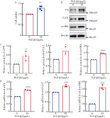
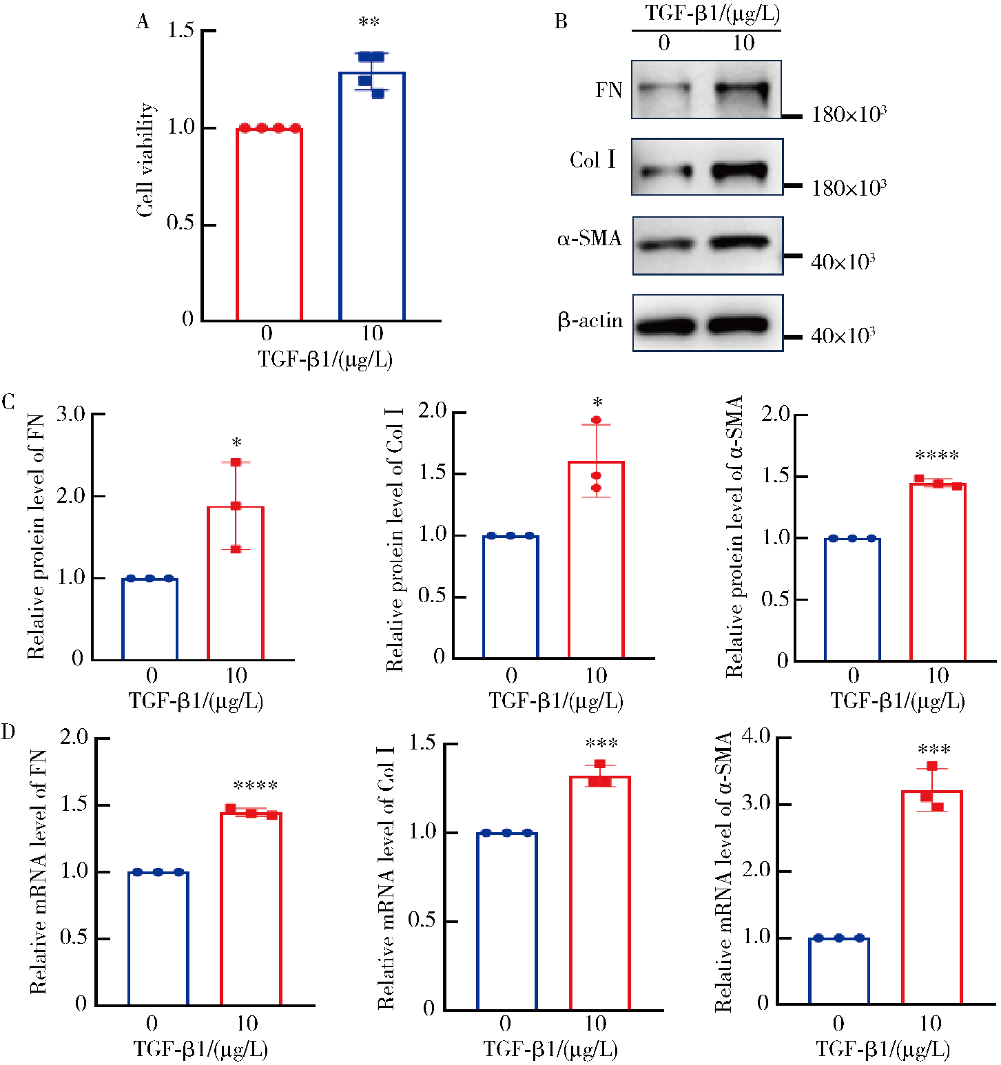
A new method for extracting adult mouse cardiac fibroblasts more efficiently and stably

Xiaojuan MA1, Hao WANG1, Xueqin MA2, Ying SONG1, Jiahui YU1, Yan SUN1, Yanfang LI1, Lixiang XUE1, Xianlong LI1, Jianling YANG1,*(), Yan WANG1,*()   

  1. 1. Institute of Medical Innovation and Research, Peking University Third Hospital, Beijing 100191, China
    2. Department of Endocrinology, The Eighth People' s Hospital of Jinan, Jinan 271100, China
  • Received:2024-11-12 Online:2026-06-18 Published:2026-04-07
  • Contact: Jianling YANG, Yan WANG
  • Supported by:
    the National Natural Science Foundation of China(82471329)

RICH HTML


Abstract:

Objective: Cardiac fibroblasts (CFs) play a central role in myocardial remodeling and fibrosis. Efficient isolation of CFs is a prerequisite for investigating related mechanisms. However, current methods for isolating primary adult mouse CFs suffer from prolonged processing time, low yield, and poor viability. This study aims to establish a rapid, high-yield, and stable isolation protocol for adult mouse CFs by optimizing the synergistic effect of enzymatic digestion and mechanical dissociation parameters. Methods: Using the gentleMACS® Octo Dissociator with Heaters, we selected different types and concentrations of collagenase, trypsin, and nuclease as the enzymatic digestion system for CFs extraction. We explored the optimal extraction conditions and compared the results with the commercial Multi Tissue Dissociation Kit 2. The cell yield was quantified using a high-content imaging analysis system by counting the number of adherent cells per field after 72 h of culture. The CFs purity was assessed using immunofluorescence staining for vimentin. The trans-differentiation activity of the CFs was evaluated with transforming growth factor β1 (TGF-β1). Results: Omitting any component of the digestion solution (collagenase Ⅱ/Ⅳ, trypsin or DNaseⅠ), significantly prolonged extraction time and reduced cell yield. In contrast, the optimized protocol outperformed the commercial kit, reducing digestion time by 32.2 min and significantly increasing cell yield, and with comparable obtained CFs purity. After TGF-β1 stimulation, CFs exhibited enhanced proliferative capacity and upregulated expression of α-smooth muscle actin (α-SMA), collagen type Ⅰ (ColⅠ), and fibronectin (FN), confirming the differentiation potential of CFs isolated via the optimized method. Conclusion: This study systematically optimized an enzymatic digestion method combining collagenase, trypsin, and nuclease in conjunction with mechanical dissociation using a tissue dissociator, leading to the efficient and stable isolation of adult mouse CFs. By fine-tuning enzyme concentrations and digestion conditions, we successfully reduced processing time, improved cell yield, and enhanced cell viability compared with conventional isolation methods. These findings validate the physiological relevance of the isolated CFs and demonstrate that the optimized protocol provides a reliable and reproducible method for studying myocardial fibrosis and remodeling. This protocol can serve as a valuable tool for researchers investigating CFs biology and its role in cardiovascular diseases.

Key words: Mice, Cardiac fibroblasts, Primary cell culture, Tissue dissociation

CLC Number: 

  • R392-33

Table 1

Sequences of the primers for qPCR"

Name Primer sequence (5′-3′)
GAPDH Forward: AGGTCGGTGTGAACGGATTTG
Reverse: TGTAGACCATGTAGTTGAGGTCAA
α-SMA Forward: CTTCCAGCCATCTTTCATTGG
Reverse: GTTCTGGAGGGGCAATGAT
ColⅠ Forward: CCTCAGGGTATTGCTGGACAAC
Reverse: CAGAAGGACCTTGTTTGCCAGG
FN Forward: CCGGTGGCTGTCAGTCAGA
Reverse: CCGTTCCCACTGCTGATTTATC

Table 2

Formulation conditions for collagenase Ⅱ and Ⅳ combination"

Enzyme concentrations Collagenase Ⅱ group Collagenase Ⅳ group
Collagenase Ⅱ/(g/L) 1.2 -
Collagenase Ⅳ/(g/L) - 1.2
Trypsin/(g/L) 0.8 0.8
DNaseⅠ/(g/L) 0.2 0.2
Red blood cell lysis + +

Table 3

Combined collagenase Ⅱ and Ⅳ digestion efficacy comparison"

Group Digestion time/min Cell yield (cell number/field), median (minimum, maximum)
Collagenase Ⅱ 22 479 (419, 653)
Collagenase Ⅳ 22 474 (435, 669)

Table 4

Treatment conditions for different groups"

Enzyme concentrations Optimized group No collagenase group No trypsin group No DNaseⅠ group No red blood cell lysis group
Collagenase Ⅱ/(g/L) 1.2 - 1.2 1.2 1.2
Trypsin/(g/L) 0.8 0.8 - 0.8 0.8
DNaseⅠ/(g/L) 0.2 0.2 0.2 - 0.2
Red blood cell lysis + + + + -

Figure 1

Comparison of digestion time and cell yield (cell number/field) A, comparison of digestion time among multiple treatment groups (n=3); B, quantification of viable cells isolated using different methods following 72-hour culture (n=3). * P < 0.05, * * P < 0.01, * * * P < 0.001, optimized group vs. other groups; ### P < 0.001, #### P < 0.000 1, multiple comparisons."

Table 5

Concentration of digestive enzymes and treatment condition in each group"

Enzyme concentrations Optimized group 0.5×concentration group 2×concentration group
Collagenase Ⅱ/(g/L) 1.2 0.6 2.4
Trypsin/(g/L) 0.8 0.4 1.6
DNaseⅠ/(g/L) 0.2 0.1 0.4
Red blood cell lysis + + +

Figure 2

Comparison of digestion time and cell yield (cell number/field) among the optimized group, 0.5× and 2× enzyme concentration group A, comparison of digestion time among the optimized group, 0.5× and 2× enzyme concentration group (n=3); B and C, after incubation for 72 h, the cells were photographed and analyzed by Operetta CLS High Content Imaging Analysis System (×10), and the number of cells in each field was counted (n=3). * P < 0.05, * * P < 0.01, optimized group vs. other groups; # P < 0.05, ## P < 0.01, multiple comparisons."

Figure 3

Comparison of digestion time and cell yield (cell number/field) between the optimized group and commercial kit group A, comparison of digestion time between the optimized group and the commercial kit group (n=3, * * * * P < 0.000 1); B, after 72 h of incubation, the cell yield of the optimized group and the commercial kit group was analyzed (n=3, * * P < 0.01); C, the obtained cells were photographed and analyzed by Operetta CLS High Content Imaging Analysis System (×10)."

Figure 4

Comparison of CFs purity between the optimized group and the commercial kit group A, immunofluorescence analysis of vimentin expression in CFs. Vimentin protein levels in CFs were assessed by immunofluorescence staining, with representative images captured using a high-content screening (HCS) system at 20× magnification. B, quantitative comparison of vimentin fluorescence intensity. Randomly selected microscopic fields (n=20 per group) with comparable cellular densities were subjected to HCS-based fluorescence quantification. No statistically significant intergroup differences were observed in vimentin intensity profiles (P>0.05). C, threshold-based vimentin-positive cell analysis. The percentage of DAPI (+) nuclei exhibiting vimentin fluorescence intensities exceeding 2 000 was calculated for each field. This threshold analysis revealed comparable proportions of vimentin (+) cells between experimental groups (P>0.05). CFs, cardiac fibroblasts; ns, no significance."

Figure 5

Evaluation of the trans-differentiation ability of CFs after TGF-β1 treatment A, after stimulation with TGF-β1, the viability of P1 generation CFs was evaluated through the CCK-8 assay (n=4); B and C, the protein expression levels of ColⅠ, α-SMA and FN were analyzed by Western blotting; D, the mRNA levels of ColⅠ, α-SMA and FN were detected by qPCR (n=3). * P < 0.05, * * P < 0.01, * * * P < 0.001, * * * * P < 0.000 1. CFs, cardiac fibroblasts; TGF-β1, transforming growth factor β1; α-SMA, α-smooth muscle actin; ColⅠ, collagen typeⅠ; FN, fibronectin; qPCR, quantitative real-time PCR."

1
Frangogiannis NG . Fibroblast: Extracellular matrix interactions in tissue fibrosis[J]. Curr Pathobiol Rep, 2016, 4 (1): 11- 18.

doi: 10.1007/s40139-016-0099-1
2
Khalil H , Kanisicak O , Vagnozzi RJ , et al. Cell-specific ablation of Hsp47 defines the collagen-producing cells in the injured heart[J]. JCI Insight, 2019, 4 (15): e128722.

doi: 10.1172/jci.insight.128722
3
Davis J , Molkentin JD . Myofibroblasts: Trust your heart and let fate decide[J]. J Mol Cell Cardiol, 2014, 70, 9- 18.

doi: 10.1016/j.yjmcc.2013.10.019
4
Gourdie RG , Dimmeler S , Kohl P . Novel therapeutic strategies targeting fibroblasts and fibrosis in heart disease[J]. Nat Rev Drug Discov, 2016, 15 (9): 620- 638.

doi: 10.1038/nrd.2016.89
5
Shinde AV , Frangogiannis NG . Fibroblasts in myocardial infarction: A role in inflammation and repair[J]. J Mol Cell Cardiol, 2014, 70, 74- 82.

doi: 10.1016/j.yjmcc.2013.11.015
6
Li L , Zhao Q , Kong W . Extracellular matrix remodeling and cardiac fibrosis[J]. Matrix Biol, 2018, 68/69, 490- 506.

doi: 10.1016/j.matbio.2018.01.013
7
Aghajanian H , Kimura T , Rurik JG , et al. Targeting cardiac fibrosis with engineered T cells[J]. Nature, 2019, 573 (7774): 430- 433.

doi: 10.1038/s41586-019-1546-z
8
Henderson NC , Rieder F , Wynn TA . Fibrosis: From mechanisms to medicines[J]. Nature, 2020, 587 (7835): 555- 566.

doi: 10.1038/s41586-020-2938-9
9
冯姗, 徐文丽, 吴济民, 等. 血管紧张素Ⅱ促进心脏成纤维细胞转分化的实验条件优化[J]. 中国病理生理杂志, 2022, 38 (1): 167- 177.
10
张毅, 孟祥雯. 小鼠乳鼠心脏成纤维细胞的原代培养及鉴定[J]. 细胞与分子免疫学杂志, 2020, 36 (4): 344- 348.
11
张聪聪, 王晓, 程乃萱, 等. 原代小鼠心脏成纤维细胞的衰老模型建立[J]. 心肺血管病杂志, 2022, 41 (7): 821- 826.
12
孙赫, 涂彬, 宋凯, 等. DNMT3A对高糖环境下小鼠心肌成纤维细胞增殖与迁移的影响[J]. 中国药理学通报, 2023, 39 (3): 555- 560.
13
陈泽润, 李艺, 梁俣, 等. 环状RNA MYO9A_043通过下调CPEB4表达发挥抑制小鼠心脏成纤维细胞纤维化表型的作用[J]. 中国病理生理杂志, 2023, 39 (7): 1225- 1232.
14
梁春宝, 李立林, 蔡冬青. 单细胞核测序揭示成年小鼠心脏构成细胞的分群及占比[J]. 暨南大学学报(自然科学与医学版), 2023, 44 (2): 124- 136.
15
Almazloum A , Khalil H . Isolation of adult mouse cardiac fibroblasts[J]. Curr Protoc, 2023, 3 (7): e840.

doi: 10.1002/cpz1.840
16
李如君, 龚开政, 张振刚. 成年小鼠心脏成纤维细胞的分离、纯化和原代培养[J]. 细胞与分子免疫学杂志, 2017, 33 (1): 67- 71.
17
师雷, 金振晓, 谭延振, 等. 简易灌注装置同时提取成年小鼠心肌细胞和心脏成纤维细胞[J]. 中国体外循环杂志, 2023, 21 (3): 172- 178.
18
肖晗, 朴成实, 陈瑞飞, 等. AMPK激活通过负性调控C/EBPβ抑制小鼠心脏成纤维细胞TGFβ1表达[J]. 生理学报, 2017, 69 (2): 123- 128.
19
陈洪群, 郑梅, 晏乘曦, 等. 改良联合酶消化法提取成年小鼠原代心脏成纤维细胞[J]. 山东医药, 2019, 59 (21): 38- 41.
20
Leask A . Potential therapeutic targets for cardiac fibrosis: TGF-beta, angiotensin, endothelin, CCN2, and PDGF, partners in fibroblast activation[J]. Circ Res, 2010, 106 (11): 1675- 1680.

doi: 10.1161/CIRCRESAHA.110.217737
21
Liu M , López de Juan Abad B , Cheng K . Cardiac fibrosis: Myofibroblast-mediated pathological regulation and drug delivery strategies[J]. Adv Drug Deliv Rev, 2021, 173, 504- 519.
22
Tallquist MD . Cardiac fibroblast diversity[J]. Annu Rev Physiol, 2020, 82, 63- 78.

doi: 10.1146/annurev-physiol-021119-034527
23
Hinz B , Phan SH , Thannickal VJ , et al. The myofibroblast: One function, multiple origins[J]. Am J Pathol, 2007, 170 (6): 1807- 1816.
[1] Ting-ting YUAN,Shen LI,Yan WU,Hai-tao WU. Establishment and behavioral evaluation of a mouse model of long-term free-choice alcohol drinking [J]. Journal of Peking University (Health Sciences), 2023, 55(2): 315-323.
[2] JIAO Cui,WANG Jian-mei,KUANG Hai-xia,WU Zhi-hong,LIU Tao. Effects of CACNA1H gene knockout on autistic-like behaviors and the morphology of hippocampal neurons in mice [J]. Journal of Peking University (Health Sciences), 2022, 54(2): 209-216.
[3] ZHU Yi-ying,MIN Sai-nan,YU Guang-yan. Effect of topical injection of cyclosporine A on saliva secretion and inflammation in the submandibular gland of non-obese diabetic mice [J]. Journal of Peking University (Health Sciences), 2021, 53(4): 750-757.
[4] YANG Rong,LI Qing-xiang,WANG Yi-fei,ZHOU Wen,WANG Wen,GUO Chuan-bin,LIU Hao,GUO Yu-xing. Application of iodine staining technique for tumor identification in Micro-CT of mouse model with skull base-infratemporal fossa tumor [J]. Journal of Peking University (Health Sciences), 2021, 53(3): 598-601.
[5] YANG Liu,CHU Xiao-yu,ZHAO Qi. Effects of RhoA on the adherens junction of murine ameloblasts [J]. Journal of Peking University(Health Sciences), 2018, 50(3): 521-526.
[6] KANG Lei, HUO Yan, WANG Rong-fu, ZHANG Chun-li, YAN Ping, XU Xiao-jie. In vivo imaging of breast tumors by a 99mTc radiolabeled probe targeting microRNA-155 in mice models [J]. Journal of Peking University(Health Sciences), 2018, 50(2): 326-330.
[7] ZHANG Wei, PANG Chun-yan,WANG Yong-fu. Adipose tissue derived stem cells’ treatment effects in MRL/lpr mice and its effects on the imbalance of Th17/Treg cells [J]. Journal of Peking University(Health Sciences), 2017, 49(6): 974-978.
[8] SUN Rui, GAO Bo, GUO Chuan-bin. Anatomy and histology characteristics of lymph node in nude mice [J]. Journal of Peking University(Health Sciences), 2017, 49(5): 893-898.
[9] YAN Xin-xing,ZHANG Wei,WANG Jing,KE Xiao-yan. B7-H3 silencing inhibits human hematological malignancy xenograft tumor tumorigenesis and metastasis in nude mice [J]. Journal of Peking University(Health Sciences), 2017, 49(2): 286-294.
[10] GAO Li, YU Xiao-qian, CAI Yu. Effect of molar ligation and local Porphyromonas gingivalis inoculation on alveolar  bone loss in the mouse [J]. Journal of Peking University(Health Sciences), 2017, 49(1): 31-035.
[11] CHEN Xiao-mei, LI Fu-qiang, YAN Su, WU Xiao-cui, TANG Cui-lan. Nicotine alleviates the liver inflammation of non-alcoholic steatohepatitis induced by high-fat and high-fructose in mice [J]. Journal of Peking University(Health Sciences), 2016, 48(5): 777-782.
[12] WANG Zhi-hua, ZHANG Wei, ZHANG Yan-qing, PANG Chun-yan,WANG Yong-fu. Effect of CD40 siRNA on inflammatory response of MRL/Lpr mice [J]. Journal of Peking University(Health Sciences), 2016, 48(5): 771-776.
[13] YU Jing-ting,MENG Huan-xin, LIU Kai-ning. An modified culture method of primary human gingival epithelial cells [J]. Journal of Peking University(Health Sciences), 2016, 48(4): 733-737.
[14] CHEN Juan, WANG Yi-chao, CUI Rong, LIU Xiao-xiao, ZHANG Bao-xu. Effects of 1,3-diphenyl-1,3-propanedione on neurotransmitter contents of brain in mice administered with cocaine [J]. Journal of Peking University(Health Sciences), 2016, 48(3): 398-402.
[15] WANG Yu, XIA Chao-ming. Dynamic alteration of CD154/CD40 and its effects on Th1/Th2 polarization in indu-cible co-stimulator ligand knockout mice infected with Schistosoma japonicum [J]. Journal of Peking University(Health Sciences), 2015, 47(6): 898-904.
Viewed
Full text


Abstract

Cited

  Shared   
  Discussed   
No Suggested Reading articles found!