Development and validation of a multicenter nomogram predicting the risk of liver metastasis after curative resection of colorectal cancer

  • Nannan WANG 1, 2 ,
  • Dajin YUAN 1 ,
  • Yubing ZHU 1 ,
  • Lei DING , 1, *
Expand
  • 1. Department of Gastrointestinal Surgery, Beijing Shijitan Hospital, Capital Medical University, Beijing 100038, China
  • 2. Department of Colorectal Surgery, The Affiliated Cancer Hospital of Nanjing Medical University & Jiangsu Cancer Hospital & Jiangsu Institute of Cancer Research, Nanjing 210009, China
DING Lei, e-mail,

Received date: 2025-11-25

  Online published: 2026-02-25

Supported by

the Youth Research Fund of Beijing Shijitan Hospital(2023-q19)

Copyright

All rights reserved. Unauthorized reproduction is prohibited.

Abstract

Objective: To identify independent clinicopathological and molecular risk factors for metachronous liver metastasis and to construct a novel multicenter nomogram for predicting 1-, 3-, and 5-year liver metastasis-free survival (LMFS). Methods: In this multicenter retrospective cohort study, we analyzed clinical data from 865 patients with stages Ⅰ-Ⅲ CRC who underwent curative resection between January 2020 and December 2024. The population was derived from two institutions: Beijing Shijitan Hospital (n=746) and Jiangsu Cancer Hospital (n=119). Patients from the primary center were randomly assigned to a training cohort (n=523) and an internal validation cohort (n=223) at a 7 ∶3 ratio, while patients from the second center served as an independent external validation cohort (n=119). Candidate variables included demographics, tumor markers, pathological features, and molecular biomarkers [KRAS/BRAF mutation and microsatellite instability (MSI)]. Multivariable Cox proportional hazards regression analyses were utilized to identify independent predictors. Model performance was evaluated using the concordance index (C-index), time-dependent area under the receiver operating characteristic curve (AUC), calibration curves, and decision curve analysis (DCA). Results: Baseline characteristics were balanced across cohorts (P>0.05). Multivariable analysis identified nine independent prognostic factors: age, differentiation, T stage, N stage, vascular invasion, perineural invasion, and molecular markers. Notably, KRAS mutation (HR=1.42, 95%CI: 1.27-1.63) and BRAF mutation (HR=1.53, 95%CI: 1.29-1.84) were associated with significantly increased risk, whereas micro-satellite instability-high (MSI-H) status (HR=0.71, 95%CI: 0.54-0.92) served as a protective factor. The nomogram demonstrated robust discrimination with C-indices of 0.85 (95%CI: 0.82-0.89) in the training cohort, 0.81 (95%CI: 0.77-0.83) in the internal validation cohort, and 0.75 (95%CI: 0.71-0.79) in the external validation cohort. In the training set, AUCs for predicting 1-, 3-, and 5-year LMFS were 0.81 (95%CI: 0.77-0.86), 0.83 (95%CI: 0.80-0.89), and 0.85 (95%CI: 0.78-0.92), respectively. Calibration curves showed excellent agreement, and DCA indicated higher net clinical benefit than the American Joint Committee on Cancer (AJCC) tumor node metastasis (TNM) staging system. Conclusion: We established and externally validated a nomogram integrating clinicopathological features with KRAS, BRAF, and MSI status. This model exhibited enhanced predictive accuracy and generalizability compared with conventional staging systems. It serves as a valuable tool for identifying high-risk patients and guiding individualized postoperative surveillance strategies to improve long-term survival outcomes.

Cite this article

Nannan WANG , Dajin YUAN , Yubing ZHU , Lei DING . Development and validation of a multicenter nomogram predicting the risk of liver metastasis after curative resection of colorectal cancer[J]. Journal of Peking University(Health Sciences), 2026 , 58(2) : 290 -300 . DOI: 10.19723/j.issn.1671-167X.2026.02.011

结直肠癌(colorectal cancer, CRC)是全球最常见的恶性肿瘤之一,2020年新发病例约占全球癌症总数的10%[1-2]。尽管以手术为主的综合治疗策略不断进步,但仍有约50%的CRC患者在初诊时或后续治疗中发生远处转移,其中肝是最常见的转移靶器官,约有25%的根治术后患者发生异时性肝转移,且多数因发现较晚而预后不良[3-4]。因此,实现术后异时性肝转移的早期预测与风险分层,对于改善患者预后具有重要的临床意义。目前,临床实践中广泛采用的AJCC(American Joint Committee on Cancer)第8版TNM分期系统主要依据肿瘤浸润深度(tumor,T)、淋巴结转移(node,N)及远处转移(metastasis,M)等解剖学指标进行预后评估[5-6]。然而,该体系难以充分反映肿瘤的生物学异质性。随着精准医疗的发展,KRASBRAF基因突变及错配修复功能(deficient mismatch repair,dMMR)/微卫星不稳定性(microsatellite instability,MSI)状态等分子标志物,已被证实与靶向及免疫治疗效果密切相关,并成为预后判断的重要依据[7-8]。这表明,单纯依赖TNM分期已无法满足异时性转移风险个体化预测的需求。列线图作为一种基于多因素回归模型的可视化预测工具,能够整合多种临床病理特征,实现风险的定量评估,具有良好的临床适用性[9-10]。然而,既往关于结直肠癌术后肝转移预测的研究多局限于单中心、小样本回顾性数据,其普适性有待验证[11-12]。为此,本研究联合首都医科大学附属北京世纪坛医院与江苏省肿瘤医院两家医疗中心的患者资料,旨在构建并验证一项可用于个体化预测结直肠癌根治术后无肝转移生存期(liver metastasis-free survival,LMFS)的列线图模型,以期为术后风险分层与随访策略的优化提供量化工具。

1 资料与方法

1.1 研究对象

本研究为多中心回顾性队列研究,研究开始前已经首都医科大学附属北京世纪坛医院伦理委员会[审批号:sjtky11-1x-2021 (106)]和江苏省肿瘤医院伦理委员会(审批号:2020科-054)审查批准。
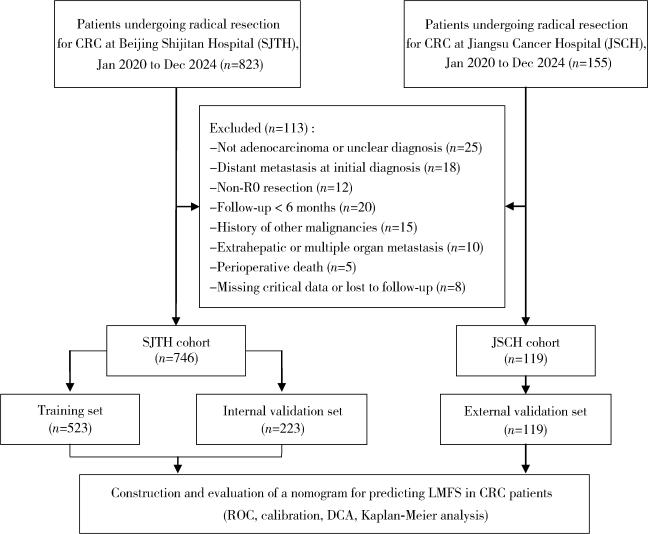
连续收集2020年1月1日至2024年12月31日首都医科大学附属北京世纪坛医院和江苏省肿瘤医院接受结直肠癌根治性切除术的患者的病例资料进行回顾性分析。病例筛选流程及具体排除原因见图 1。纳入标准如下:(1)术前经病理学确诊为结肠或直肠腺癌;(2)初次诊断时影像学检查证实无远处转移;(3)接受结直肠癌原发灶根治性切除术(R0切除);(4)术后随访时间≥6个月,且临床病理资料完整。排除标准包括:(1)既往或同时合并其他恶性肿瘤病史者;(2)随访期间在主要研究终点前发生肝外转移或多器官广泛转移者;(3)关键临床病例资料缺失或随访失访者。最终共纳入865例患者,其中首都医科大学附属北京世纪坛医院746例(按7 ∶ 3比例随机划分为训练集523例与内部验证集223例),江苏省肿瘤医院119例作为独立外部验证集。
图1 患者筛选与研究设计的CONSORT流程图

CRC, colorectal cancer; LMFS, liver metastasis-free survival; ROC, receiver operating characteristic; DCA, decision curve analysis.

Figure 1 CONSORT flow diagram of patient selection and study design

1.2 数据收集

通过医院的电子病历系统收集患者的病例资料,收集变量包括:(1)一般资料:年龄、性别、体重指数(body mass index,BMI,依据中国成人标准划分为 < 18.5 kg/m2、18.5~23.9 kg/m2、24.0~27.9 kg/m2、≥28.0 kg/m2四组);(2)肿瘤标志物:癌胚抗原(carcinoembryonic antigen,CEA,界值5.0 μg/L)、糖类抗原19-9(carbohydrate antigen 19-9,CA199,界值37 U/mL);(3)病理特征:肿瘤原发部位(细分为升结肠、横结肠、降结肠、乙状结肠、直肠)、分化程度(高、中、低分化)、肿瘤最大径(< 5 cm或≥5 cm)、T分期(T1~T4)、N分期(N0~N2)、脉管侵犯、神经侵犯;(4)分子特征:KRAS/NRAS/BRAF基因状态(突变型或野生型)、微卫星状态[微卫星高度不稳定(microsatellite instability-high, MSI-H)或微卫星稳定(microsatellite stable, MSS)];(5)治疗信息:术后辅助化疗及靶向药物治疗史。本研究的主要观察终点为无肝转移生存期(liver metastasis-free survival, LMFS),定义为从手术日起至首次影像学(CT/MRI)确认发生肝转移或末次随访的时间。

1.3 统计学分析

采用R软件(v4.5.2)进行统计分析,主要使用“survival”“rms”“timeROC”“dcurves”“ggplot2”等程序包。对连续变量采用Shapiro-Wilk法进行正态性检验,符合正态分布的数据以${\bar x}$>±s表示;不符合正态分布的数据以M(P25, P75)表示,分类变量以n(%)表示,组间比较使用卡方检验或Fisher精确检验。LMFS相关因素的分析采用单因素与多因素Cox比例风险回归模型,首先在单因素分析中筛选P < 0.05的变量,再纳入多因素模型计算风险比及其95%置信区间。基于多因素分析结果构建预测术后1、3、5年LMFS的列线图模型。模型性能通过以下方法评估:采用Bootstrap重复抽样1 000次绘制校准曲线,评估预测概率与实际观察概率的一致性;绘制时间-受试者工作特征曲线并计算曲线下面积评价区分度;采用一致性指数综合评价模型判别能力;通过决策曲线分析比较列线图与AJCC第8版TNM分期系统的临床净获益。使用“survminer”包确定列线图评分的最佳截断值,将患者划分为高风险组与低风险组,采用Kaplan-Meier法绘制生存曲线,组间差异采用Log-rank检验。对于临床病理资料中的缺失数据,鉴于各变量的缺失比例均低于5%且经检验符合完全随机缺失假设,本研究采用了行删除法进行处理。此外,本研究的预测模型构建与报告规范严格遵循《个体预后或诊断多变量预测模型透明报告(TRIPOD)》声明要求[13]。所有假设检验均为双侧,P < 0.05认为差异具有统计学意义。

2 结果

2.1 多中心CRC患者的基线临床病理特征比较

经筛选后,本研究共纳入865例CRC患者,其中,首都医科大学附属北京世纪坛医院(SJTH)队列纳入746例,按7 ∶ 3比例随机划分为训练集(523例)与内部验证集(223例);江苏省肿瘤医院(JSCH)队列纳入119例,作为外部验证集,患者基线资料见表 1
表1 多中心CRC患者的基线临床病理特征比较

Table 1 Comparison of baseline clinicopathologic characteristics of CRC patients across multiple centers

Variables SJTH cohort JSCH cohort P
Training set (n=523) Internal validation set (n=223) External validation set (n=119)
Age/years, n (%) 0.246
   < 65 298 (57.0) 138 (61.9) 77 (64.7)
  ≥65 225 (43.0) 85 (38.1) 42 (35.3)
Gender, n (%) 0.867
  Male 325 (62.1) 136 (61.0) 71 (59.7)
  Female 198 (37.9) 87 (39.0) 48 (40.3)
BMI/(kg/m2), n (%) 0.115
   < 18.5 43 (8.2) 17 (7.6) 15 (12.6)
  18.5 - < 24 251 (48.0) 109 (48.9) 55 (46.2)
  24 - < 28 206 (39.4) 87 (39.0) 44 (37.0)
  ≥28 23 (4.4) 10 (4.5) 5 (4.2)
CEA/(μg/L), n (%) 0.372
   < 5.0 240 (45.9) 95 (42.6) 53 (44.5)
  ≥5.0 283 (54.1) 128 (57.4) 66 (55.5)
CA199/(U/mL), n (%) 0.391
   < 37 245 (46.8) 97 (43.5) 47 (39.5)
  ≥37 278 (53.2) 126 (56.5) 72 (60.5)
Tumor site, n (%) 0.092
  Ascending colon 103 (19.7) 38 (17.0) 31 (26.1)
  Transverse colon 13 (2.5) 13 (5.8) 4 (3.4)
  Descending colon 29 (5.5) 14 (6.3) 12 (10.1)
  Sigmoid colon 155 (29.6) 61 (27.4) 28 (23.5)
  Rectum 223 (42.6) 97 (43.5) 44 (37.0)
Differentiation grade, n (%) 0.238
  High 78 (14.9) 26 (11.7) 21 (17.6)
  Moderate 333 (63.7) 151 (67.7) 66 (55.5)
  Low 112 (21.4) 46 (20.6) 32 (26.9)
Tumor size/cm, n (%) 0.155
   < 5 352 (67.3) 148 (66.3) 82 (68.9)
  ≥5 171 (32.7) 75 (33.6) 37 (31.1)
T stage, n (%) 0.054
  T1 47 (9.0) 18 (8.1) 6 (5.0)
  T2 93 (17.8) 31 (13.9) 15 (12.6)
  T3 348 (66.5) 166 (74.4) 85 (71.4)
  T4 35 (6.7) 8 (3.6) 13 (10.9)
N stage, n (%) 0.875
  N0 321 (61.4) 133 (59.6) 74 (62.2)
  N1 128 (24.5) 58 (26.0) 32 (26.9)
  N2 74 (14.1) 32 (14.3) 13 (10.9)
KRAS, n (%) 0.779
  Mutant type 256 (48.9) 112 (50.2) 55 (46.2)
  Wild type 267 (51.1) 111 (49.8) 64 (53.8)
NRAS, n (%) 0.425
  Mutant type 85 (16.3) 45 (20.2) 20 (16.8)
  Wild type 438 (83.7) 178 (79.8) 99 (83.2)
BRAF, n (%) 0.309
  Mutant type 79 (15.1) 40 (17.9) 14 (11.8)
  Wild type 444 (84.9) 183 (82.1) 105 (88.2)
MSI status, n (%) 0.287
  MSI-H 110 (21.0) 49 (22.0) 18 (15.1)
  MSS 413 (79.0) 174 (78.0) 101 (84.9)
Vascular invasion, n (%) 0.296
  Yes 170 (32.5) 71 (31.8) 30 (25.2)
  No 353 (67.5) 152 (68.2) 89 (74.8)
Nerve invasion, n (%) 0.313
  Yes 174 (33.3) 71 (31.8) 31 (26.1)
  No 349 (66.7) 152 (68.2) 88 (73.9)
Chemotherapy, n (%) 0.197
  Yes 258 (49.3) 120 (53.8) 52 (43.7)
  No 265 (50.7) 103 (46.2) 67 (56.3)
Targeted therapy, n (%) 0.112
  Yes 46 (8.8) 21 (9.4) 4 (3.4)
  No 477 (91.2) 202 (90.6) 115 (96.6)

CRC, colorectal cancer; SJTH, Beijing Shijitan Hospital; JSCH, Jiangsu Cancer Hospital; BMI,body mass index;CEA,carcinoembryonic antigen;CA199,carbohydrate antigen 19-9;KRAS,kirsten rat sarcoma viral oncogene homolog;NRAS,neuroblastoma RAS viral oncogene homolog;BRAF,b-raf proto-oncogene, serine/threonine kinase;MSI-H,microsatellite instability-high;MSS,microsatellite stability.

本研究收集了患者的人口学特征(年龄、性别、BMI)、肿瘤标志物(CEA、CA199)、病理特征(肿瘤部位、分化程度、大小、TNM分期、脉管/神经侵犯)、基因及微卫星状态(KRAS/NRAS/BRAF、MSI状态)以及治疗情况。本研究的随访时间为38.0 (24.0, 52.0)个月。随访期间,共有38例患者失访,失访率为4.4%。全组患者术后1年、3年及5年的肝转移累积发生率分别为3.5%、9.2% 和16.5%。统计分析显示,两中心队列患者的各项基线临床病理特征分布均衡,差异无统计学意义(P>0.05)。

2.2 CRC患者术后肝转移风险因素分析

采用Cox比例风险回归模型分析CRC患者原发灶根治性切除术后LMFS的预测因素(表 2)。单因素分析筛选出年龄、分化程度、T分期、N分期、KRAS/NRAS/BRAF/MSI状态、脉管/神经侵犯及治疗史等显著相关变量(P < 0.05)。将上述变量纳入多因素Cox回归分析,发现MSI-H状态是CRC患者术后LMFS的独立保护因素(HR < 1.00,P=0.035);而高龄(≥65岁)、肿瘤低分化、T分期进展(T2~T4)、淋巴结转移(N1~N2)、KRAS突变、BRAF突变、脉管侵犯、神经侵犯以及接受化疗或靶向治疗均为影响LMFS的独立危险因素(HR>1.00,P < 0.05)。
表2 CRC患者术后无肝转移生存期的Cox回归分析

Table 2 Cox regression for liver metastasis-free survival in patients with CRC

Variables Univariate analysis Multivariate analysis
P HR (95%CI) P HR (95%CI)
Age/years
   < 65 Reference Reference
  ≥65 0.039 1.55 (1.12-1.95) 0.041 1.52 (1.22-1.74)
Gender
  Female Reference
  Male 0.610 1.12 (0.72-1.73)
BMI/(kg/m2)
   < 18.5 Reference
  18.5 - < 24 0.571 1.00 (0.94-1.05)
  24 - < 28 0.173 1.23 (0.94-1.45)
  ≥28 0.119 1.35 (0.95-1.59)
CEA/(μg/L)
   < 5.0 Reference
  ≥5.0 0.422 1.19 (0.78-1.82)
CA199/(U/mL)
   < 37 Reference
  ≥37 0.237 1.35 (0.82-2.27)
Tumor site
  Ascending colon Reference
  Transverse colon 0.352 0.38 (0.05-2.90)
  Descending colon 0.751 0.84 (0.27-2.54)
  Sigmoid colon 0.420 1.30 (0.69-2.47)
  Rectum 0.652 1.15 (0.63-2.11)
Differentiation grade
  High Reference Reference
  Moderate 0.019 1.85 (1.68-2.11) 0.005 1.19 (1.04-1.28)
  Low 0.001 2.41 (2.24-2.70) 0.005 2.54 (2.30-2.97)
Tumor size/cm
   < 5 Reference
  ≥5 0.119 1.26 (0.95-1.54)
T stage
  T1 Reference Reference
  T2 0.005 2.89 (1.97-3.42) 0.038 1.59 (1.03-2.38)
  T3 0.001 4.75 (3.55-5.97) 0.021 3.55(2.20-4.68)
  T4 < 0.001 8.22 (7.41-10.24) < 0.001 5.21(3.58-6.04)
N stage
  N0 Reference Reference
  N1 0.001 2.17 (1.69-2.8) 0.003 1.62 (1.18-2.02)
  N2 < 0.001 3.96 (2.55-4.80) < 0.001 2.88 (1.96-3.14)
KRAS
  Wild type Reference Reference
  Mutant type 0.012 1.56 (1.35-1.88) 0.005 1.42 (1.27-1.63)
NRAS
  Wild type Reference Reference
  Mutant type 0.105 1.24 (0.89-1.45) 0.275 1.27 (0.93- 1.43)
BRAF
  Wild type Reference Reference
  Mutant type 0.031 1.43 (1.20-1.93) 0.029 1.53 (1.29-1.84)
MSI status
  MSS Reference Reference
  MSI-H 0.024 0.60 (0.47-0.81) 0.035 0.71 (0.54-0.92)
Vascular invasion
  No Reference Reference
  Yes < 0.001 2.78 (1.82-4.25) 0.017 1.90 (1.67-2.28)
Nerve invasion
  No Reference Reference
  Yes < 0.001 2.80 (1.83-4.28) 0.046 1.12 (1.08-1.38)
Chemotherapy
  No Reference Reference
  Yes < 0.001 4.29 (2.55-7.21) < 0.001 2.65 (1.49-4.72)
Targeted therapy
  No Reference Reference
  Yes < 0.001 9.26 (5.16-15.43) < 0.001 8.35 (4.91-14.20)

Abbreviations as in Table 1.

2.3 LMFS列线图预测模型的构建及验证

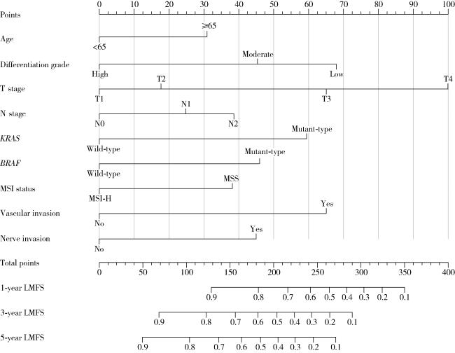
基于上述9个独立影响因素,构建了用于预测LMFS的列线图模型(图 2)。各变量依据其对结局的影响程度被赋予不同分值,其中T分期、肿瘤分化程度及脉管侵犯的贡献权重最高。在使用该模型进行个体化预测时,首先根据患者各变量的具体取值,在模型上方相应的评分轴线上确定其单项得分;随后将各项得分累加,并在模型的总分轴线上定位该累计总分;最终,通过该总分位置向下引垂线,与底部表示1年、3年及5年无肝转移生存概率的轴线相交,交点所对应的读数即为患者的个体化预测概率。例如一位75岁直肠癌患者,根治术后病理分期为T3N0M0,肿瘤中分化,无脉管及神经侵犯,KRASBRAF均为野生型,微卫星状态为MSI-H,该模型预测其术后1年无肝转移生存概率为82%。
图2 预测CRC患者1年、3年和5年LMFS的列线图模型

Abbreviations as in Table 1.

Figure 2 Nomogram predicting 1-, 3- and 5-year probabilities for LMFS in patients with CRC

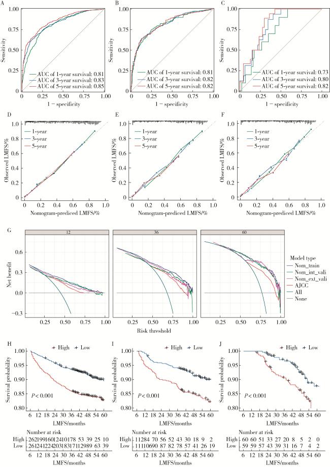
为量化模型的判别能力,本研究采用C指数与时间依赖性ROC曲线进行了综合评估(图 3A~C)。在训练集中,模型表现出良好的区分度,C指数为0.85 (95%CI: 0.82~0.89);其预测1年、3年及5年LMFS的AUC值分别为0.81 (95%CI: 0.77~0.86)、0.83 (95%CI: 0.80~0.89) 和0.85 (95%CI: 0.78~0.92)。在内部验证集中,模型性能保持稳健,C指数为0.81 (95%CI: 0.77~0.83),相应时间点的AUC值分别为0.81 (95%CI: 0.77~0.86)、0.82 (95%CI: 0.79~0.85)和0.82 (95%CI: 0.75~0.89)。值得注意的是,在独立外部验证集中,模型仍展现出可靠的泛化能力,C指数为0.75 (95%CI: 0.71~0.79),预测1年、3年和5年LMFS的AUC值分别为0.73 (95%CI: 0.69~0.78)、0.80 (95%CI: 0.74~0.83) 和0.82 (95%CI: 0.78~0.86)。
图3 预测CRC患者LMFS列线图模型的综合评估

A,B,C,ROC analysis of the nomogram for the LMFS in the training, internal validation, and external validation cohorts;D,E,F,calibration curves of the nomogram for predicting 1-, 3-, and 5-year LMFS in the training, internal validation, and external validation cohorts;G,decision curves of the nomogram for predicting 1-, 3-, and 5-year LMFS;H,I,J,Kaplan-Meier curves of LMFS for patients in the low-and high-risk groups in the training set, internal validation set, and external validation set, respectively. AJCC, American Joint Committee on Cancer.

Figure 3 Comprehensive evaluation of the nomogram for predicting liver metastasis-free survival (LMFS) in patients with colorectal cancer (CRC)

在模型校准度与临床获益评估方面,校准曲线显示,在各队列中模型预测的LMFS概率与经Bootstrap法校正后的实际观察概率曲线均高度吻合,表明模型具有良好的校准度(图 3D~F)。决策曲线分析进一步证实,在绝大多数阈值概率范围内,该列线图模型的临床净获益均高于第8版AJCC TNM分期系统(图 3G)。此外,为量化本模型相较于传统解剖学分期的预测增益,计算了重分类改善指数(net reclassification index, NRI)和综合判别改善指数(integrated discrimination improvement, IDI)。外部验证集数据显示,引入分子标志物的新模型在风险重分类及判别能力上均实现了显著提升:NRI为0.15 (95%CI: 0.05~0.25, P < 0.05),IDI为0.12 (95%CI: 0.04~0.20, P < 0.05);同时,该模型的Brier分数(0.11)低于TNM分期系统(0.16),客观证实了其整体预测准确性的提高。
基于“survminer”R包确定的最佳风险截断值,将所有患者划分为高风险组与低风险组。Kaplan-Meier生存分析显示,在训练集、内部验证集及外部验证集中,高风险组患者的无肝转移生存率均显著低于低风险组,组间差异具有统计学意义(Log-rank检验,P < 0.001,图 3H~J),表明该模型能有效识别术后发生异时性肝转移的高危人群。为提升临床实用性,研究基于该模型构建了交互式在线动态列线图,可用于个体化风险评估,访问链接为 https://crclmfs.shinyapps.io/CRC_LMFS_Tool/
本研究构建的LMFS列线图预测模型在区分度、校准度及临床实用性方面均表现良好,可作为结直肠癌根治术后个体化肝转移风险评估的有效工具。

3 讨论

本研究基于两家医疗中心的临床数据,构建并验证了结直肠癌根治术后无肝转移生存期的列线图预测模型,该模型整合了临床病理特征与分子标志物,在多中心验证中表现出良好的预测性能,为术后个体化风险评估提供了量化工具。
总结本研究与近十年代表性预测模型的对比分析结果见表 3[10, 12, 14-17]。与既往同类研究相比,本模型在预测视角、变量整合及验证策略上呈现出特定优势。首先,在预测终点方面,Yan等[10]的模型侧重于对初诊时同步肝转移风险进行横断面评估;而本研究聚焦于根治术后患者的异时性肝转移风险,通过无肝转移生存期提供时间依赖性的预后信息,这对制定术后长期监测策略具有实际参考价值。其次,在变量选择方面,Li等[12]及Yan等[10]的研究多受限于SEER(Surveillance, Epidemiology, and End Results)数据库记录,主要依赖解剖学分期;本研究则进一步整合了KRASBRAF基因突变及MSI状态,填补了传统临床模型在分子生物学层面的缺失。最后,在模型性能与验证方面,尽管Xiao等[15]亦纳入了部分分子指标,但其模型区分度相对有限(C指数为0.657)。相比之下,本研究采用了严格的多中心外部验证策略,结果显示引入分子标志物的模型在外部队列中C指数仍保持在0.75。这一数据表明,结合解剖学分期与分子特征的综合评估模式,在反映肿瘤生物学行为及保证模型泛化能力方面具有潜在优势。对风险因素的深入分析揭示了分子标志物的重要预测价值。多因素分析表明MSI-H状态(HR=0.71)为保护性因素,这与该亚型通常具有更强的免疫浸润和更好的预后报道一致[16-17]。值得注意的是,本研究中KRAS(HR=1.42)和BRAF突变(HR=1.53)表现出高风险效应,可能与特定突变亚型导向的转移模式差异有关[18-19],但其具体机制仍需进一步探讨。在治疗因素方面,化疗史(HR=2.65)和靶向治疗史(HR=8.35)与较高转移风险相关,这很可能反映了疾病严重程度而非独立的预测因素[20-21]。从临床应用角度,该模型的主要目标是在术后早期识别高危患者,而此时辅助治疗方案尚未完全确定。将治疗史纳入模型会限制其在术前或术后即刻的风险评估价值。因此,本课题组选择构建一个基于基线临床病理和分子特征的模型,使其能够在治疗决策前就提供风险预测,更具临床实用性和普适性。
表3 2015—2025年发表的代表性结直肠癌肝转移预测模型特征对比

Table 3 Comparison of representative prediction models for colorectal cancer liver metastasis published from 2015 to 2025

Study Data source Sample size Prediction endpoint Methodology Variables Model performance Validation strategy
Yan, et al. (2019)[10] SEER database 32 819 LM & LNM Logistic regression Age, CEA, tumor size, grade, N stage AUC: 0.74-0.81 Internal random split
Li, et al. (2020)[12] SEER database 9 958 OS & CSS Cox regression Age, marital status, race, tumor location, pathological grade, histological type, T stage, N stage, colectomy, hepatic surgery, CEA C-index: 0.749 Internal validation
Hao, et al. (2022)[14] Single left 623 Metachronous LM Lasso regression Age, CEA, vascular invasion, T stage, N stage, family history of cancer, KRAS mutation C-index: 0.787 Internal validation
Xiao, et al. (2022)[15] Single left 611 Metachronous LM Deep learning (ResNet-50)+Cox HE image risk score, VELIPI, pT, pN C-index: 0.807 Internal validation split (7 ∶3)
Shao, et al. (2024)[16] SEER database 4 981 OS & CSS Cox & competing risk models Age, race, grade, T stage, N stage, surgery, chemotherapy, CEA, tumor deposits, lung metastasis, tumor size C-index: 0.74- 0.79 Internal random split (7 ∶3)
Jing, et al. (2025)[17] Single left 212 Postoperative liver metastasis Random forest (fusion model) CT radiomics features + Clinical data AUC: 0.751 Internal random split

SEER, Surveillance, Epidemiology, and End Results; OS,overall survival;CSS,cancer-specific survival;LM,liver metastasis;LNM,lymph node metastasis; CEA, carcinoembryonic antigen.

本研究尚存在一定的局限性。首先,鉴于回顾性研究的设计特点,本研究可能存在潜在的选择偏倚,但已通过严格的多中心纳入排除标准加以控制;其次,样本仅来源于国内两家医院,模型在不同族群中的普适性仍需广泛验证;最后,本模型目前主要依赖临床病理特征,尚未纳入ctDNA等液体活检及高维影像学指标[22-23]。展望未来,随着人工智能技术的进步,单纯的临床预测模型正向多模态深度融合演进。近期研究表明,基于CT/MRI的影像组学及深度学习框架在捕捉微观病理特征方面潜力巨大[24],而整合18F-FDG PET/CT代谢参数及炎症-营养评分的机器学习方法亦能显著提升预测效能[25-26]。因此,未来研究应致力于将上述分子、影像及代谢特征与现有列线图深度融合,构建更加立体化、智能化的全周期风险预测体系,以实现更精准的风险分层。
综上所述,本研究采用多中心回顾性队列设计,通过多因素Cox回归分析筛选出结直肠癌根治术后LMFS的独立预测指标,并据此构建了整合临床病理参数与分子标志物的列线图模型。内外部验证结果表明,该模型在不同队列中均保持了良好的预测精度与泛化能力。作为一种可视化的风险评估工具,该模型可用于辅助临床医师进行术后风险分层,从而为个体化随访监测方案的制定提供量化依据。

利益冲突  所有作者均声明不存在利益冲突。

作者贡献说明  王楠楠、袁大晋:提出研究思路,收集、整理、分析数据,撰写论文;朱昱冰:设计研究方案;丁磊:总体把关和审定论文。所有作者均参与论文修改,并对最终文稿进行审读和确认。

1
Siegel RL , Miller KD , Wagle NS , et al. Cancer statistics, 2023[J]. CA Cancer J Clin, 2023, 73 (1): 17- 48.

2
Zheng R , Zhang S , Zeng H , et al. Cancer incidence and mortality in China, 2016[J]. J Natl Cancer Cent, 2022, 2 (1): 1- 9.

3
Morris VK , Kennedy EB , Baxter NN , et al. Treatment of metastatic colorectal cancer: ASCO guideline[J]. J Clin Oncol, 2023, 41 (3): 678- 700.

DOI

4
Reboux N , Jooste V , Goungounga J , et al. Incidence and survival in synchronous and metachronous liver metastases from colorectal cancer[J]. JAMA Netw Open, 2022, 5 (10): e2236666.

DOI

5
Carconi C , Cerreti M , Roberto M , et al. The management of oligometastatic disease in colorectal cancer: Present strategies and future perspectives[J]. Crit Rev Oncol, 2023, 186, 103990.

DOI

6
Benson AB , Venook AP , Al-Hawary MM , et al. Colon cancer, version 2. 2021, NCCN clinical practice guidelines in oncology[J]. J Natl Compr Cancer Netw, 2021, 19 (3): 329- 359.

DOI

7
Tan EK , Ooi LL . Colorectal cancer liver metastases-understanding the differences in the management of synchronous and metachronous disease[J]. Ann Acad Med Singap, 2010, 39 (9): 715- 719.

8
Horie T , Kanemitsu Y , Takamizawa Y , et al. Prognostic differences between oligometastatic and polymetastatic disease after resection in patients with colorectal cancer and hepatic or lung metastases: Retrospective analysis of a large cohort at a single institution[J]. Surgery, 2023, 173 (2): 328- 334.

DOI

9
Balachandran VP , Gonen M , Smith JJ , et al. Nomograms in oncology: More than meets the eye[J]. Lancet Oncol, 2015, 16 (4): e173- e180.

DOI

10
Yan Y , Liu H , Mao K , et al. Novel nomograms to predict lymph node metastasis and liver metastasis in patients with early colon carcinoma[J]. J Transl Med, 2019, 17 (1): 193.

DOI

11
Li G , Li S , Cao L , et al. Nomogram development and validation for predicting minimally invasive step-up approach failure in infected necrotizing pancreatitis patients: A retrospective cohort study[J]. Int J Surg, 2023, 109 (6): 1677- 1687.

DOI

12
Li Y , Liu W , Zhao L , et al. Nomograms predicting overall survival and cancer-specific survival for synchronous colorectal liver-limited metastasis[J]. J Cancer, 2020, 11 (21): 6213.

DOI

13
Collins GS , Reitsma JB , Altman DG , et al. Transparent reporting of a multivariable prediction model for individual prognosis or diagnosis (TRIPOD): The TRIPOD statement[J]. BMJ, 2014, 350, g7594.

14
Hao M , Li H , Wang K , et al. Predicting metachronous liver metastasis in patients with colorectal cancer: Development and assessment of a new nomogram[J]. World J Surg Oncol, 2022, 20 (1): 80.

DOI

15
Xiao C , Zhou M , Yang X , et al. Accurate prediction of metachronous liver metastasis in stage Ⅰ-Ⅲ colorectal cancer patients using deep learning with digital pathological images[J]. Front Oncol, 2022, 12, 844067.

DOI

16
Shao S, Tian D, Li M, et al. Survival prediction in sigmoid-colon-cancer patients with liver metastasis: A prospective cohort study[J/OL]. JNCI Cancer Spectr, 2024, 8(5): pkae080[2025-11-01]. https://doi.org/10.1093/jncics/pkae080.

17
Jing HH , Hao D , Liu XJ , et al. Development and validation of a radiopathomics model for predicting liver metastases of colorectal cancer[J]. Eur Radiol, 2025, 35 (6): 3409- 3417.

18
Zhang Q , Zhou X , Cheng S , et al. Prognostic nomogram for colorectal cancer liver metastasis treated with tumor resection and chemotherapy based on SEER database[J]. J Gastrointest Oncol, 2025, 16 (5): 2067- 2083.

DOI

19
Pitroda SP , Khodarev NN , Huang L , et al. Integrated molecular subtyping defines a curable oligometastatic state in colorectal liver metastasis[J]. Nat Commun, 2018, 9, 1793.

DOI

20
Bi F , Dong J , Jin C , et al. Iparomlimab (QL1604) in patients with microsatellite instability-high (MSI-H) or mismatch repair-deficient (dMMR) unresectable or metastatic solid tumors: A pivotal, single-arm, multicenter, phase Ⅱ trial[J]. J Hematol Oncol, 2024, 17 (1): 109.

DOI

21
Margonis GA , Buettner S , Andreatos N , et al. Association of BRAF mutations with survival and recurrence in surgically treated patients with metastatic colorectal liver cancer[J]. JAMA Surg, 2018, 153 (7): e180996.

DOI

22
Li Y , Solis-Ruiz J , Yang F , et al. NGS-defined measurable residual disease (MRD) after initial chemotherapy as a prognostic biomarker for acute myeloid leukemia[J]. Blood Cancer J, 2023, 13, 59.

DOI

23
Hamfjord J , Guren TK , Dajani O , et al. Total circulating cell-free DNA as a prognostic biomarker in metastatic colorectal cancer before first-line oxaliplatin-based chemotherapy[J]. Ann Oncol, 2019, 30 (7): 1088- 1095.

DOI

24
Luo X , Deng H , Xie F , et al. Prognostication of colorectal cancer liver metastasis by CE-based radiomics and machine learning[J]. Transl Oncol, 2024, 47, 101997.

DOI

25
Lee HS , Kwon HW , Lim SB , et al. FDG metabolic parameter-based models for predicting recurrence after upfront surgery in synchronous colorectal cancer liver metastasis[J]. Eur Radiol, 2023, 33 (3): 1746- 1756.

26
Okimoto S , Kobayashi T , Tashiro H , et al. Significance of the Glasgow Prognostic Score for patients with colorectal liver metastasis[J]. Int J Surg, 2017, 42, 209- 214.

DOI

Outlines

/