Endoscopic characteristics of primary gastric lymphoma and prediction of treatment response

  • Jingyao WEI 1 ,
  • Juxiang YE 2 ,
  • Meiling ZHOU 1 ,
  • Weiwei FU 1 ,
  • Xin LIU 1 ,
  • Kangle ZHAI 1 ,
  • Yanyan SHI 3 ,
  • Shigang DING , 1, * ,
  • Jing ZHANG , 1, *
Expand
  • 1. Department of Gastroenterology, Peking University Third Hospital, Beijing 100191, China
  • 2. Department of Pathology, Peking University Third Hospital, Beijing 100191, China
  • 3. Clinical Epidemiology Research Center, Peking University Third Hospital, Beijing 100191, China
DING Shigang, e-mail,
ZHANG Jing, e-mail,

Received date: 2025-11-03

  Online published: 2026-02-25

Copyright

All rights reserved. Unauthorized reproduction is prohibited.

Abstract

Objective: Primary gastric lymphoma (PGL) is a rare form of lymphoma that arises within the gastric mucosa-associated lymphoid tissue (MALT), often linked to Helicobacter pylori (Hp) infection. The endoscopic features of PGL are heterogeneous, and understanding these characteristics could help distinguish between different lymphoma subtypes. This study aims to systematically assess the endoscopic features of PGL and explore the role of complement receptor type 2/B-cell lymphoma 6 protein (CD21/BCL6)-based grading of lymphoid follicular disruption in predicting the effectiveness of Hp eradication (HPE) treatment in gastric MALT lymphoma. Methods: A retrospective study was conducted involving 100 patients diagnosed with PGL at Peking University Third Hospital between January 2010 and January 2025. Patients were divided into two groups based on histopathological findings: indolent and aggressive lymphoma. The clinical and endoscopic characteristics of these two groups were compared. Survival analysis, including overall survival (OS) and progression-free survival (PFS), was performed using Kaplan-Meier curves and Log-rank tests. A subgroup of 25 patients with gastric MALT lymphoma and known HPE outcomes was selected for further analysis. Diagnostic biopsies were immunohistochemically stained with CD21 and BCL6 and graded from G0 to G4 based on follicular disruption. Logistic regression analysis was used to identify factors associated with HPE failure. Results: Among the 100 patients, the average age was 63.0 (55.8, 71.0) years, with 47 men and 53 women. Aggressive lymphoma showed a significantly higher incidence of B symptoms compared with indolent lymphoma (49.0% vs. 19.6%, P= 0.004). Endoscopically, aggressive lymphoma presented more frequently with ulcerative or mixed morphologies (P < 0.001) and exhibited higher rates of mucosal erosion, ulceration with white slough, lesion friability, bleeding tendency, gastric stenosis, and impaired peristalsis (P < 0.001 for all). Aggressive lymphoma also had significantly worse OS and PFS (OS: P=0.009; PFS: P=0.003). In the subgroup of 25 MALT lymphoma patients, those with ineffective HPE were more likely to be Hp-negative (P=0.049) and had a significantly higher degree of follicular disruption (P=0.015). Multivariable Logistic regression revealed that follicular disruption grading was independently associated with HPE failure (AOR=3.63, 95%CI: 1.14-11.58, P=0.021), while Hp infection status was not (P=0.240). Conclusion: PGL demonstrates considerable variability in its endoscopic presentation. Features, such as ulcerative/mixed morphology, friability, bleeding tendency, stenosis, and impaired peristalsis are indicative of more aggressive disease and correlate with poorer survival outcomes. The CD21/BCL6-based grading of lymphoid follicular disruption provides a valuable tool for identifying patients at high risk of HPE failure, supporting early intervention and risk stratification for gastric MALT lymphoma treatment.

Cite this article

Jingyao WEI , Juxiang YE , Meiling ZHOU , Weiwei FU , Xin LIU , Kangle ZHAI , Yanyan SHI , Shigang DING , Jing ZHANG . Endoscopic characteristics of primary gastric lymphoma and prediction of treatment response[J]. Journal of Peking University(Health Sciences), 2026 , 58(2) : 342 -350 . DOI: 10.19723/j.issn.1671-167X.2026.02.019

原发性胃淋巴瘤(primary gastric lymphoma, PGL)指起源于胃黏膜下层的淋巴系统恶性肿瘤,是仅次于胃腺癌的第二大常见胃恶性肿瘤,其病理类型以胃黏膜相关淋巴组织(mucosa associated lymphoid tissue, MALT)淋巴瘤和弥漫大B细胞淋巴瘤(diffuse large B-cell lymphoma, DLBCL)最为常见[1],二者在生物学行为、临床表现与治疗策略上差异显著。针对胃MALT淋巴瘤,美国国家综合癌症网络(National Comprehensive Cancer Network, NCCN)指南[2]指出,仅对幽门螺杆菌(Helicobacter pylori, Hp)阳性且t(11;18)阴性或未知的早期患者仅推荐根除Hp(Helicobacter pylori eradication, HPE),同期欧洲肿瘤内科学会(European Society for Medical Oncology, ESMO)指南[3]推荐对所有早期患者均试行HPE,因此寻找简单可靠的HPE疗效预测因子便极为关键,对根除有效患者避免过度治疗和根除无效患者避免延误治疗均具有重要意义。本研究旨在系统总结北京大学第三医院PGL患者的内镜特征并探索HPE疗效预测因子,为PGL患者内镜诊断与治疗决策提供实证依据。

1 资料与方法

1.1 病例选择

本研究为单中心回顾性队列研究,连续纳入2010年1月至2025年1月在北京大学第三医院就诊、经胃镜活检或手术切除病理确诊为PGL的患者共100例。病例纳入标准(PGL的判定参照Dawson标准[4]):①病理证实病灶原发于胃,以胃(包括胃壁及胃周淋巴结)为首发或主要受累部位,伴/不伴局部引流区淋巴结受累;②全身浅表及纵隔淋巴结无明确肿大;③肝、脾无受累;④外周血白细胞计数及分类基本正常(急性感染除外)。排除标准:①继发于系统性淋巴瘤或非胃原发淋巴瘤胃受累者;②既往已接受系统抗肿瘤治疗且无法准确获得基线评估者;③关键临床变量或随访信息严重缺失,无法完成主要结局分析者。关键临床变量和随访信息包括Hp状态、主要内镜分型/部位、Lugano分期[5]、治疗方案、随访结局;缺失≥2项或无法判断主要结局者被排除。病理学诊断由北京大学第三医院病理科两名有资质的病理医师独立阅片并达成一致诊断,病理分型参考第5版世界卫生组织淋巴组织肿瘤分类[6]
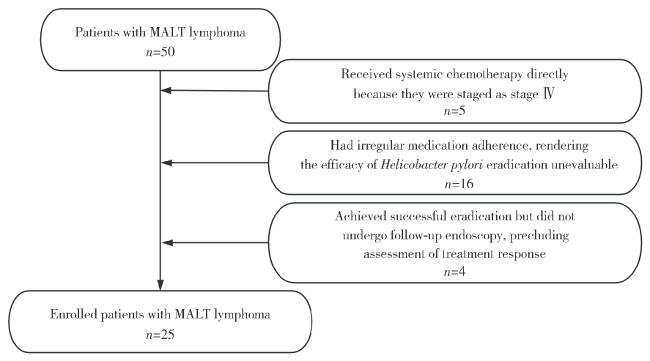
选择2010年1月至2025年1月在北京大学第三医院(门诊/住院)就诊,并经内镜活检及病理检查确诊为胃MALT淋巴瘤的25例患者,用于淋巴滤泡破坏程度的半定量评估。入组标准为:①病理明确诊断为胃MALT淋巴瘤,经两名有资质的病理医师独立阅片并达成一致诊断,组织学亚型符合WHO淋巴造血组织肿瘤分类标准;②选取初诊活检标本,即入组前未接受HPE或放疗、化疗等治疗;③初诊后均接受HPE,并于疗程结束后3~6个月复查胃镜,且经病理综合判定根除疗效(有效或无效)明确。入组筛选流程详见图 1
图1 入组患者筛选流程图

Figure 1 Flow diagram of patient selection

MALT, mucosa associated lymphoid tissue.

本研究遵循了《赫尔辛基宣言》中确立的人体实验道德标准,研究开始前已经北京大学第三医院医学科学研究伦理委员会审查批准(编号:M20260013)。

1.2 研究方法

查阅医院电子病历系统和内镜管理系统,收集患者的基本信息、临床表现、胃镜报告、病理报告和治疗方案,对于接受HPE的患者,在疗程结束后3~6个月复查胃镜并活检,通过病理结果综合评估疗效,疗效分级参考GELA分级[7]。对于患者的Hp感染状态,需采用多种方法(快速尿素酶试验、组织学Warthin-Starry染色/免疫组化、13C/14C呼气试验、粪便抗原)综合判定,多项检查结果中至少有1项为阳性即记录为阳性,否则记录为阴性,并记录既往根除Hp史或抗生素使用史。
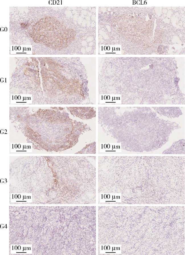
内镜活检石蜡切片在免疫组化染色[显微镜下控制显色时间:补体受体2(complement receptor type 2, CD21)组为50 s,B细胞淋巴瘤-6蛋白(B-cell lymphoma 6 protein,BCL6)组为60 s]后,预先设置阳性对照(已知表达组织)及阴性对照参考,每张切片至少取3个淋巴滤泡/视野;若多个滤泡等级不一致,则以最高等级作为其最终等级,评价有无淋巴滤泡结构及其破坏程度。分级标准见表 1[8],每张切片均由两名有资质的病理医师双盲阅片评估。
表1 淋巴滤泡破坏程度分级

Table 1 Grading of lymphoid follicular architecture disruption

Grade Name Histomorphological features
G0 Architecture-intact type A continuous, dense CD21 meshwork surrounds a well-defined BCL6-positive germinal center; The follicular contour is intact, with no disruption
G1 Marginal thinning/eccentric type The CD21 meshwork remains continuous but shows peripheral “crescent-shaped” condensation toward the outer margin or focal thinning; The germinal center remains intact
G2 Disrupted with preserved outline type The CD21 meshwork shows breaks and/or gaps; The follicular outline is still preserved. Features suggestive of tumor cell “follicular colonization” are present (BCL6-positive areas are intercalated by non-germinal-center cells)
G3 Outline-loss type The follicular contour is indistinct or largely lost; Only scattered fragmentary CD21 meshwork persists, and BCL6-positive germinal-center cells are no longer detectable
G4 Absent/disintegrated type The CD21 meshwork is almost completely unrecognizable/absent; Germinal centers are lost, or only rare scattered BCL6-positive cells are seen

CD21, complement receptor type 2; BCL6, B-cell lymphoma 6 protein.

1.3 统计学分析

采用SPSS软件(version 26.0,IBM Corp., Armonk, NY, USA),对于计量资料先进行Shapiro-Wilk正态性检验及Levene方差齐性检验,符合正态分布者以${\bar x}$±s表示,两组间比较采用独立样本t检验;不符合正态分布者以M(P25, P75)表示,两组间比较采用Mann-Whitney U检验。计数资料以n(%)表示,组间采用χ2检验或校正χ2检验;当理论频数 < 5或样本量较小时采用Fisher精确检验。等级资料采用秩和检验进行组间比较。在生存分析中,时间结局采用Kaplan-Meier法绘制生存曲线,并使用Log-rank检验比较组间差异。此外,采用二元Logistic回归分析(必要时进行Firth校正以处理样本量较小或完全分离问题),以根除治疗“无效”为结局变量评估候选临床/病理指标对根除疗效的预测作用,并报告OR及其95%CIP值。除特别说明外均为双侧检验,P < 0.05即认为差异具有统计学意义。资料将按病理类型(惰性淋巴瘤和侵袭性淋巴瘤)分组。

2 结果

2.1 基本信息

本研究纳入了符合标准的100例PGL患者的病例资料,其中男性47例,女性53例,男女比例约0.89 ∶ 1,首诊年龄为18~84岁,平均年龄为63.0(55.8, 71.0)岁。根据患者首诊内镜活检病理结果,将其分为惰性淋巴瘤和侵袭性淋巴瘤两组,其中最常见的病理类型为胃MALT淋巴瘤和DLBCL,分别有50例和40例,此外惰性淋巴瘤组中有滤泡淋巴瘤1例,侵袭性淋巴瘤组中还有MALT伴大B细胞转化型4例、外周T细胞淋巴瘤3例和套细胞淋巴瘤2例。

2.2 基线资料

在两组患者中,除B症状外,其余资料在两组间的差异均无统计学意义,而在侵袭性淋巴瘤组中,24例患者有B症状,明显多于惰性淋巴瘤组中的11例,差异有统计学意义(P=0.004)。值得注意的是,惰性淋巴瘤组中有近30%的患者无明显临床表现(表 2)。
表2 100例PGL患者的基线资料

Table 2 Baseline data of 100 primary gastric lymphma (PGL) patients

Indicator Indolent lymphoma (n=51) Aggressive lymphoma (n=49) P
Gender 0.680
  Male 25 (49.0) 22 (44.9)
  Female 26 (51.0) 27 (55.1)
Age/years
  Male 63.52±8.71a 60.68±14.34a 0.410
  Female 59.88±12.90a 65.26±11.84a 0.120
Presenting symptoms 0.137
  Abdominal pain 13 (25.5) 22 (44.9)
  Abdominal distension 8 (15.7) 5 (10.2)
  Acid reflux/heartburn 3 (5.9) 3 (6.1)
  Nausea/vomiting 4 (7.8) 4 (8.2)
  Hematemesis/melena 8 (15.7) 10 (20.4)
  Asymptomatic 15 (29.4) 5 (10.2)
Hypertension 0.790
  No 32 (62.7) 32 (65.3)
  Yes 19 (37.3) 17 (34.7)
Diabetes mellitus 0.716
  No 44 (86.3) 41 (83.7)
  Yes 7 (13.7) 8 (16.3)
Smoking 0.722
  No 38 (74.5) 38 (77.6)
  Yes 13 (25.5) 11 (22.4)
Alcohol consumption 0.315
  No 41 (80.4) 43 (87.8)
  Yes 10 (19.6) 6 (12.2)
B symptoms 0.004
  No 40 (80.4) 25 (51.0)
  Yes 11 (19.6) 24 (49.0)

Data are n(%) or ${\bar x}$±s. a,normality was assessed using the Shapiro-Wilk test, and the data were consistent with a normal distribution.

2.3 内镜资料

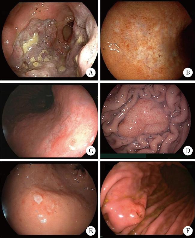
本研究纳入的100例患者均于北京大学第三医院行胃镜检查,其内镜下表现多样(图 2),包括溃疡型病变、浸润型病变、结节型病变、息肉型病变和混合型病变(指包含至少两种前述类型)。
图2 PGL在内镜下的表现

Figure 2 Endoscopic appearance of PGL

A, ulcer type; B, infiltrative type; C, nodular type; D, polypoid type; E and F, mixed type in sequence; PGL, primary gastric lymphma.

在惰性淋巴瘤组中,病变集中在胃体部,以前壁侧和大弯侧居多,且病变多发,而在侵袭性淋巴瘤组中,病变集中在胃体、胃窦部,以小弯侧和大弯侧居多,病变亦多发,但两组间的差异无统计学意义。在惰性淋巴瘤组中,病变分型以溃疡型、浸润型、结节型为主,质地以质软为主,胃腔均通畅,少有蠕动差;而在侵袭性淋巴瘤组中,病变分型以溃疡型和混合型为主,质地以质脆为主,部分胃腔狭窄,近半数蠕动差,组间差异均有统计学意义(表 3)。
表3 100例PGL患者的内镜结果

Table 3 Endoscopic characteristics of 100 PGL patients

Indicator Indolent lymphoma (n=51) Aggressive lymphoma (n=49) P
Lesion locationb 0.104
  Cardia 0 (0.0) 1 (1.2)
  Fundus 7 (8.8) 5 (6.0)
  Upper gastric body 18 (22.5) 9 (10.8)
  Middle gastric body 22 (27.5) 19 (22.9)
  Lower gastric body 22 (27.5) 27 (32.5)
  Antrum 11 (13.7) 22 (26.6)
Lesion orientation 0.103
  Anterior wall 13 (25.5) 8 (16.3)
  Posterior wall 6 (11.8) 9 (18.4)
  Lesser curvature 7 (13.7) 15 (30.6)
  Greater curvature 25 (49.0) 17 (34.7)
Gross morphology < 0.001
  Ulcerative type 11 (21.6) 35 (71.4)
  Infiltrative type 12 (23.5) 0 (0.0)
  Nodular type 19 (37.3) 1 (2.0)
  Polypoid type 4 (7.8) 1 (2.0)
  Mixed type 5 (9.8) 12 (24.6)
Number of lesions 0.568
  Solitary 19 (37.3) 21 (42.9)
  Multiple 32 (62.7) 28 (57.1)
Long diameter/cmc 0.178a
  <1 7 (23.3) 4 (12.9)
  1-<3 16 (53.4) 13 (41.9)
  ≥3 7 (23.3) 14 (45.2)
Short diameter/cmc 0.131a
  <1 11 (36.7) 8 (25.8)
  1-<3 15 (50.0) 12 (38.7)
  ≥3 4 (13.3) 11 (35.5)
Erosion < 0.001
  Absent 26 (51.0) 1 (2.0)
  Present 25 (49.0) 48 (98.0)
Ulceration/white slough < 0.001
  Absent 32 (62.7) 2 (4.0)
  Present 19 (37.3) 47 (96.0)
Consistency < 0.001
  Soft 32 (62.7) 8 (16.3)
  Hard 4 (7.8) 5 (10.2)
  Tough/firm 9 (17.6) 13 (26.5)
  Brittle 6 (11.9) 23 (47.0)
Bleeding 0.008
  Active bleeding 4 (7.8) 9 (18.4)
  Easy bleeding on biopsy 40 (78.4) 40 (81.6)
  Not prone to bleeding 7 (13.8) 0 (0.0)
Perilesional mucosa 0.550
  Normal mucosa 1 (2.0) 0 (0.0)
  Erythema/edema 21 (41.2) 16 (32.7)
  Converging folds 6 (11.8) 5 (10.2)
  Uneven mucosa 23 (45.0) 28 (57.1)
Background mucosa 0.259
  Normal mucosa 4 (7.8) 2 (4.1)
  Erythema/edema 5 (9.8) 5 (10.2)
  Mottled mucosa 32 (62.7) 24 (49.0)
  Uneven mucosa 10 (19.7) 18 (36.7)
Gastric lumen < 0.001
  Patent 51 (100.0) 30 (61.2)
  Stenosis 0 (0.0) 19 (38.8)
Peristalsis < 0.001
  Good 46 (90.2) 25 (51.0)
  Poor 5 (9.8) 24 (49.0)

Data are n(%). a, ordinal variables were analyzed using the Wilcoxon rank-sum test; b, classified according to the primary lesion location; c, data were missing for 21 patients in the indolent lymphoma group and 18 patients in the aggressive lymphoma group; PGL, primary gastric lymphma.

2.4 分期/治疗资料

在惰性淋巴瘤组中,Lugano分期Ⅰ期患者占比达72.5%,在全部患者中,有16例仅做HPE,22例做了HPE和放/化疗,有31例(占60.7%)患者在治疗后达到缓解;而在侵袭性淋巴瘤组中,Lugano分期Ⅳ期患者占比达61.2%,在全部患者中,有42例仅做放疗/化疗/手术,有30例(占61.2%)患者在治疗后达到缓解,两组间的差异均具有统计学意义(P < 0.001,P < 0.001,P=0.007,表 4)。
表4 100例PGL患者的分期/治疗资料

Table 4 Staging and treatment data of 100 PGL patients

Indicator Indolent lymphoma (n=51) Aggressive lymphoma (n=49) P
Lugano classification < 0.001a
  Stage Ⅰ 37 (72.5) 8 (16.3)
  Stage Ⅱ 9 (17.6) 9 (18.4)
  Stage Ⅲ 0 (0.0) 2 (4.1)
  Stage Ⅳ 5 (9.9) 30 (61.2)
Therapeutic regimen < 0.001
  Untreated 6 (11.8) 4 (8.2)
  HPE only 16 (31.4) 0 (0.0)
  Radiotherapy/chemotherapy/surgery 7 (13.7) 42 (85.7)
  HPE+Radiotherapy/chemotherapy 22 (43.1) 3 (6.1)
Follow-up effect 0.007
  Remission after treatment 31 (60.7) 30 (61.2)
  Residual/ineffective 8 (15.7) 1 (2.0)
  Progress/death 6 (11.8) 16 (32.7)
  Untreated and alive 6 (11.8) 2 (4.1)

Data are n(%). a,ordinal variables were analyzed using the Wilcoxon rank-sum test; PGL, primary gastric lymphma.

2.5 生存资料

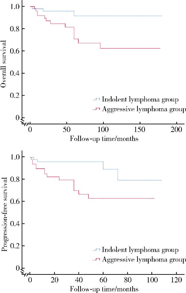
惰性淋巴瘤组患者随访时间为3~180个月,共5例患者出现进展/复发,3例患者因病死亡;在侵袭性淋巴瘤组患者随访时间为6~178个月,共17例患者出现进展/复发,14例患者因病死亡。两组患者在总生存率(overall survival, OS)和无进展生存率(progression free survival, PFS)方面的差异均有统计学意义,即侵袭性淋巴瘤组患者的总生存时间和无病生存时间较惰性淋巴瘤组患者均明显更短(P1=0.009,P2=0.003,图 3)。
图3 原发性胃淋巴瘤患者的生存曲线

Figure 3 Survival curve of primary gestric lymphoma (PGL) patients

2.6 淋巴滤泡破坏程度分级

根据患者在接受规范HPE后复查内镜的病理结果,按疗效将25例患者分为根除有效组(共10例)和根除无效组(共15例),由两名病理医师独立阅片分级并汇总结果,发现根除有效组中G0~G4级分别有3、1、4、0、2例,根除无效组中G0~G4级分别有0、0、3、8、4例,两组间差异有统计学意义(P=0.015),即根除有效组初诊病理组织中淋巴滤泡的破坏程度要明显轻于根除无效组(图 4)。
图4 G0~G4级免疫组织化学示意图(×20)

Figure 4 Schematic diagram of G0-G4 level immunohistochemistry (×20)

CD21, complement receptor type 2; BCL6, B-cell lymphoma 6 protein.

2.7 危险因素分析

经Shapiro-Wilk正态性检验,连续变量并非均满足正态分布,故以中位数(四分位距)描述。根除有效组(n=10)与根除无效组(n=15)组间比较提示无效组Hp感染阴性患者比例显著更高[73.3%(11/15) vs. 30.0%(3/10)],差异具有统计学意义(P=0.049)。同时,两组淋巴滤泡破坏程度分级的分布存在显著差异(P=0.015):有效组以G0~G2为主(G0 30.0%,G1 10.0%,G2 40.0%),而无效组以G3和G4为主(G3 53.3%,G4 26.7%),其余临床及内镜相关指标在两组间差异均无统计学意义(表 5)。
表5 根除有效组和根除无效组的组间比较

Table 5 Inter group comparison of HPE effective group and HPE ineffective group

Indicator HPE effective (n=10) HPE ineffective (n=15) P
Gender 0.099
  Male 2 (20.0) 9 (60.0)
  Female 8 (80.0) 6 (40.0)
Age/years 61.1 (52.8, 68.0) 62.9 (59.0, 69.0) 0.453
Hypertension >0.999
  No 6 (60.0) 9 (60.0)
  Yes 4 (40.0) 6 (40.0)
Diabetes mellitus >0.999
  No 8 (80.0) 13 (86.7)
  Yes 2 (20.0) 2 (13.3)
Smoking 0.179
  No 9 (90.0) 9 (60.0)
  Yes 1 (10.0) 6 (40.0)
Alcohol 0.615
  No 9 (90.0) 11 (73.3)
  Yes 1 (10.0) 4 (26.7)
B symptoms 0.499
  No 7 (70.0) 13 (86.7)
  Yes 3 (30.0) 2 (13.3)
Lesion orientation >0.999
  Anterior wall 2 (20.0) 2 (13.3)
  Posterior wall 1 (10.0) 1 (6.7)
  Lesser curvature 2 (20.0) 3 (20.0)
  Greater curvature 5 (50.0) 9 (60.0)
Gross morphology 0.912
  Ulcerative type 1 (10.0) 2 (13.3)
  Infiltrative type 2 (20.0) 2 (13.3)
  Nodular type 6 (60.0) 8 (53.5)
  Polypoid type 0 (0.0) 2 (13.3)
  Mixed type 1 (10.0) 1 (6.7)
Number of lesions >0.999
  Solitary 3 (30.0) 5 (33.3)
  Multiple 7 (70.0) 10 (66.7)
  Erosion >0.999
  Absent 6 (60.0) 9 (60.0)
  Present 4 (40.0) 6 (40.0)
Ulceration >0.999
  Absent 7 (70.0) 11 (73.3)
  Present 3 (30.0) 4 (26.7)
Consistency 0.510
  Soft 5 (50.0) 11 (73.3)
  Hard 1 (10.0) 0 (0.0)
  Tough 1 (10.0) 2 (13.3)
  Brittle 3 (30.0) 2 (13.3)
Peristalsis 0.400
  Good 9 (90.0) 15 (100.0)
  Poor 1 (10.0) 0 (0.0)
Hp infection status 0.049
  Positive 7 (70.0) 4 (26.7)
  Negative 3 (30.0) 11 (73.3)
Disruption grading 0.015
  G0 3 (30.0) 0 (0.0)
  G1 1 (10.0) 0 (0.0)
  G2 4 (40.0) 3 (20.0)
  G3 0 (0.0) 8 (53.3)
  G4 2 (20.0) 4 (26.7)

Data are n(%) or M (P25, P75). Not all variables were normally distributed in both groups.

进一步行二元Logistic回归分析,单因素结果提示:与Hp阳性相比,Hp阴性与根除无效风险升高相关(OR=6.42,95%CI: 1.09~37.74,P=0.040);破坏程度分级每升高1级,根除无效风险显著增加(OR=3.20,95%CI: 1.17~8.78,P=0.024)。将Hp感染状态与破坏程度分级同时纳入多因素回归后,破坏程度分级仍与根除无效独立相关(AOR=3.63,95%CI: 1.14~11.58,P=0.021),而Hp感染状态与疗效的关联不再具有统计学意义(AOR=4.63,95%CI: 0.52~41.09,P=0.240)。

3 讨论

PGL是最常见的结外非霍奇金淋巴瘤之一,病理谱系以胃MALT淋巴瘤与DLBCL为主,既往有研究提示两者在生物学行为、分期分布、治疗策略及预后方面差异显著[2-3]。本研究基于单中心回顾性队列共纳100例经病理确诊的PGL患者,并在其中进一步构建“初诊后仅接受规范HPE且在预设随访窗口可判定根除疗效”的胃MALT淋巴瘤亚队列,用于探索以组织形态学为基础的HPE疗效预测工具。本研究得到三点主要发现:第一,惰性与侵袭性PGL在内镜形态、黏膜脆性/出血倾向及病灶负荷方面存在差异;第二,两亚组的生存结局差异显著,且这一差异与分期及治疗路径密切相关;第三,在胃MALT淋巴瘤中,基于CD21标记的滤泡树突状细胞(follicular dendritic cells, FDC)网与BCL6标记的生发中心(germinal center, GC)结构完整性所建立的“淋巴滤泡破坏程度”分级,可用于识别HPE不敏感人群,为首诊分层治疗与随访策略优化提供了可及的病理学依据。
在基线资料方面,研究人群的性别比例接近,年龄以中老年为主,与既往PGL流行病学特征一致。惰性组与侵袭性组在多数基线资料上差异无统计学意义,但侵袭性组B症状更常见,提示其更高的全身炎症水平/肿瘤负荷与更强的生物学侵袭性,这与DLBCL等亚型的临床表型相吻合[2-3, 9]。需要强调的是,PGL的临床表现与内镜形态均存在高度异质性,单一特征往往不足以完成可靠的亚型推断,因此“临床-内镜-病理”一体化评估仍是诊断与分层的核心框架。
在内镜表现方面,本研究提示惰性组更常见浸润型/结节型表现,病变质地以“质软”为主,溃疡、白苔、黏膜脆性与活动性出血的比例较低;侵袭性组更倾向溃疡型/混合型,常伴“质脆”、糜烂/出血、较大病灶及腔内狭窄等提示更深层组织破坏与更高风险的内镜征象。既往研究也指出,胃MALT淋巴瘤的内镜表现可从近正常黏膜、斑片样充血、皱襞增粗到结节样改变不等,而明显深溃疡与出血倾向相对更常见于侵袭性病例或发生转化的患者[10]。然而,内镜形态的提示价值并非“决定性”的,不同病理亚型之间存在重叠,同一患者亦可呈现多种形态。因此,提高早期检出与分型准确度的关键仍在于规范多点取材、必要时联合放大/超声内镜,并在病理层面完成可靠分型与分子风险评估[11-12]
在治疗结局与生存方面,本研究显示惰性组以局限期为主,治疗策略多以HPE作为首选;侵袭性组晚期的比例更高,系统治疗(含利妥昔单抗方案)或联合放疗/手术的比例显著升高[13]。两亚组的OS与PFS存在显著差异[14-15],这一差异在真实世界队列中并不意外,但其解释需要谨慎,分期与治疗路径本身是强混杂因素,可能在很大程度上决定长期结局。然而,鉴于本研究为单中心回顾性设计,仍需在更大样本、多中心数据的基础上进一步验证该关联的稳定性与外推性。
胃MALT淋巴瘤的HPE疗效预测是本研究的重点。既往指南与队列研究均指出,局限期、Hp阳性MALT对HPE更敏感,但病灶退缩具有缓慢性,部分患者在早期复查窗口可能呈现残留或未达完全缓解,随后仍可持续退缩[16]。因此,本研究在预设的随访窗口内采用可复现的疗效判定规则,以降低“随访时点差异/临床决策差异”带来的错分风险。本研究观察到HPE无效组在初诊活检中呈现更重的淋巴滤泡结构破坏,提示“肿瘤对Hp抗原驱动的依赖性降低”可能是根除治疗不应答的重要组织学线索。
从机制角度看,淋巴滤泡结构是抗原依赖性B细胞反应的关键微环境单元,FDC以CD21等标记构成网架,负责抗原捕获与递呈、维持GC反应与选择,BCL6则是GC B细胞的核心转录调控因子,其表达与GC结构完整性高度相关。既往研究提示,MALT淋巴瘤发生与进展过程中常出现滤泡殖入、FDC网破碎/消失及GC结构紊乱,反映出肿瘤细胞与淋巴滤泡微环境的相互重塑[17]。本研究采用CD21与BCL6双指标对滤泡结构从“完整-边缘变薄/偏心-轮廓尚存但断裂-轮廓丧失-完全缺失/崩解”的层面进行半定量分级,并在阅片流程中采用双盲独立评估,以最大限度降低主观性,且在定义最高等级时采用更严格标准(多部位、多切片、双指标共同提示滤泡结构缺失/崩解),旨在减少取材不足或偶然未取到代表性滤泡导致的误判。该分级体系所捕捉到的是“反应性GC微环境瓦解”的程度,随着滤泡结构被破坏,肿瘤细胞生长从“Hp抗原驱动”逐步转向“Hp抗原非依赖”的自主生长状态,从而降低HPE的应答概率。这一组织学观察与既往关于NF-κB持续活化、染色体易位[如t(11;18)(q21;q21)/API2-MALT1]、BCL10异常定位等与Hp非依赖、HPE抵抗相关的分子事件方向一致[18-19],提示“形态-免疫表型”可作为分子检测不全条件下的互补风险分层工具[20-21]
在单因素分析中,Hp感染与HPE有效呈正向关联,符合经典的抗原驱动模型,但在纳入滤泡破坏分级等关键病理变量后,Hp状态的独立效应在减弱,提示其作用可能受到“肿瘤-微环境结构破坏程度”的调节或影响。换言之,Hp是上游驱动因素,但当肿瘤获得抗原非依赖性分子事件并伴滤泡结构瓦解时,即使根除Hp后上游刺激被切断也可能不足以逆转肿瘤生长,从而表现为HPE抵抗[22-24]。这一解释不仅与本研究观察一致,也与真实世界队列中“Hp阴性/HPE抵抗者往往需要放疗或系统治疗以获得稳定缓解”的临床经验相符[24]
值得注意的是,除Hp状态与滤泡破坏分级外,部分临床与内镜指标虽呈差异趋势,但差异未达到有统计学意义。这一现象可能与:(1)疗效预测队列样本量较小导致β错误上升;(2)指标间相关性/共线性(如病灶大小与黏膜脆性、出血、狭窄等共现);(3)测量误差与随访时点差异;(4)多重比较背景下的偶然波动等因素有关。因此,本课题组在小样本建模中采用更稳健的策略(如Firth惩罚Logistic回归),并将结果定位为风险分层信号而非对所有因素的否定性结论。本课题组未来仍需扩大样本量,并纳入分子学事件(如MALT1相关易位、NF-κB通路标志物)与微环境因子(APRIL/BAFF等)以提升模型的解释力与可迁移性。
本研究的优势在于:其一,提出了一个基于常规免疫组化即可实现的、可重复的滤泡结构分级体系,为胃MALT根除疗效预测提供便捷工具;其二,以“惰性-侵袭性”分层系统比较其内镜特征与临床结局,有助于形成更可操作的首诊风险识别框架;其三,数据来自同一中心,内镜与病理评估流程较一致,减少了跨中心操作带来的差异。
本研究存在一定的局限性:单中心回顾性队列研究不可避免存在有选择偏倚;部分指标存在缺失与测量不一致;疗效预测队列样本量较小而导致存在模型不稳定的风险;分子学检测不全限制了对“形态-分子-疗效”链条的强因果推断。基于上述局限,本研究结论更适合作为临床实践中的分层提示,并可为多中心前瞻性验证提供假设与方法学基础。
临床实践层面,本研究支持“临床-内镜-病理”整合的分层策略,对临床负荷较轻、内镜呈浸润/结节样改变、黏膜相对不脆且无明显出血风险的患者,应在明确Hp感染状态后优先实施HPE,并严格按预设随访计划接受胃镜及活检病理复评;若病理提示滤泡结构相对保留(低等级破坏),可适当延长观察时间并避免过早治疗;相反,如初诊提示滤泡结构重度破坏(高等级破坏)或存在明确的侵袭性内镜征象(深溃疡、易出血、狭窄等),应提高对Hp非依赖/根除抵抗的警惕,尽早启动二线治疗评估(放疗或系统治疗),以减少无效等待与疾病进展风险[25]
综上所述,惰性PGL更常呈浸润/结节型、质软、低出血风险的内镜表型,并具有更好的生存结局;侵袭性PGL更常表现为溃疡/混合型、质脆出血及更高病灶负荷,需更积极的系统治疗与随访管理。对于胃MALT淋巴瘤,基于CD21与BCL6评估的淋巴滤泡破坏程度分级可作为识别HPE无效高风险人群的简单工具,并有望与分子事件互补,推动更精细的个体化治疗路径选择。

利益冲突  所有作者均声明不存在利益冲突。

作者贡献声明  魏竞尧:设计研究方案,收集、整理与分析数据,撰写论文;叶菊香:病理阅片,统筹诊断;周美玲:收集数据,进行免疫组化染色;付伟伟:设计研究方案;刘鑫:协助内镜分型;翟康乐:收集数据;石岩岩:指导统计分析;丁士刚、张静:设计研究方案、论文审定修改。所有作者均参与论文修改,并对最终文稿进行审读和确认。

1
Dizengof V , Levi I , Etzion O , et al. Incidence rates and clinical characteristics of primary gastrointestinal non-Hodgkin lymphoma: A population study[J]. Eur J Gastroenterol Hepatol, 2020, 32 (5): 569- 574.

DOI

2
National Comprehensive Cancer Network (NCCN). NCCN Guidelines Navigator: B-cell lymphomas. Version 3.2025[EB/OL]. [2025-10-01]. https://www.nccn.org/guidelines/guidelines-detail?category=1&id=1480.

3
Eyre TA , Cwynarski K , D'Amore F , et al. Lymphomas: ESMO clinical practice guideline for diagnosis, treatment and follow-up[J]. Ann Oncol, 2025, 36 (11): 1263- 1284.

DOI

4
Ghimire P . Primary gastrointestinal lymphoma[J]. World J Gastroenterol, 2011, 17 (6): 697.

DOI

5
Rohatiner A , D'Amore F , Coiffier B , et al. Report on a workshop convened to discuss the pathological and staging classifications of gastrointestinal tract lymphoma[J]. Ann Oncol, 1994, 5 (5): 397- 400.

DOI

6
Alaggio R , Amador C , Anagnostopoulos I , et al. The 5th edition of the World Health Organization classification of haematolymphoid tumours: lymphoid neoplasms[J]. Leukemia, 2022, 36 (7): 1720- 1748.

DOI

7
Copie-Bergman C , Wotherspoon AC , Capella C , et al. Gela histological scoring system for post-treatment biopsies of patients with gastric MALT lymphoma is feasible and reliable in routine practice[J]. Br J Haematol, 2013, 160 (1): 47- 52.

DOI

8
Kurshumliu F , Sadiku-Zehri F , Qerimi A , et al. Divergent immunohistochemical expression of CD21 and CD23 by follicular dendritic cells with increasing grade of follicular lymphoma[J]. World J Surg Oncol, 2019, 17 (1): 115.

DOI

9
Herlevic V, Reynolds SB, Morris JD. Gastric Lymphoma[M/OL]. In: StatPearls[Internet]. Treasure Island (FL): StatPearls Publishing, 2024[2025-10-01]. https://www.ncbi.nlm.nih.gov/books/NBK567799/.

10
Watanabe M , Nonaka K , Kishino M , et al. Endoscopic features of gastric mucosa-associated lymphoid tissue lymphoma without Helicobacter pylori[J]. Diagnostics, 2024, 14 (6): 607.

DOI

11
Matysiak-Budnik T , Priadko K , Bossard C , et al. Clinical management of patients with gastric MALT lymphoma: A gastroen-terologist' s point of view[J]. Cancers, 2023, 15 (15): 3811.

DOI

12
Yang BC , Yan HL , Luo XY , et al. Endoscopic morphology of gastric MALT lymphoma correlate with API2/MALT1 fusion and predict treatment response after Helicobacter pylori eradication[J]. BMC Gastroenterol, 2024, 24 (1): 388.

DOI

13
Walewska R , Eyre TA , Barrington S , et al. Guideline for the diagnosis and management of marginal zone lymphomas: A British Society of Haematology Guideline[J]. Br J Haematol, 2024, 204 (1): 86- 107.

DOI

14
Fischbach W , Herold J , Eck M . Watch-and-Wait und Nachsorge Bei gastralen MALT-lymphomen nach alleiniger Helicobacter pylori-eradikation unter besonderer Berücksichtigung der Patientencompliance[J]. Z Gastroenterol, 2025, 63 (12): 1246- 1252.

DOI

15
Lewis CS , Joy G , Jensen P , et al. Primary gastric diffuse large B-cell lymphoma: A multicentre retrospective study[J]. Br J Haematol, 2024, 205 (2): 534- 541.

DOI

16
Lu SN , Huang C , Li LL , et al. Synchronous early gastric and intestinal mucosa-associated lymphoid tissue lymphoma in a Helicobacter pylori-negative patient: A case report[J]. World J Clin Cases, 2022, 10 (33): 12447- 12454.

DOI

17
Salama ME , Lossos IS , Warnke RA , et al. Immunoarchitectural patterns in nodal marginal zone B-cell lymphoma[J]. Am J Clin Pathol, 2009, 132 (1): 39- 49.

DOI

18
Iwamuro M , Takenaka R , Miyahara K , et al. Long-term monitoring of gastric mucosa-associated lymphoid tissue lymphoma in patients with extra copies of the MALT1 gene[J]. Sci Rep, 2024, 14, 4953.

DOI

19
Blosse A , Peru S , Levy M , et al. APRIL-producing eosinophils are involved in gastric MALT lymphomagenesis induced by Helicobacter sp infection[J]. Sci Rep, 2020, 10, 14858.

DOI

20
Liu H , Ye H , Ruskone-Fourmestraux A , et al. T(11;18) is a marker for all stage gastric MALT lymphomas that will not respond to H. pylori eradication[J]. Gastroenterology, 2002, 122 (5): 1286- 1294.

DOI

21
Yeh KH . Nuclear expression of BCL10 or nuclear factor kappa B helps predict Helicobacter pylori-independent status of low-grade gastric mucosa-associated lymphoid tissue lymphomas with or without t(11;18)(q21;q21)[J]. Blood, 2005, 106 (3): 1037- 1041.

DOI

22
Tsai HJ , Tai JJ , Chen LT , et al. A multicenter prospective study of first-line antibiotic therapy for early-stage gastric mucosa-associated lymphoid tissue lymphoma and diffuse large B-cell lymphoma with histological evidence of mucosa-associated lymphoid tissue[J]. Haematologica, 2020, 105 (7): e349- e354.

DOI

23
Ishikawa E , Nakamura M , Satou A , et al. Mucosa-associated lymphoid tissue (MALT) lymphoma in the gastrointestinal tract in the modern era[J]. Cancers, 2022, 14 (2): 446.

DOI

24
Min GJ , Kang D , Lee HH , et al. Long-term clinical outcomes of gastric mucosa-associated lymphoid tissue lymphoma in real-world experience[J]. Ann Hematol, 2023, 102 (4): 877- 888.

DOI

25
Jeon SH , Chang JH , Kim IH , et al. Reduced-dose radiation therapy for stage IE gastric mucosa-associated lymphoid tissue lymphoma: A multi-institutional prospective study (KROG 16-18)[J]. Int J Radiat Oncol, 2025, 121 (4): 1000- 1005.

DOI

Outlines

/