
痛风是嘌呤代谢异常和/或尿酸排泄障碍所致的代谢性疾病,以单钠尿酸盐结晶沉积于关节或软组织引发的复发性急性关节炎为特征,根据病因可分为原发性和继发性。多数病例与尿酸排泄障碍相关,其自然病程包括无症状期、急性关节炎期与间歇期、痛风石及慢性关节炎期。急性痛风性关节炎常表现为突发性关节剧痛,伴受累关节及周围软组织红肿、发热、压痛,大关节受累时可出现关节积液,发作常于夜间或清晨,具有自限性,多在两周内自行缓解。急性痛风发作最常见的部位是第一跖趾关节,其次为中足、踝、膝、腕、指和肘关节,极少累及中轴关节[1]。疼痛常伴有活动受限,严重影响患者的日常生活与生活质量。若累及罕见部位或血尿酸正常时易致误诊,因此需结合病史、查体、实验室及影像学检查综合判断。
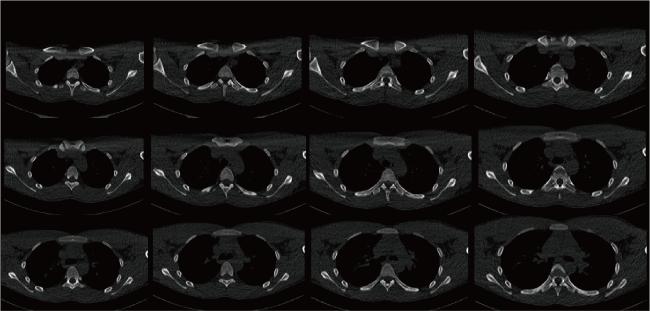
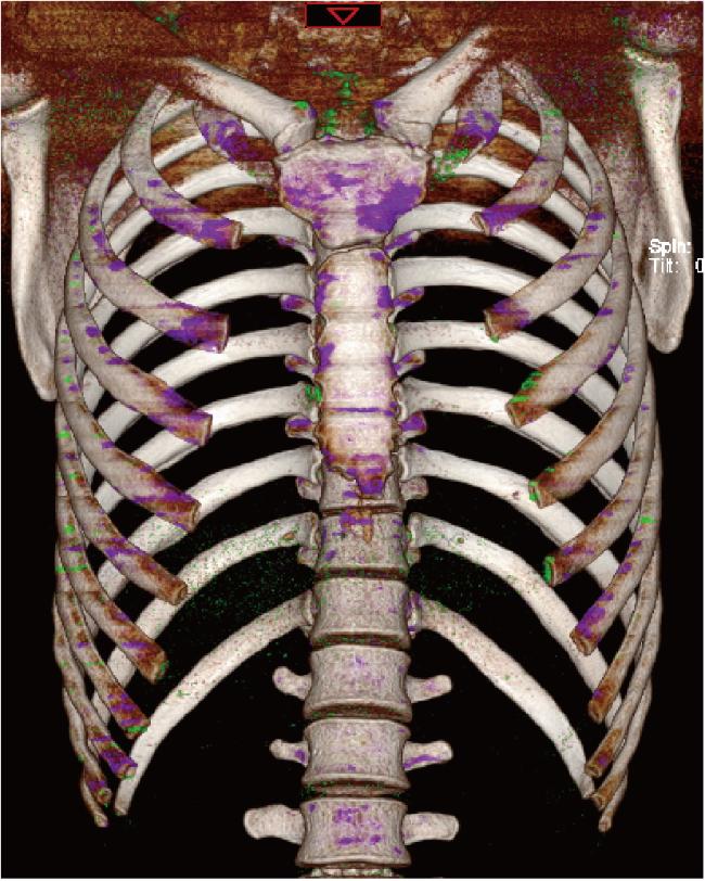
本文报道1例反复前胸痛的19岁男性,血清尿酸水平正常,行胸部双能量CT检查显示胸骨、双侧锁骨、肩胛骨、多根肋骨及胸骨边缘多处痛风石,而得以确诊为痛风的患者,现将诊疗过程报道如下。
1 临床资料
患者,男性,19岁,主因“反复胸前区疼痛2月”就诊于南华大学附属第一医院风湿免疫科,患者于2个月前无明显诱因出现胸前区疼痛,伴有疼痛部位肿胀,无发热、畏寒,无第一跖趾关节等关节肿痛,无对称性多关节肿痛,无口干、眼干,无腰痛,无反复口腔溃疡,无光过敏,无面部红斑,无雷诺现象(Raynaud phenomenon),无皮肤黏膜出血点。患者否认既往高尿酸血症、冠心病、高血压、糖尿病、慢性肾病、乙型肝炎、结核等病史,否认既往长期使用药物史。患者曾于外院检查人类白细胞抗原B27(human leukocyte antigen B27, HLA-B27)阳性,诊断考虑强直性脊柱炎,给予止痛药治疗后,疼痛症状较前好转,但病情仍有反复。患者为求进一步诊治,就诊于我院风湿免疫科门诊。
体格检查:双侧胸锁关节明显压痛、红肿,局部皮温升高,活动轻度受限,脊柱无畸形,活动可,骶髂关节无压痛,“4字征”阴性。
实验室辅助检查:红细胞沉降率34 mm/h,C反应蛋白5.73 mg/L,血尿酸282.6 μmol/L,血常规、肝肾功能、抗核抗体谱、免疫球蛋白、补体、抗链球菌溶血素O等未见异常。
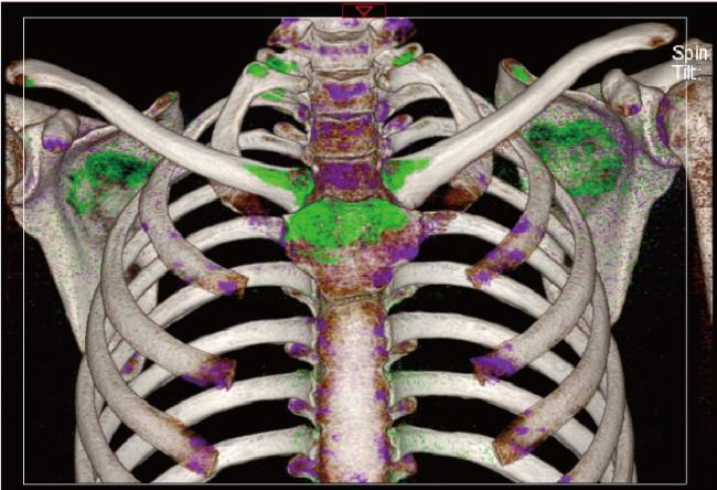
图1 双能量CT显示胸骨、双侧锁骨、肩胛骨、双侧多根肋骨、胸骨边缘多处有尿酸盐结晶沉积(绿色显示)Figure 1 Dual-energy CT demonstrated the urate crystal deposition (shown in green) in the sternum, bilateral clavicles, scapulae, multiple ribs on both sides, and along the edges of the sternum |


2 分析与讨论
本例患者的血尿酸水平并不高,由于患者受累关节的特殊性,无法行关节腔穿刺抽取关节液,排除其他相关疾病后,根据2015年美国风湿病学会/欧洲抗风湿病联盟发布的痛风分类标准:(1)患者受累关节不能忍受触摸、按压(2分);(2)反复典型症状发作,且发作间期完全缓解(2分);(3)双能量CT证实尿酸盐沉积(4分);患者总得分为8分,符合痛风分类诊断标准,故该患者诊断为痛风。
目前,难治性痛风越来越受到临床医生的重视,根据2019年中华医学会内分泌学会发布的相关指南,难治性痛风定义为:(1)单用或联用常规降尿酸药物足量、足疗程,但血尿酸仍≥360 μmol/L;(2)接受规范化治疗,痛风仍发作≥2次/年;(3)存在多发性和/或进展性痛风石[15]。长期的高尿酸血症如果不能得到控制,最终会导致关节骨质破坏、痛风性肾病甚至是急性肾衰竭,目前该指南建议使用普瑞凯希来控制难治性痛风。普瑞凯希是一种尿酸氧化酶,能催化尿酸盐氧化为尿囊素。一项重复、随机、双盲、安慰剂对照试验提示,在患有慢性痛风、血清尿酸水平升高和别嘌呤醇不耐受或难治性的患者中,普瑞凯希能降低尿酸水平[16]。近些年,白细胞介素1(interleukin 1, IL-1)拮抗剂(如阿纳白滞素、卡那单抗和利纳西普)也被批准用于痛风的治疗与预防。
综上所述,随着目前诊断与治疗手段的增多,对于临床医生而言,需要精准地做出诊断,避免误诊和漏诊一些不常见部位的痛风以及血尿酸水平正常的痛风。