Journal of Peking University (Health Sciences) ›› 2026, Vol. 58 ›› Issue (3): 670-673. doi: 10.19723/j.issn.1671-167X.2026.03.030

Previous Articles     Next Articles

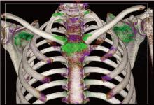
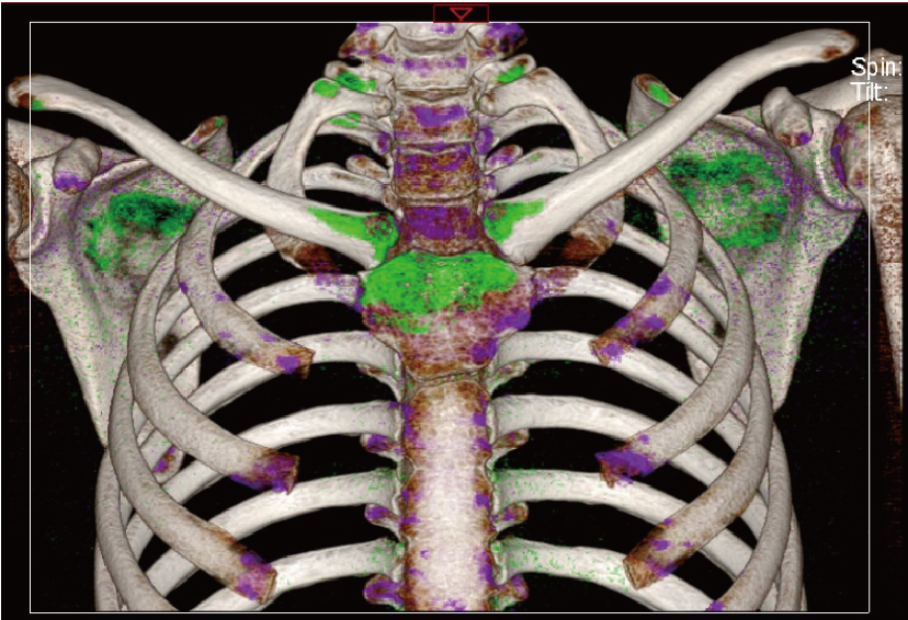
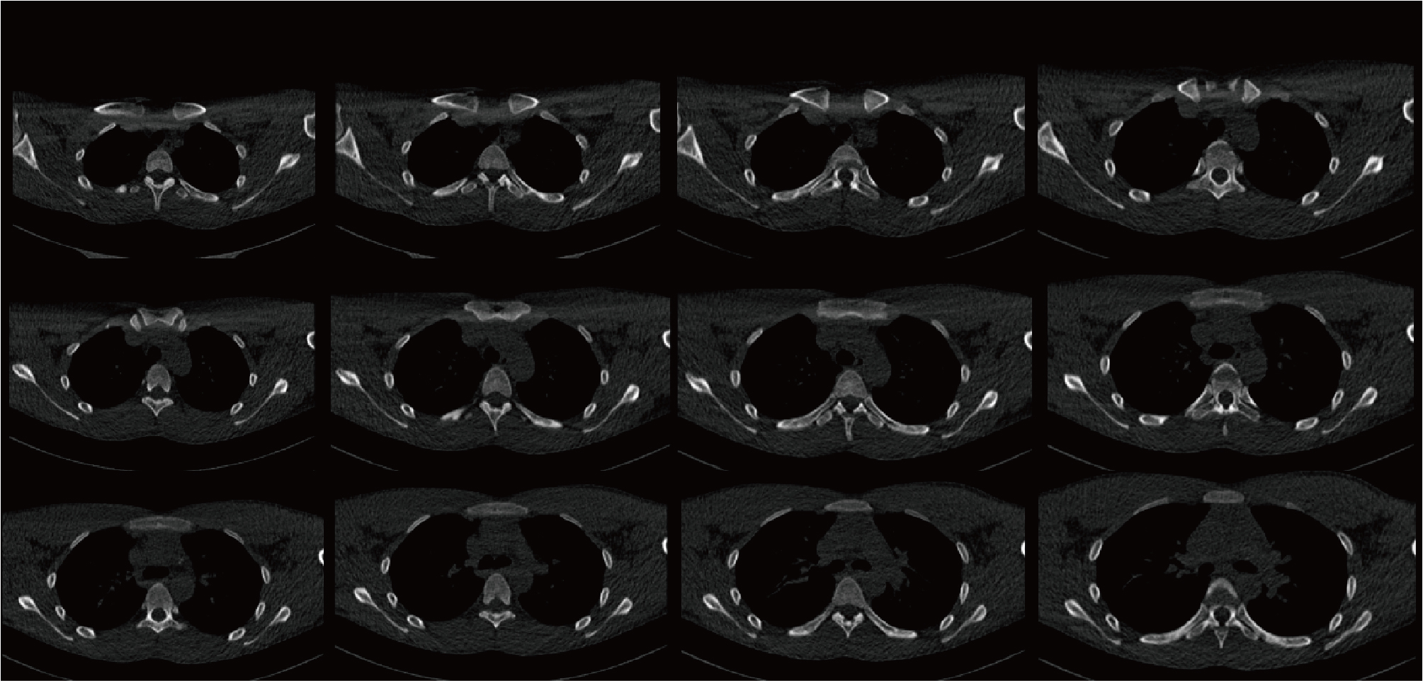
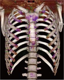
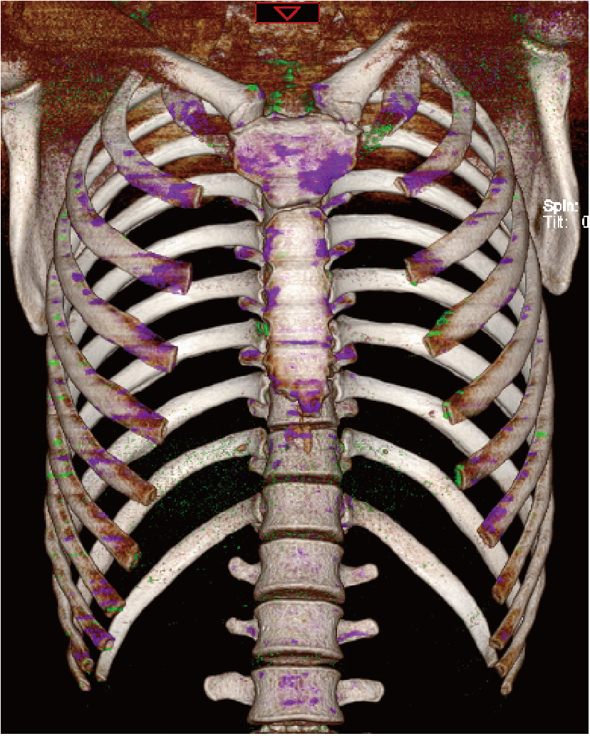
Gout of the manubriosternal joints: A case report

Di GAN1, Qiang FU1, Xiaohui TANG1, Chuwei LI1, Zhaoping SHU2,*()   

  1. 1. Department of Rheumatology and Immunology, The First Affiliated Hospital, Hengyang Medical School, University of South China, Hengyang 421001, Hunan, China
    2. Department of Healthcare-associated Infection Management, The First Affiliated Hospital, Hengyang Medical School, University of South China, Hengyang 421001, Hunan, China
  • Received:2024-07-31 Online:2026-06-18 Published:2026-01-07
  • Contact: Zhaoping SHU
  • Supported by:
    the Scientific Research Fund of Hunan Provincial Education Department(22C0214); the Hunan Provincial Natural Science Foundation(2023JJ40582)

RICH HTML


Abstract:

Gout is a metabolic disorder caused by abnormalities in purine metabolism and/or impaired uric acid excretion. It is characterized by the deposition of monosodium urate crystals in joints or connective tissues, leading to recurrent episodes of acute arthritis. Gout can be categorized into two major types based on its etiology: primary and secondary. The majority of gout cases are associated with impaired uric acid excretion. The natural course of gout can be divided into three stages: (1) asymptomatic phase; (2) acute arthritis phase and intercritical phase; (3) chronic gouty arthritis and tophaceous phase. Acute gouty arthritis typically presents with sudden, severe joint pain, accompanied by redness, swelling, warmth, and tenderness of the affected joint and surrounding soft tissues. When large joints are involved, joint effusion may occur. Acute gouty arthritis often develops at night or in the early morning, is self-limiting, and usually resolves within two weeks. The most common site of acute gout flare-ups is the first metatarsophalangeal joint, followed by the midfoot, ankle, knee, wrist, fingers, and elbow joints. Acute gout attacks rarely affect the axial joints. Pain in these affected areas is often accompanied by limited function, significantly impacting a patient' s daily activities and quality of life. Uncommon sites of gout involvement, as well as gout with normal serum uric acid levels, can easily lead to misdiagnosis. Therefore, a comprehensive diagnosis requires a detailed medical history, physical examination, laboratory tests, and imaging studies. This case report describes a 19-year-old male who was admitted with recurrent anterior chest pain. The diagnosis at another hospital suggested ankylosing spondylitis. Physical examination revealed significant tenderness, redness, and swelling of the bilateral sternoclavicular joints, with local warmth and mildly restricted movement. MRI of the sacroiliac joints showed no significant abnormalities, and serum uric acid levels were normal. Chest dual-energy CT scans revealed multiple tophi in the sternum, bilateral clavicles, scapulae, multiple ribs, and along the edges of the sternum. Based on the patient' s medical history, physical findings, and imaging studies, a diagnosis of gout was confirmed. Treatment included urate-lowering therapy, pain management, and dietary adjustments. During a two-year follow-up period, the patient did not experience recurrent anterior chest pain, and multiple serum uric acid tests consistently showed levels below 300 μmol/L. Repeat chest dual-energy CT scans indicated significant reduction of tophi in the sternoclavicular joints and other affected areas.

Key words: Gout, Tophus, Dual-energy computed tomography, Sternoclavicular joint

CLC Number: 

  • R589.7

Figure 1

Dual-energy CT demonstrated the urate crystal deposition (shown in green) in the sternum, bilateral clavicles, scapulae, multiple ribs on both sides, and along the edges of the sternum"

Figure 2

Chest CT of the patient"

Figure 3

Follow-up after two years with dual-energy CT showed significantly fewer urate crystal deposits compared to before"

1
Dalbeth N , Gosling AL , Gaffo A , et al. Gout[J]. Lancet, 2021, 397 (10287): 1843- 1855.

doi: 10.1016/S0140-6736(21)00569-9
2
Neogi T , Jansen TLTA , Dalbeth N , et al. 2015 Gout classification criteria: An American College of Rheumatology/European League Against Rheumatism collaborative initiative[J]. Ann Rheum Dis, 2015, 74 (10): 1789- 1798.

doi: 10.1136/annrheumdis-2015-208237
3
Richette P , Doherty M , Pascual E , et al. 2018 updated European League Against Rheumatism evidence-based recommendations for the diagnosis of gout[J]. Ann Rheum Dis, 2020, 79 (1): 31- 38.

doi: 10.1136/annrheumdis-2019-215315
4
Schlesinger N , Norquist JM , Watson DJ . Serum urate during acute gout[J]. J Rheumatol, 2009, 36 (6): 1287- 1289.

doi: 10.3899/jrheum.080938
5
Singh JA , Budzik JF , Becce F , et al. Dual-energy computed tomography vs ultrasound, alone or combined, for the diagnosis of gout: A prospective study of accuracy[J]. Rheumatology, 2021, 60 (10): 4861- 4867.

doi: 10.1093/rheumatology/keaa923
6
McCollough CH , Leng S , Yu L , et al. Dual- and multi-energy CT: Principles, technical approaches, and clinical applications[J]. Radiology, 2015, 276 (3): 637- 653.

doi: 10.1148/radiol.2015142631
7
Ng W , Sin CH , Wong CH , et al. Unusual presentation of spinal gout: 2 cases report and literature review[J]. J Orthop Case Rep, 2017, 7 (6): 50- 54.
8
Park EH , Yoo WH , Choi Y . Tophaceous gout: An unusual cause of cervical cord compression[J]. Rheumatology (Oxford), 2021, 61 (1): 467.

doi: 10.1093/rheumatology/keab301
9
Adamson R , Lacy JM , Cheng AM , et al. Tophus causing bronchial obstruction[J]. Am J Respir Crit Care Med, 2013, 188 (12): e72- e73.

doi: 10.1164/rccm.201301-0097IM
10
Montelongo-Rodríguez FA , Madero-Morales PA , Martínez-Fernández AM , et al. Gouty tophus in the scrotum: An unusual extra-articular manifestation of gout[J]. Ochsner J, 2021, 21 (4): 413- 415.

doi: 10.31486/toj.20.0143
11
Rohani A , Chamanian S , Hosseinzade P , et al. A case of mitral valve tophus in a patient with severe gout tophaceous arthritis[J]. J Clin Imaging Sci, 2012, 2, 68.

doi: 10.4103/2156-7514.103058
12
Varinot J , Cazejust J , Wendum D . A gouty tophus appearing as an atypical liver nodule in a cirrhotic patient[J]. Clin Res Hepatol Gastroenterol, 2011, 35 (12): 855- 856.

doi: 10.1016/j.clinre.2011.09.003
13
Mutlu A , Dündar E , İşeri M , et al. An unusual presentation of gout: Tophi in the middle ear[J]. J Int Adv Otol, 2016, 12 (2): 216- 218.
14
Green R , Sensarma K , Jain M , et al. An unusual breast lump presenting as a malignancy found to be a mammary gouty tophus[J]. Breast J, 2011, 17 (5): 528- 529.

doi: 10.1111/j.1524-4741.2011.01129.x
15
中华医学会内分泌学分会. 中国高尿酸血症与痛风诊疗指南(2019)[J]. 中华内分泌代谢杂志, 2020, 36 (1): 1- 13.
16
Sundy JS , Baraf HSB , Yood RA , et al. Efficacy and tolerability of pegloticase for the treatment of chronic gout in patients refractory to conventional treatment: Two randomized controlled trials[J]. JAMA, 2011, 306 (7): 711- 720.

doi: 10.1001/jama.2011.1169
[1] Wei LIU, Wen GUO, Zhe GUO, Chunyan LI, Yunlong LI, Siqi LIU, Liang ZHANG, Hui SONG. Risk factors associated with non-radiographic bone erosion in patients with gout [J]. Journal of Peking University (Health Sciences), 2025, 57(4): 735-739.
[2] Hong DONG,Li-min WANG,Zhi-qiang WANG,Yan-qing LIU,Xiao-gang ZHANG,Ming-ming ZHANG,Juan LIU,Zhen-bin LI. Diurnal differences in acute gout attacks: A clinical study of male gout patients [J]. Journal of Peking University (Health Sciences), 2023, 55(5): 915-922.
[3] Yi-lin YE,Heng LIU,Li-ping PAN,Wei-bing CHAI. Periprosthetic gout flare after total knee arthroplasty: A misdiagnostic case report [J]. Journal of Peking University (Health Sciences), 2023, 55(2): 362-365.
[4] Yu WANG,Hui-min ZHANG,Xue-rong DENG,Wei-wei LIU,Lu CHEN,Ning ZHAO,Xiao-hui ZHANG,Zhi-bo SONG,Yan GENG,Lan-lan JI,Yu WANG,Zhuo-li ZHANG. Diagnostic values of urinary citrate for kidney stones in patients with primary gout [J]. Journal of Peking University (Health Sciences), 2022, 54(6): 1134-1140.
[5] Xiao-wei XIE,Fen LI,Guang-hui LING,Xi XIE,Su-qing XU,Yi-yue CHEN. Development and clinical application of the health education knowledge assessment questionnaire for gout [J]. Journal of Peking University (Health Sciences), 2022, 54(4): 699-704.
[6] PENG Zhe,DING Ya-min,PEI Lin,YAO Hai-hong,ZHANG Xue-wu,TANG Su-mei. Clinical characteristics of crystal deposits in joints and tendons in patients with gout [J]. Journal of Peking University (Health Sciences), 2021, 53(6): 1067-1071.
[7] WANG Gui-hong,ZUO Ting,LI Ran,ZUO Zheng-cai. Effect of rebamipide on the acute gouty arthritis in rats induced by monosodium urate crystals [J]. Journal of Peking University (Health Sciences), 2021, 53(4): 716-720.
[8] XIE Yi-fan,WANG Yu,DENG Xue-rong,GENG Yan,JI Lan-lan,ZHANG Zhuo-li. Analysis of risk factors influencing the detection rate of urate crystal by dual energy computed tomography [J]. Journal of Peking University (Health Sciences), 2021, 53(2): 261-265.
[9] WANG Yu,DENG Xue-rong,JI Lan-lan,ZHANG Xiao-hui,GENG Yan,ZHANG Zhuo-li. Risk factors and diagnostic value for ultrasound-detected tendon monosodium urate crystal deposition in patients with gout [J]. Journal of Peking University (Health Sciences), 2021, 53(1): 143-149.
[10] Lan-lan JI,Yan-jie HAO,Zhuo-li ZHANG. A case of gout secondary to primary myelofibrosis [J]. Journal of Peking University(Health Sciences), 2018, 50(6): 1117-1119.
[11] ZHANG Qian-ru, WANG Yu, ZHANG Zhuo-li. Performance of the 2015 ACR/EULAR classification criteria compared with other classification criteria for diagnosis of gout in Chinese patients [J]. Journal of Peking University(Health Sciences), 2017, 49(6): 979-984.
Viewed
Full text


Abstract

Cited

  Shared   
  Discussed   
No Suggested Reading articles found!