
1 病例资料
1.1 患者一般情况
患者男性,42岁,因发现阴茎肿物30余年于2023年12月7日入院。患者30余年前发现阴茎根部肿物,大小约2 cm×2 cm,无其他不适,未诊治。近半年来肿物增长明显加快,查体阴茎根部偏左侧可及1个约7 cm×4 cm×5 cm的囊性肿物,挤压无明显缩小,透光试验阳性,双侧睾丸、附睾及精索触诊无明显异常,与肿物无明显关联。患者已育2子,均体健,暂无生育诉求。检验及检查:患者病程极其漫长,且肿物位置及性质均不提示睾丸来源,因此, 未行睾丸肿瘤标志物检验,仅完善术前常规检验。超声及磁共振成像提示阴茎根部囊性肿物(图 1),考虑为淋巴管囊肿,拟行囊性肿物切除术。
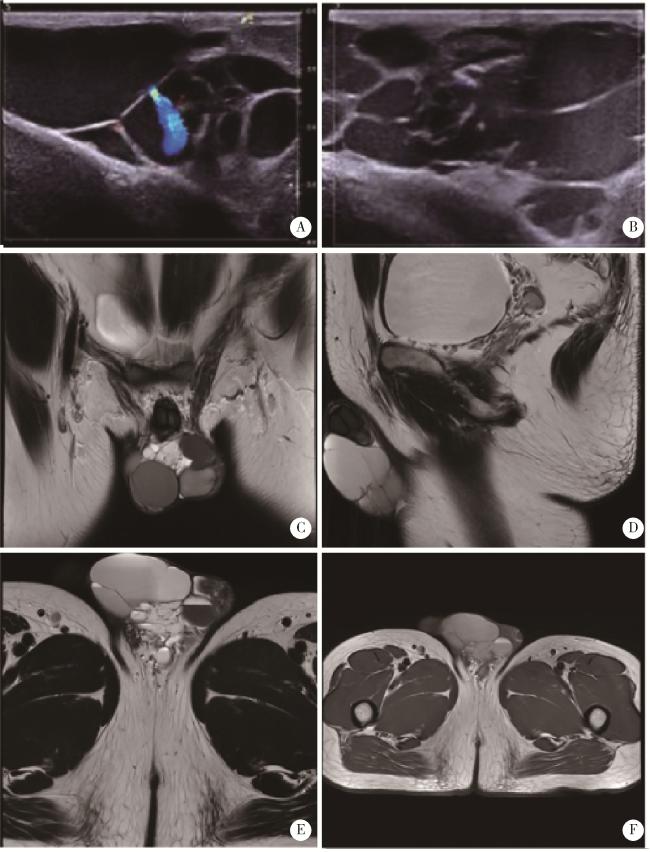
图1 囊性淋巴管瘤超声及磁共振成像Figure 1 Ultrasonography and MRI of cystic lymphangioma A, B, ultrasound revealed an irregular cystic anechoic area at the penoscrotal junction with clear margins; C-F, MRI revealed a multilocular cystic lesion at the scrotal root with clear boundaries. MRI, magnetic resonance imaging. |
1.2 入院诊断
临床诊断:(1)阴茎肿物;(2)淋巴管囊肿可能。
1.3 治疗经过
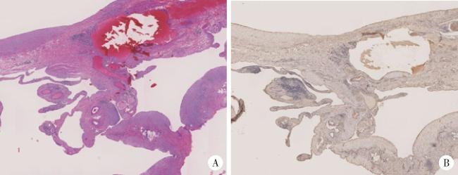
图2 囊性淋巴管瘤的组织学特征Figure 2 Histological features of cystic lymphangioma. A, cystic spaces lined by endothelial cells in dermis (HE ×100); B, CD31 immunostaining highlights the endothelial lining of the cysts (IHC ×100). HE, immunostaining; IHC, immunohistochemistry. |


2 分析与讨论
淋巴管瘤是由淋巴系统发育异常所致的先天性脉管畸形,发病率约1/4 000[1],多见于儿童,头颈部为好发部位,成人阴茎部位发病极为罕见,临床易误诊漏诊。在胚胎时期,淋巴内皮细胞自胚胎静脉中发育迁移而来,形成原始淋巴结构,脉管系统通过响应邻近间充质组织表达的血管内皮生长因子C,以离心出芽方式进一步扩张[2-3]。淋巴丛成熟过程中受到血管生成素2、转录因子FOXC2和GATA2等多种信号通路调控[4-7],上述分子或通路异常会导致淋巴发育成熟受阻,进而引起淋巴管结构功能异常[8]。根据国际脉管异常研究学会(International Society for the Study of Vascular Anomalies, ISSVA)分类,可以将淋巴管瘤分为普通型淋巴管瘤、泛发性淋巴管异常如卡波西样淋巴管瘤病(Kaposiform lymphangiomatosis,KLA)、戈谢病伴淋巴管瘤、通道型淋巴管瘤、“获得性”进行性淋巴管异常(淋巴管瘤)、原发性淋巴管水肿,其中原发性淋巴管水肿又可以分为若干亚型:如Nonne-Milroy综合征(致病基因FLT4/VEGFR3);原发性遗传型淋巴水肿(致病基因VEGF-C/GJC2/Connexin 47)-双行睫(致病基因FOXC2);少毛症-淋巴水肿-毛细血管扩张症(致病基因SOX18);原发性淋巴水肿伴骨髓发育不良(致病基因GATA2);Hennekam淋巴管扩张-淋巴水肿综合征(致病基因CCBE1);小头畸形伴或不伴脉络膜视网膜病变,淋巴水肿,智力障碍综合征(致病基因KIF11);淋巴水肿-后鼻孔闭锁(致病基因PTPN14)[9]。
2.1 病因及临床表现
2.2 诊断
影像学检查是诊断囊性淋巴管瘤的主要手段。超声是首选的辅助检查,有助于判断淋巴管瘤的大小、性质、部位、深度及与周围血管的关系,可以为进一步的治疗提供依据。MRI可在超声的基础上进一步明确瘤体特征与周围组织的关系。也可进行诊断性穿刺,抽出液体若为淡黄色清亮液体提示为淋巴管瘤,结合病理若为血性液体,提示淋巴管瘤伴出血。本文患者超声提示阴茎阴囊交界囊性无回声区,边界清晰形态不规则,MRI提示阴茎根部多房囊性病变,边界清晰,范围约5.6 cm×3.8 cm×6.3 cm,影像学均符合囊性淋巴管瘤的特征。
2.3 鉴别诊断
患者病史极为漫长,且肿物生长缓慢,考虑为慢性良性疾病,因形态不规则呈囊状,透光试验阳性,且肿物位于阴囊阴茎交界处,需要与以下疾病相鉴别:(1)腹股沟斜疝:可见腹股沟可复性包块,包块可进入阴囊,平卧时可还纳,嵌顿时无法还纳,透光试验阴性; (2)鞘膜积液:透光试验阳性,触之有波动感,但是无囊性感,超声提示液性暗区,但是无分隔或囊性结构; (3)精索静脉曲张:平卧时包块缩小或消失,站立位时蚯蚓状团块明显,触之为静脉丛,多见于青年男性,超声显示精索内静脉扩张(内径≥2 mm),进行瓦尔萨尔瓦(Valsalva)动作时血流信号增强。
2.4 治疗
一般情况下,淋巴管瘤的生长较为缓慢,不危及生命,但是患者的病情也会根据病变生长部位的不同出现不同情况,倘若出现压迫、感染等症状也会危及生命,病变生长在面部、颈部也会影响容貌,因此,针对不同部位、不同类型的淋巴管瘤往往需要医务人员选择不同的治疗方式。
药物治疗:目前使用最广泛的药物为西罗莫司,该药物为哺乳动物雷帕霉素受体抑制剂,动物实验表明,西罗莫司可以有效抑制PIK3CA驱动的淋巴管瘤及复杂性淋巴管异常小鼠模型的淋巴管异常增殖[14-15],既往大量研究表明,西罗莫司可以有效缓解多种类型的淋巴管瘤[1]。外用型西罗莫司在毛细淋巴管瘤及浅表微囊性淋巴管瘤中具有较好疗效。曲美替尼可与西罗莫司发挥协同作用[8]。阿培利司是特异性磷脂酰肌醇3-激酶抑制剂,被美国食品药品监督管理局批准用作PIK3CA突变介导的乳腺癌的治疗,研究表明该药物针对CLOVES(congenital lipomatous overgrowth, vascular malformations, epidermal nevi, skeletal/scoliosis and spinal anomalies)综合征患者,可使病灶体积减小37%[8]。
硬化治疗:适用于大囊型和混合型淋巴管瘤,创伤小,与手术相比操作简单,瘢痕小,同时复发率也较低,目前常用的硬化剂有博来霉素、平阳霉素、溶血性链球菌制剂OK-432、无水乙醇等[1]。一篇针对儿童囊性淋巴管瘤硬化治疗的荟萃分析指出,临床上常用的硬化剂为溶血性链球菌制剂OK-432,其次还有博来霉素、玉米蛋白乙醇溶液(alcoholic solution of zein, ASZ)及多西环素。经皮硬化治疗的患者中,48%反应率极佳。针对不同病变类型的统计中,大囊型患者治疗效果(66%)优于微囊性患者(23%),上述常用药物针对大囊型病变的成功率相近,均为80%以上,而针对微囊型ASZ成功率为77%。同时,上述药物均具有一定的副作用,如发热、局部炎症、疼痛等[16]。另一篇仅聚焦于溶血性链球菌制剂OK-432硬化治疗的研究提示,70%患者经过硬化治疗后有良好或极好的效果,既往接受过治疗、病变位置较差(例如纵隔),以及注射次数多,是预后不良的主要因素[17]。
手术治疗:通过手术切除淋巴管囊肿需要考虑病变部位周围重要血管神经的浸润情况,手术指征如下:病灶较小可完全切除,且对外观和功能影响小;有症状的微囊性淋巴管瘤;硬化治疗无法消除,且仍有症状的大囊型及混合型淋巴管瘤;有危及生命的并发症;对外观影响较大[1]。本文中患者的淋巴管囊肿位于阴茎阴囊交界处,且瘤体相对较大,严重影响外观及功能,因此,选择手术切除。
淋巴管瘤的病因主要为体细胞PIK3CA突变,其下游通路可促进淋巴内皮细胞增殖,临床表现因囊型、部位而异。诊断以影像学检查为主,需与腹股沟斜疝等疾病鉴别。治疗需个体化,包括药物治疗、硬化治疗及手术切除,其中手术切除适用于瘤体较大、影响外观功能等情况。