北京大学学报(医学版) ›› 2019, Vol. 51 ›› Issue (4): 678-683. doi: 10.19723/j.issn.1671-167X.2019.04.014

• 论著 • 上一篇    下一篇

肾癌伴下腔静脉瘤栓合并血栓的多种影像学比较

李丽伟1,刘茁2,王国良2,张华3,陈文1,马静1,张丽1,何为4,马潞林2,(),王淑敏1,()   

  1. 1. 北京大学第三医院 超声科, 北京 100191
    2. 北京大学第三医院 泌尿外科, 北京 100191
    3. 北京大学第三医院 临床流行病研究中心, 北京 100191
    4. 北京大学第三医院 放射科, 北京 100191
  • 收稿日期:2019-03-18 出版日期:2019-08-18 发布日期:2019-09-03
  • 通讯作者: 马潞林,王淑敏 E-mail:malulin@medmail.com.cn;shuminwang2014@163.com
  • 基金资助:
    国家重点研发计划专项(2016YFC0104700)

Comparison of various imaging in the diagnosis of renal cell carcinoma with inferior vena cava tumor thrombus combined with bland thrombus

Li-wei LI1,Zhuo LIU2,Guo-liang WANG2,Hua ZHANG3,Wen CHEN1,Jing MA1,Li ZHANG1,Wei HE4,Lu-lin MA2,(),Shu-min WANG1,()   

  1. 1. Department of Ultrasound, Peking University Third Hospital, Beijing 100191, China
    2. Department of Urology, Peking University Third Hospital, Beijing 100191, China
    3. Research Center of Clinical Epidemiology, Peking University Third Hospital, Beijing 100191, China
    4. Department of Radiology, Peking University Third Hospital, Beijing 100191, China
  • Received:2019-03-18 Online:2019-08-18 Published:2019-09-03
  • Contact: Lu-lin MA,Shu-min WANG E-mail:malulin@medmail.com.cn;shuminwang2014@163.com
  • Supported by:
    Supported by National Key Research and Development Program of China(2016YFC0104700)

摘要:

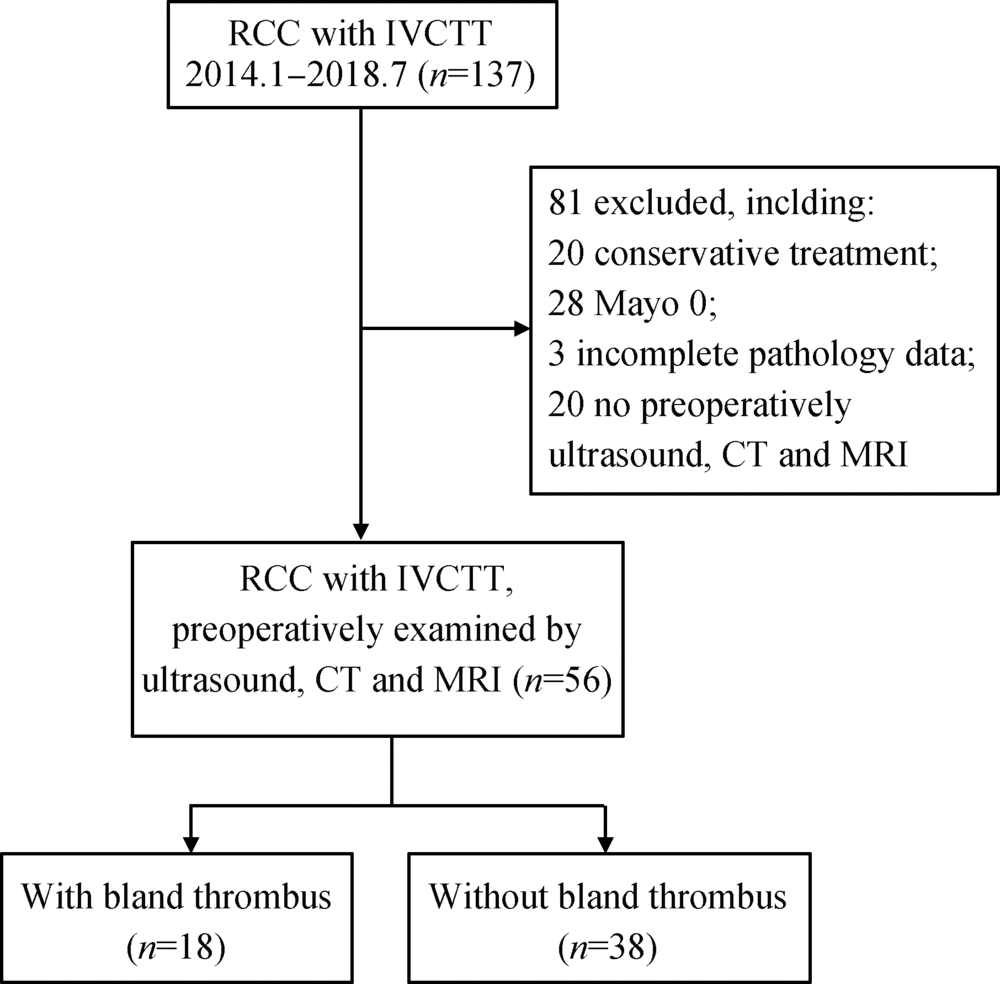
目的:研究肾癌伴下腔静脉瘤栓合并血栓患者的临床及影像资料,分析下腔静脉超声、泌尿系增强CT及增强磁共振3种影像学检查方法对下腔静脉瘤栓伴血栓的诊断效能。方法:选择北京大学第三医院泌尿外科2014年1月至2018年7月的肾癌伴瘤栓病例56例进行回顾性分析,所有患者术前均同时行下腔静脉超声、泌尿系增强CT及增强磁共振检查并完成手术治疗,且术后病理诊断证实为肾癌伴下腔静脉瘤栓。结果:根据术中观察及术后病理诊断证实下腔静脉瘤栓是否合并血栓为标准,将56例患者分为合并血栓组(n=18)及不合并血栓组(n=38)。比较发现,瘤栓合并血栓的患者,瘤栓长度更长[(10.50 ± 5.55) cm vs.(6.66 ± 3.73) cm,P = 0.014];瘤栓直径/下腔静脉(inferior vena cava, IVC)冠状最大径比值更接近1[1.0(0.7,1.0) vs. 0.9 (0.2,1.0), P= 0.004];出现下肢水肿的比例更高[66.7%(12/18) vs.5.3%(2/36),P = 0.005];行下腔静脉节段性切除或下腔静脉横断术的比例更高[66.7%(12/18) vs.15.8%(6/38), P<0.001]。对比下腔静脉超声、泌尿系增强CT及增强磁共振3种影像检查方法,鉴别瘤栓合并血栓,灵敏度最高的是增强磁共振(77.8%),特异性最高的是下腔静脉超声和增强CT(97.4%), 准确性最高的是增强CT及增强磁共振(83.9%),阳性预测值最高的是增强CT(90.9%),阴性预测值最高的是增强磁共振(89.2%)。结论:肾癌伴下腔静脉瘤栓合并血栓的患者,下腔静脉瘤栓长度更长,瘤栓直径/IVC冠状最大径比值更接近1,更易出现下肢水肿。术前需综合多种影像方法,提高诊断的准确率。

关键词: 肾癌, 下腔静脉瘤栓, 血栓, 影像诊断, 超声

Abstract:

Objective: To analyse the clinical and imaging data of patients with renal cell carcinoma (RCC) with inferior vena cava tumor thrombus (IVCTT), and to assess the diagnostic efficacy of ultrasound, enhanced computed tomography (CT) and enhanced magnetic resonance imaging (MRI) in the diagnosis of RCC with IVCTT combined with bland thrombus was assessed.Methods: We retrospectively analyzed the clinical and imaging data of 56 RCC patients with IVCTT who underwent radical nephrectomy and IVC thrombectomy between January 2014 and July 2018 in Department of Urology, Peking University Third Hospital. All the patients underwent US, enhanced CT and enhanced MRI preoperatively, and all the cases were confirmed with RCC with IVCTT by histological evaluation.Results: The criteria of RCC with IVCTT combined with bland thrombus was confirmed by intraoperative observation and postoperative pathology. The 56 patients were divided into bland thrombus group (n=18) and non bland thrombus group (n=38). Compared the two groups, it was found that the length of IVCTT was longer [(10.50 ± 5.55) cm vs.(6.66 ± 3.73) cm, P=0.014]; the ratio of diameter of IVCTT to maximum coronal diameter of IVC was closer to 1 [1.0 (0.7, 1.0) vs. 0.9 (0.2, 1.0), P=0.004]; the proportion of lower limb edema was higher [66.7(12/8)% vs.5.3%(2/36), P=0.005];the proportion of segmental resection or interrupt of IVC was higher [66.7%(12/18) vs.15.8%(6/38), P<0.001], with statistical significance. Compared with the three imaging methods of US, enhanced CT and MRI, the highest sensitivity was MRI (77.8%), the highest specificity was enhanced MRI and enhanced CT (97.4%), the highest accuracy was enhanced CT and enhanced MRI (83.9%), the highest positive predictive value was enhanced CT (90.9%) and the highest negative predictive value was enhanced MRI (89.2%).Conclusion: For the patients that RCC with IVCTT combined with bland thrombus, the length of IVCTT is longer, and the ratio of the diameter of IVCTT to the maximum corona diameter of IVC is closer to 1, and more likely to cause lower limb edema. Preoperative comprehensive evaluation of multiple images is needed to improve the accuracy of diagnosis.

Key words: Renal cell carcinoma, Inferior vena cava tumor thrombus, Bland thrombus, Imaging diagnosis, Ultrasound

中图分类号: 

  • R737.1

图1

研究流程及排除标准"

表1

两组患者临床及影像特征"

Features n With bland thrombus
(n=18)
Without bland thrombus
(n=38)
P
Age/years, x?±s 56 57.37±8.78 60.21±13.33 0.369
Tumor diameter/cm, x?±s 56 8.82±3.75 9.04±3.34 0.263
IVCTT length/cm, x?±s 56 10.50±5.55 6.66±3.73 0.014*
IVCTT diameter/cm, x?±s 56 3.54±0.68 2.76±1.04 0.105
Maximum coronal IVC diameter/cm, x?±s 56 3.63±0.60 3.22±0.65 0.950
IVCTT diameter/maximum coronal IVC diameter, median (min, max) 56 1.0 (0.7,1.0) 0.9 (0.2,1.0) 0.004*
Gender, n (%) 56 0.773
Male 13 (72.2) 26 (68.4)
Female 5 (27.8) 12 (31.6)
Renal tumor side, n (%) 56 0.419
Right 11 (61) 28 (73.7)
Left 7 (38.9) 9 (23.7)
Blateral 0 (0) 1 (2.6)
Mayo classification, n (%) 56 0.203
Mayo Ⅰ 1 (5.6) 10 (26.3)
Mayo Ⅱ 6 (33.3) 14 (36.8)
Mayo Ⅲ 6 (33.3) 9 (23.7)
Mayo Ⅳ 5 (27.8) 5 (13.2)
Clinical symptoms, n (%) 56 0.005*
Lower limb edema 12 (66.7) 2 (5.3)
No lower limb edema 6 (33.3) 36 (97.4)
Pathological type, n (%) 56 0.447
ccRCC 16 (88.9) 30 (78.9)
pRCC 2 (11.1) 5 (13.2)
Other 0 (0) 3 (7.9)
Rhabdomyosis or sarcomatoid degeneration, n (%) 50 0.639
Yes 5 (27.8) 7 (21.9)
No 13 (72.2) 25 (78.1)
Nuclear grading, n (%) 53 0.829
4 (23.6) 8 (22.9)
9 (52.9) 16 (45.7)
4 (23.5) 11 (31.4)
Segmental resection or interuption IVC, n (%) 58 <0.001*
Yes 12 (66.7) 6 (15.8)
No 6 (33.3) 32 (84.2)

表2

肾癌伴下腔静脉瘤栓合并血栓患者临床、病理及影像资料"

No. Gender Age/
years
Renal
tumor
side
Renal
tumor
diameter/cm
Pathology Nuclear
grading
IVCTT
length
Mayo IVC
resection
Bland thrombus
location
length in
imaging/
cm
Imaging diagnosis
US MRI CT
1 M 62 L 5.80 ccRCC 3 11.30 No Proximal - 0 0 0
2 M 67 R 3.60 ccRCC 2 16.40 Interruption Distal 12.94 0 1 1
3 M 53 R 7.70 ccRCC 4 18.60 Segmental Distal 8.50 0 1 0
4 M 53 R 6.40 ccRCC 4 20.00 Segmental Distal 2.00 0 1 1
5 F 62 R 10.30 ccRCC 3 20.00 Interruption Distal,contralateral 12.25 0 1 1
6 M 50 R 5.80 ccRCC 3 6.60 Segmental Distal,contralateral 2.70 1 1 1
7 M 71 R 7.20 ccRCC 3 15.50 Interruption Distal 8.80 0 1 1
8 M 45 R 10.30 ccRCC 2 10.00 Interruption Distal 2.13 0 1 1
9 M 58 L 13.00 ccRCC 3 6.25 Interruption Distal 3.30 0 1 0
10 M 47 L 15.50 pRCC 3 6.20 No Contralateral, - 0 0 0
11 M 58 L 14.50 ccRCC 4 5.00 Interruption Distal 1.30 0 1 1
12 F 69 L 10.90 ccRCC 3 14.30 No Distal 1.30 0 1 0
13 F 67 R 3.80 pRCC 3 9.40 Segmental Distal,contralateral 11.75 1 1 1
14 M 62 R 4.50 ccRCC 2 5.48 Interruption Distal, contralatera 7.84 0 1 1
15 M 48 R 12.00 ccRCC 4 6.90 Segmental Distal 2.00 0 1 0
16 F 41 R 6.80 ccRCC 3 3.20 No Contralateral - 0 0 0
17 M 57 L 13.30 ccRCC 4 5.80 No Contralateral - 0 0 0
18 F 63 R 7.30 ccRCC 2 8.00 No Distal 1.13 1 1 1

表3

超声、增强磁共振及增强CT诊断肾癌伴下腔静脉瘤栓合并血栓与临床病理诊断的对比"

Imaging diagnosis result Surgical pathologic diagnosis
With bland thrombus (n=18) Without bland thrombus (n=38)
US Positive 3 1
Negative 15 37
Enhanced MRI Positive 14 5
Negative 4 33
Enhanced CT Positive 10 1
Negative 8 37

表4

超声、增强磁共振及增强CT诊断肾癌伴下腔静脉瘤栓合并血栓的诊断效能"

Imaging diagnosis Sensitivity Specificity Accuracy Positive predictive value Negative predictive value
US 16.8 97.4* 71.4 75.0 71.2
Enhanced MRI 77.8* 86.8 83.9* 73.7 89.2*
Enhanced CT 55.7 97.4* 83.9* 90.9* 82.2
[1] 刘茁, 马潞林, 田晓军 , 等. 根治性肾切除术+下腔静脉癌栓取出术治疗Mayo 0~Ⅳ 级下腔静脉癌栓的临床经验[J]. 中华泌尿外科杂志, 2017,38(11):842-847.
[2] 刘茁, 马潞林, 田晓军 , 等. 肾癌根治性切除加癌栓取出术治疗Mayo Ⅲ 级下腔静脉癌栓的手术技术及临床经验[J]. 北京大学学报(医学版), 2017,49(4):597-602.
[3] 刘茁, 田晓军, 马潞林 . 根治性肾切除术联合Mayo 0-Ⅱ 级静脉癌栓取出术的临床麻醉管理[J]. 现代肿瘤医学, 2017,25(16):2672-2677.
[4] 刘茁, 马潞林, 田晓军 , 等. 腹腔镜和开放肾癌根治性切除+Mayo Ⅱ 级下腔静脉癌栓取出术11例临床分析[J]. 现代泌尿外科杂志, 2017,22(8):603-607.
[5] 马潞林, 刘茁 . 肾癌并肝段和肝以上下腔静脉癌栓的诊治体会[J]. 中华泌尿外科杂志, 2017,38(7):481-484.
[6] Blute ML, Leibovich BC, Lohse CM , et al. The mayo clinic experience with surgical management, complications and outcome for patients with renal cell carcinoma and venous tumour thrombus[J] . BJU Int, 2004,94(1):33-41.
[7] Wang M, Ping H, Niu Y , et al. Pure conventional laparoscopic radical nephrectomy with level II vena cava tumor thrombectomy[J]. Int Braz J Urol, 2014,40(2):266-273.
[8] 程艳, 蔡欣, 刘基巍 . 恶性肿瘤与血栓形成[J]. 临床肿瘤学杂志, 2010,15(4):376-379.
[9] Ayyathurai R, Garciaroig M, Gorin MA , et al. Bland thrombus association with tumour thrombus in renal cell carcinoma: Analysis of surgical significance and role of inferior vena caval interruption[J]. BJU Int, 2013,110(11b):E449-E455.
[10] Hutchinson R, Rew C, Chen G , et al. The adverse survival implications of bland thrombus in renal cell carcinoma with venous tumor thrombus[J]. Urology, 2018,115:119-124.
[11] Blute ML, Boorjian SA, Leibovich BC , et al. Results of inferior vena caval interruption by greenfield filter, ligation or resection during radical nephrectomy and tumor thrombectomy[J]. J Urol, 2007,178(2):440-445.
[12] 马鑫 . 机器人腹腔镜腔静脉瘤栓取出术:新的思考新的策略[J]. 中华腔镜外科杂志: 电子版, 2017,10(5):272-273.
[13] Quencer KB, Friedman T, Sheth R , et al. Tumor thrombus: Incidence, imaging, prognosis and treatment[J]. Cardiovasc Diagn Ther, 2017,7(Suppl 3):S165-S177.
[14] Mukai M, Oka T . Mechanism and management of cancer-associated thrombosis[J]. J Cardiol, 2018,72(2):89-93.
[15] 肾癌伴静脉瘤栓北京专家共识[J]. 微创泌尿外科杂志, 2017: 6(6):321-327.
[16] 宋奕宁, 赵艺超, 李建国 . 下腔静脉肿瘤的超声影像诊断与鉴别[J]. 中国超声医学杂志, 2018,34(1):37-39.
[17] Tarantino L . Contrast-enhanced ultrasound in differentiating malignant from benign portal vein thrombosis in hepatocellular carcinoma[J]. World J Gastroenterol, 2015,21(32):9457-9460.
[18] Sonavane SN, Malhotra G, Asopa R , et al. Role of fluorine-18 fluorodeoxyglucose positron emission tomography in a case of renal cell carcinoma to differentiate tumor thrombus from bland thrombus[J]. Indian J Nucl Med, 2015,30(4):355-357.
[19] Sharma P, Kumar R, Jeph S , et al. 18F-FDG PET-CT in the diagnosis of tumor thrombus[J]. Nucl Med Commun, 2011,32(9):782-788.
[1] 彭喆,丁亚敏,裴林,姚海红,张学武,唐素玫. 痛风患者发生关节及肌腱内晶体沉积的临床特点[J]. 北京大学学报(医学版), 2021, 53(6): 1067-1071.
[2] 宋志博,耿研,邓雪蓉,张晓慧,张卓莉. 肌肉骨骼超声在指导银屑病关节炎临床分型中的价值[J]. 北京大学学报(医学版), 2021, 53(6): 1061-1066.
[3] 邓雪蓉,孙晓莹,张卓莉. 类风湿关节炎患者足踝部体征和超声下病变的一致性[J]. 北京大学学报(医学版), 2021, 53(6): 1037-1042.
[4] 庄金满,李天润,李选,栾景源,王昌明,冯琦琛,韩金涛. Rotarex经皮机械性血栓切除装置在急性下肢缺血中的应用[J]. 北京大学学报(医学版), 2021, 53(6): 1159-1162.
[5] 敖明昕,李学民,于媛媛,时会娟,黄红拾,敖英芳,王薇. 视觉重建对老年人行走动态足底压力的影响[J]. 北京大学学报(医学版), 2021, 53(5): 907-914.
[6] 王新宇,崔哲,和清源,邓湘宁,郭歌,冯新恒,冯杰莉. 斑点追踪技术评价中国优秀男子举重运动员心脏的改变[J]. 北京大学学报(医学版), 2021, 53(5): 832-837.
[7] 肖若陶,刘承,徐楚潇,何为,马潞林. 术前血小板参数与局部进展期肾细胞癌预后[J]. 北京大学学报(医学版), 2021, 53(4): 647-652.
[8] 庄金满,李天润,李选,栾景源,王昌明,冯琦琛,韩金涛. Rotarex 旋切导管在下肢动脉硬化闭塞症支架内再狭窄中的应用[J]. 北京大学学报(医学版), 2021, 53(4): 740-743.
[9] 洪鹏,田晓军,赵小钰,杨飞龙,刘茁,陆敏,赵磊,马潞林. 肾移植术后双侧乳头状肾癌1例[J]. 北京大学学报(医学版), 2021, 53(4): 811-813.
[10] 杨广鑫,栾景源,贾子昌. 静脉造影和超声联合引导射频闭合治疗下肢静脉曲张[J]. 北京大学学报(医学版), 2021, 53(2): 332-336.
[11] 孟圆,张丽琪,赵雅宁,柳登高,张祖燕,高岩. 67例上颌根尖周囊肿的三维影像特点分析[J]. 北京大学学报(医学版), 2021, 53(2): 396-401.
[12] 王昱,邓雪蓉,季兰岚,张晓慧,耿研,张卓莉. 超声检测痛风患者肌腱受累的危险因素和诊断价值[J]. 北京大学学报(医学版), 2021, 53(1): 143-149.
[13] 石茂静,高伟波,黄文凤,朱继红. 61例血栓性血小板减少性紫癜患者的临床分析[J]. 北京大学学报(医学版), 2021, 53(1): 210-214.
[14] 李思雨,段雪飞,曹烨. 应用超声器械改善预备体肩台的效果[J]. 北京大学学报(医学版), 2021, 53(1): 88-94.
[15] 朱冯赟智,邢晓燕,汤晓菲,李依敏,邵苗,张学武,李玉慧,孙晓麟,何菁. 肌炎合并血栓栓塞患者的临床及免疫学特征[J]. 北京大学学报(医学版), 2020, 52(6): 995-1000.
Viewed
Full text


Abstract

Cited

  Shared   
  Discussed   
[1] 赵磊, 王天龙 . 右心室舒张末期容量监测用于肝移植术中容量管理的临床研究[J]. 北京大学学报(医学版), 2009, 41(2): 188 -191 .
[2] 万有, , 韩济生, John E. Pintar. 孤啡肽基因敲除小鼠电针镇痛作用增强[J]. 北京大学学报(医学版), 2009, 41(3): 376 -379 .
[3] 张燕, 韩志慧, 钟延丰, 王盛兰, 李玲玲, 郑丹枫. 骨骼肌活组织检查病理诊断技术的改进及应用[J]. 北京大学学报(医学版), 2009, 41(4): 459 -462 .
[4] 赵奇, 薛世华, 刘志勇, 吴凌云. 同向施压测定自酸蚀与全酸蚀粘接系统粘接强度[J]. 北京大学学报(医学版), 2010, 42(1): 82 -84 .
[5] 林红, 王玉凤, 吴野平. 学校生活技能教育对小学三年级学生行为问题影响的对照研究[J]. 北京大学学报(医学版), 2007, 39(3): 319 -322 .
[6] 丰雷, 程嘉, 王玉凤. 注意缺陷多动障碍儿童的运动协调功能[J]. 北京大学学报(医学版), 2007, 39(3): 333 -336 .
[7] 李岳玲, 钱秋瑾, 王玉凤. 儿童注意缺陷多动障碍成人期预后及其预测因素[J]. 北京大学学报(医学版), 2007, 39(3): 337 -340 .
[8] . 书讯[J]. 北京大学学报(医学版), 2007, 39(3): 225 -328 .
[9] 牟向东, 王广发, 刁小莉, 阙呈立. 肺黏膜相关淋巴组织型边缘区B细胞淋巴瘤一例[J]. 北京大学学报(医学版), 2007, 39(4): 346 -350 .
[10] 张宏文, 丁洁, 王芳, 杨惠霞. 一例X连锁Alport综合征女性妊娠期随访[J]. 北京大学学报(医学版), 2007, 39(4): 351 -354 .