Journal of Peking University(Health Sciences) ›› 2019, Vol. 51 ›› Issue (1): 59-64. doi: 10.19723/j.issn.1671-167X.2019.01.011
Previous Articles Next Articles
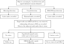
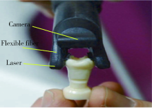
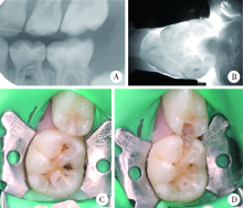
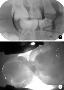
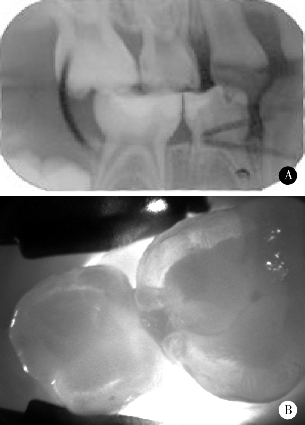
Near-infrared light transillumination for detection of incipient proximal caries in primary molars
Qiong ZHOU,Chu-fang PENG,Man QIN( )
)
- Department of Pediatric Dentistry, Peking University School and Hospital of Stomatology & National Clinical Research Center for Oral Diseases & National Engineering Laboratory for Digital and Material Technology of Stomatology & Beijing Key Laboratory of Digital Stomatology, Beijing 100081, China
CLC Number:
- R781.1


| [1] | 葛立宏 . 儿童口腔医学 [M]. 北京: 人民卫生出版社, 2012: 113. |
| [2] |
Verdonschot EH, Angmar-Månsson B, ten Bosch JJ , et al. Deve-lopments in caries diagnosis and their relationship to treatment decisions and quality of care. ORCA Saturday Afternoon Symposium 1997[J]. Caries Res, 1999,33(1):32-40.
doi: 10.1159/000016493 |
| [3] |
Wenzel A . Bitewing and digital bitewing radiography for detection of caries lesions[J]. J Dent Res, 2004,83:C72-C75.
doi: 10.1177/154405910408301S14 pmid: 15286126 |
| [4] |
Friedman J, Marcus MI . Transillumination of theoral cavity with use of fiber optics[J]. J Am Dent Assoc, 1970,80(4):801-809.
doi: 10.14219/jada.archive.1970.0117 pmid: 5264574 |
| [5] | 陈君, 岳林 . 对光纤透照法诊断后牙邻面龋的评价[J]. 现代口腔医学杂志, 1998,12(3):178-180. |
| [6] |
Ye SY, Kim JY, Ro JH , et al. Detecting incipient caries using front-illuminated infrared light scattering imaging[J]. Trans Electr Electron Mater, 2012,13(6):310-316.
doi: 10.4313/TEEM.2012.13.6.310 |
| [7] |
Astvaldsdóttir A, Ahlund K, Holbrook WP , et al. Approximal caries detection by DIFOTI: in vitro comparison of diagnostic accuracy/efficacy with film and digital radiography[J]. Int J Dent, 2012,326401. doi: 10.1155/2012/326401.
doi: 10.1155/2012/326401 pmid: 23213335 |
| [8] |
Abdelaziz M, Krejci I . DIAGNOcam—a near infrared digital imaging transillumination (NIDIT) technology[J]. Int J Esthet Dent, 2015,10(1):158-165.
pmid: 25625132 |
| [9] |
高洁, 李波, 刘月华 . 乳切牙牙体硬组织的扫描电镜观察及临床分析[J]. 实用口腔医学杂志, 2008,24(2):274-276.
doi: 10.3969/j.issn.1001-3733.2008.02.029 |
| [10] | 于世凤 . 口腔组织病理学 [M]. 北京: 人民卫生出版社, 2006: 141-147. |
| [11] |
Abogazalah N, Eckert GJ, Ando M . In vitro performance of near infrared light transillumination at 780-nm and digital radiography for detection of non-cavitated approximal caries[J]. J Dent, 2017,63:44-50.
doi: 10.1016/j.jdent.2017.05.018 pmid: 28559050 |
| [12] |
Kühnisch J, Söchtig F, Pitchika V , et. al. In vivo validation of near-infrared light transillumination for interproximal dentin caries detection[J]. Clin Oral Investig, 2016,20(4):821-829.
doi: 10.1007/s00784-015-1559-4 pmid: 26374746 |
| [13] |
Mejàre I, Gröndahl HG, Carlstedt K , et al. Accuracy at radiography and probing for the diagnosis of proximal caries[J]. Scand J Dent Res, 1985,93(2):178-184.
doi: 10.1111/j.1600-0722.1985.tb01328.x pmid: 3858967 |
| [14] |
于江利, 唐仁韬, 冯琳 , 等. 数字化光纤透照法判断龋洞深度[J]. 北京大学学报(医学版), 2017,49(1):81-85.
doi: 10.3969/j.issn.1671-167X.2017.01.014 |
| [1] | Xinxin CHEN, Zhe TANG, Yanchun QIAO, Wensheng RONG. Caries experience and its correlation with caries activity of 4-year-old children in Miyun District of Beijing [J]. Journal of Peking University (Health Sciences), 2024, 56(5): 833-838. |
| [2] | Hua ZHONG, Yuan LI, Liling XU, Mingxin BAI, Yin SU. Application of 18F-FDG PET/CT in rheumatic diseases [J]. Journal of Peking University (Health Sciences), 2024, 56(5): 853-859. |
| [3] | Zhengfang LI,Cainan LUO,Lijun WU,Xue WU,Xinyan MENG,Xiaomei CHEN,Yamei SHI,Yan ZHONG. Application value of anti-carbamylated protein antibody in the diagnosis of rheumatoid arthritis [J]. Journal of Peking University (Health Sciences), 2024, 56(4): 729-734. |
| [4] | Hai-hong YAO,Fan YANG,Su-mei TANG,Xia ZHANG,Jing HE,Yuan JIA. Clinical characteristics and diagnostic indicators of macrophage activation syndrome in patients with systemic lupus erythematosus and adult-onset Still's disease [J]. Journal of Peking University (Health Sciences), 2023, 55(6): 966-974. |
| [5] | Yan XIONG,Xin LI,Li LIANG,Dong LI,Li-min YAN,Xue-ying LI,Ji-ting DI,Ting LI. Evaluation of accuracy of pathological diagnosis based on thyroid core needle biopsy [J]. Journal of Peking University (Health Sciences), 2023, 55(2): 234-242. |
| [6] | Xue-mei HA,Yong-zheng YAO,Li-hua SUN,Chun-yang XIN,Yan XIONG. Solid placental transmogrification of the lung: A case report and literature review [J]. Journal of Peking University (Health Sciences), 2023, 55(2): 357-361. |
| [7] | Bo-han NING,Qing-xia ZHANG,Hui YANG,Ying DONG. Endometrioid adenocarcinoma with proliferated stromal cells, hyalinization and cord-like formations: A case report [J]. Journal of Peking University (Health Sciences), 2023, 55(2): 366-369. |
| [8] | Rui-jie CAO,Zhong-qiang YAO,Peng-qing JIAO,Li-gang CUI. Comparison of diagnostic efficacy of different classification criteria for Takayasu arteritis in Chinese patients [J]. Journal of Peking University (Health Sciences), 2022, 54(6): 1128-1133. |
| [9] | Zhe HAO,Shu-hua YUE,Li-qun ZHOU. Application of Raman-based technologies in the detection of urological tumors [J]. Journal of Peking University (Health Sciences), 2022, 54(4): 779-784. |
| [10] | Bo YU,Yang-yu ZHAO,Zhe ZHANG,Yong-qing WANG. Infective endocarditis in pregnancy: A case report [J]. Journal of Peking University (Health Sciences), 2022, 54(3): 578-580. |
| [11] | FENG Sha-wei,GUO Hui,WANG Yong,ZHAO Yi-jiao,LIU He. Initial establishment of digital reference standardized crown models of the primary teeth [J]. Journal of Peking University (Health Sciences), 2022, 54(2): 327-334. |
| [12] | TIAN Jing,QIN Man,CHEN Jie,XIA Bin. Early loss of primary molar and permanent tooth germ caused by the use of devitalizer during primary molar root canal therapy: Two cases report [J]. Journal of Peking University (Health Sciences), 2022, 54(2): 381-385. |
| [13] | MENG Guang-yan,ZHANG Yun-xiao,ZHANG Yu-xin,LIU Yan-ying. Clinical characteristics of central nervous system involvement in IgG4 related diseases [J]. Journal of Peking University (Health Sciences), 2021, 53(6): 1043-1048. |
| [14] | ZHAI Li,QIU Nan,SONG Hui. Multicentric reticulohistiocytosis: A case report [J]. Journal of Peking University (Health Sciences), 2021, 53(6): 1183-1187. |
| [15] | ZHAO Si-ming,ZHAO Xiao-han,ZHANG Jie,WANG Dang-xiao,WANG Xiao-yan. Preliminary evaluation of a virtual reality dental simulation system on training of caries identification ability [J]. Journal of Peking University (Health Sciences), 2021, 53(1): 139-142. |
|
||