Journal of Peking University (Health Sciences) ›› 2022, Vol. 54 ›› Issue (2): 381-385. doi: 10.19723/j.issn.1671-167X.2022.02.029

Previous Articles    

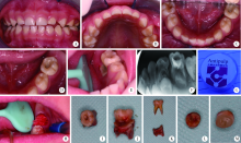
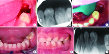
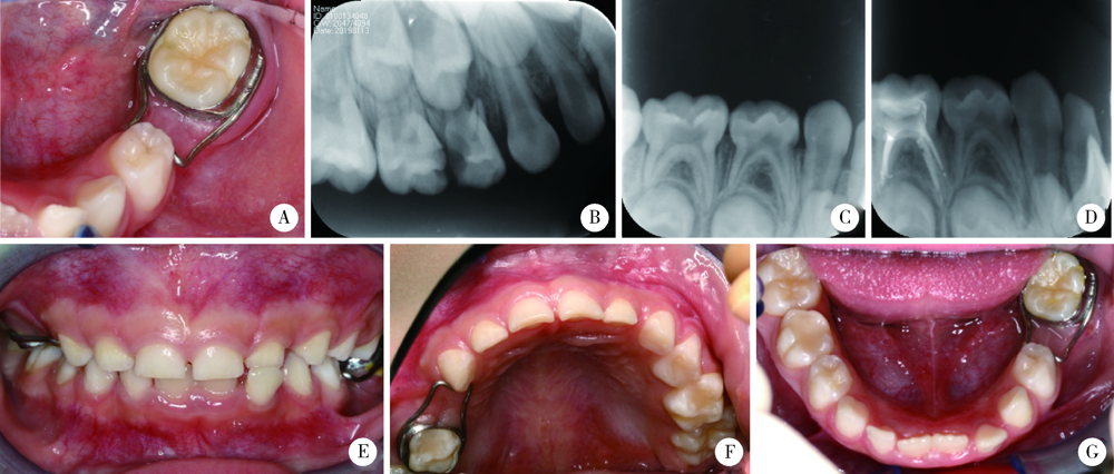
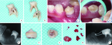
Early loss of primary molar and permanent tooth germ caused by the use of devitalizer during primary molar root canal therapy: Two cases report

TIAN Jing,QIN Man,CHEN Jie,XIA Bin()   

  1. Department of Pediatric Dentistry, Peking University School and Hospital of Stomatology & National Center of Stomatology & National Clinical Research Center for Oral Diseases & National Engineering Research Center of Oral Biomaterials and Digital Medical Devices & Beijing Key Laboratory of Digital Stomatology & NHC Research Center of Engineering and Technology for Computerized Dentistry & NMPA Key Laboratory for Dental Materials, Beijing 100081, China
  • Received:2020-03-08 Online:2022-04-18 Published:2022-04-13
  • Contact: Bin XIA E-mail:summerinbeijing@vip.sina.com

Abstract:

Devitalization has been widely used in the root canal therapy of primary and permanent teeth in China more than ten years ago. With the development of local anesthetic drugs and injection technologies, this treatment method with high potential risks has been gradually abandoned. However, a questionnaire survey targeted all the participants at the 2018 China Pediatric Dentistry Conference showed that the devitalizer utilization proportion was still as high as 38.1% (383/1 005), even though the ratio was much lower than 75.5% (105/139) in 2003. These doctors had pay more attention to tissue burn caused by devitalizer marginal leakage or direct leakage, and know how to identify and handle with devitalizer burn. Devitalizers were usually made of arsenic trioxide, metal arsenic or paraformaldehyde, which have cytotoxicity, allergenicity, mutagenicity, carcinogenicity, and teratogenic effects on animals. Marginal leakage of devitalizers have high risks of causing soft and hard tissue necrosis. Most of the dentists have an understanding of the potential damages of arsenic-containing devitalizers, so they will choose parafor-maldehyde with relatively less toxicity. Paraformaldehyde has a certain self-limitation, and there are few cases reported, so some dentists lack of vigilance. Paraformaldehyde can also causes tissue necrosis if leakage happens, and the treatment methods are similar to that of arsenic-containing devitalizers. When handling with devitalizers burn, the necrosed soft and hard tissue, for example gingiva, alveolar bone or teeth that cannot keep, must be completely removed until fresh blood appears, then rinse with large amount of saline and seal with iodoform gauze. This paper described two cases of devitalizer burn during the root canal treatment of primary molars, both of the doctors failed to identify the devitalizer burn symptoms in the early stage, thus didn’t do proper treatments immediately after burning. Resulting in the necrosis of large area of gingiva and alveolar bone, loss of primary molars and permanent tooth germs 1-2 months after devitalizer burn. This paper reported these two cases in detail in order to warn dentists the high risks of using any kind of devitalizers, help them learn how to identify and treat devitalizer burn, and remind them to stop using devitalizers as soon as possible.

Key words: Dental pulp devitalization, Deciduous tooth, Molar, Tooth germ, Radiography

CLC Number: 

  • R788

Figure 1

Intraoral image, periapical X-rays and treatment procedures of case 1 A, frontal view; B, maxillary occlusal view; C, mandibular occlusal view; D, 75 occlusal and buccal view before the treatment; E, 75 lingual view before the treatment; F, 75 periapical X-rays before the treatment; G, the devitalizer used in this case; H, iodoform gauze placed in the 75 socket during the treatment; I, occlusal view of 75; J and K, buccal view of 75 and the necrotic alveolar around it; L, occlusal view of 35 germ; M, root view of 35 germ."

Figure 2

Intraoral image and periapical X-rays during the treatment of case 1 A, 75 socket view after removing of iodoform gauze at 1 week after the treatment; B, periapical X-rays at 1 week after the treatment; C, 75 socket view at 2 weeks after the treatment; D, mandibular occlusal view at 6 months after the treatment; E, left Buccal view at 6 months after the treatment; F, periapical X-rays at 6 months after the treatment."

Figure 3

Intraoral image and periapical X-rays at 1 year and 1.5 years follow-up after the therapy of case 1 A, band-loop space maintainer was used to maintain 75 space at 1 year follow-up; B, 54, 55 periapical X-rays before treatment; C, 85 periapical X-rays before treatment; D, 85 periapical X-rays after treatment; E-G, intraoral photographs at 1.5 years follow-up."

Figure 4

Intraoral image, periapical X-rays and treatment procedures of case 2 A and B, 74 and necrotic alveolar bone; C, 34 buccal view; D, 34 lingual view; E, 34 periapical X-rays; F, 34 lateral view; G, 34 root view; H, granulation tissue during debridement; I, 34 periapical X-rays at 4 months follow-up."

[1] 高学军, 岳林. 牙体牙髓病学[M]. 2版. 北京: 北京大学医学出版社, 2013: 478.
[2] 葛立宏. 儿童口腔医学[M]. 4版. 北京: 人民卫生出版社, 2012.
[3] 葛立宏. 儿童口腔医学[M]. 2版. 北京: 北京大学医学出版社, 2013: 295.
[4] 夏斌, 秦满, 葛立宏. 我国儿童口腔科医师临床诊疗工作调查分析[J]. 现代口腔医学杂志, 2007, 21(1):80-82.
[5] 包晶, 汪奕, 姜德龙, 等. Antipulp对儿童乳磨牙牙髓失活的临床疗效观察[J]. 吉林医学, 2014, 35(25):5644-5645.
[6] 鲁伟, 高玉萍, 冯琼芬, 等. 三氧化二砷泄漏致牙周组织及牙槽骨坏死20例[J]. 中华口腔医学杂志, 2011, 46(9):575-576.
[7] 沈国华, 翟启善. 碘中和法治疗砷性根尖周炎70例临床报告: 全国第四次牙体牙髓病学术会议[C]. 上海: [出版者不详], 1995.
[8] 张敬雷, 窦金红. 氮酮多聚甲醛致牙龈严重烧伤[J]. 临床误诊误治, 2004, 17(2):143.
[1] FENG Sha-wei,GUO Hui,WANG Yong,ZHAO Yi-jiao,LIU He. Initial establishment of digital reference standardized crown models of the primary teeth [J]. Journal of Peking University (Health Sciences), 2022, 54(2): 327-334.
[2] CHEN Zhen,GU Bao-xin,TANG Yu-fang,YAN Zi-yu,NI Fang-duan,CUI Nian-hui. Constructions of the scale of difficulty in the extraction of impacted mandibular third molars by using Delphi method [J]. Journal of Peking University (Health Sciences), 2022, 54(1): 100-104.
[3] JIANG You-sheng,FENG Lin,GAO Xue-jun. Influence of base materials on stress distribution in endodontically treated maxillary premolars restored with endocrowns [J]. Journal of Peking University (Health Sciences), 2021, 53(4): 764-769.
[4] Wei QI,Jian-nan LI,Jing-ren ZHAO,Hai-xia XING,Jie PAN. Effect of triangular flap design and healing procedure on the sequelae after extraction of impacted lower third molars [J]. Journal of Peking University(Health Sciences), 2019, 51(5): 949-953.
[5] Li-ping ZHAO,Wen-jie HU,Tao XU,Ya-lin ZHAN,Yi-ping WEI,Min ZHEN,Cui WANG. Two procedures for ridge preservation of molar extraction sites affected by severe bone defect due to advanced periodontitis [J]. Journal of Peking University(Health Sciences), 2019, 51(3): 579-585.
[6] Qiong ZHOU,Chu-fang PENG,Man QIN. Near-infrared light transillumination for detection of incipient proximal caries in primary molars [J]. Journal of Peking University(Health Sciences), 2019, 51(1): 59-64.
[7] Yue LEI,Ying-ting YANG,Yuan ZHAN. Evaluation of bioceramic putty repairment in primary molars pulpotomy [J]. Journal of Peking University(Health Sciences), 2019, 51(1): 70-74.
[8] WU Ling, LIU Xiao-jing, LI Zi-li, WANG Xing. Evaluation of accuracy of virtual occlusal definition in Angle class Ⅰ molar relationship [J]. Journal of Peking University(Health Sciences), 2018, 50(1): 154-159.
[9] ZHANG Ming-ming, ZHENG Ying-dong, LIANG Yu-hong. A prognostic model for assessment of outcome of root canal treatment in teeth with pulpitis or apical periodontitis#br# [J]. Journal of Peking University(Health Sciences), 2018, 50(1): 123-130.
[10] JIA Peng-chen, YANG Gang, HU Wen-jie, ZHAO Yi-jiao, LIU Mu-qing. Preliminary study on the accuracy of infrabony root surface area of single-root teeth by periapical films [J]. Journal of Peking University(Health Sciences), 2018, 50(1): 91-97.
[11] DOU Gui-li, WU Nan, ZHAO Shuang-yun, XIA Bin. Two-year outcomes of pulpotomy in primary molars using mineral trioxide aggregate: a retrospective study [J]. Journal of Peking University(Health Sciences), 2018, 50(1): 170-175.
[12] WANG Tian-jiao, LIU Yu, GUAN Ming. Intravenous sedation with midazolam and propofol target controlled infusion on patients’ perioperative anxiety under the mandibular third molar extraction [J]. Journal of Peking University(Health Sciences), 2017, 49(6): 1044-1049.
[13] ZHAN Yuan, LIU He. Unerupted first deciduous molar located higher to the first premolar: a case report [J]. Journal of Peking University(Health Sciences), 2017, 49(1): 181-183.
[14] ZHU Jie,OUYANG Xiang-ying. Observation of bone morphology in furcation defects of mandibular molars using cone beam computed tomography [J]. Journal of Peking University(Health Sciences), 2017, 49(1): 67-070.
[15] LI Dan, GUO Chuan-bin, LIU Yu, WANG En-bo. Applicational evaluation of split tooth extractions of upper molars using piezosurgery [J]. Journal of Peking University(Health Sciences), 2016, 48(4): 709-713.
Viewed
Full text


Abstract

Cited

  Shared   
  Discussed   
[1] Xin-yu WANG,Zhe CUI,Qing-yuan HE,Xiang-ning DENG,Ge GUO,Xin-heng FENG,Jie-li FENG. Assessment of heart’s changes of elite Chinese male weightlifter by speckle tracking echocardiography[J]. Journal of Peking University (Health Sciences), 2021, 53(5): 832 -837 .
[2] . [J]. Journal of Peking University (Health Sciences), 2022, 54(1): 1 -6 .
[3] . [J]. Journal of Peking University (Health Sciences), 2021, 53(6): 1017 -1019 .
[4] LOU Xue,LIAO Li,LI Xing-jun,WANG Nan,LIU Shuang,CUI Ruo-mei,XU Jian. Methylation status and expression of TWEAK gene promoter region in peripheral blood of patients with rheumatoid arthritis[J]. Journal of Peking University (Health Sciences), 2021, 53(6): 1020 -1025 .
[5] ZHONG Hua,XU Li-ling,BAI Ming-xin,SU Yin. Effect of chemokines CXCL9 and CXCL10 on bone erosion in patients with rheumatoid arthritis[J]. Journal of Peking University (Health Sciences), 2021, 53(6): 1026 -1031 .
[6] . [J]. Journal of Peking University (Health Sciences), 2021, 53(4): 633 -634 .
[7] . [J]. Journal of Peking University (Health Sciences), 2021, 53(5): 828 -831 .
[8] . [J]. Journal of Peking University (Health Sciences), 2021, 53(4): 635 -639 .
[9] YU Yan-fei,HE Shi-ming,WU Yu-cai,XIONG Sheng-wei,SHEN Qi,LI Yan-yan,YANG Feng,HE Qun,LI Xue-song. Clinicopathological features and prognosis of fumarate hydratase deficient renal cell carcinoma[J]. Journal of Peking University (Health Sciences), 2021, 53(4): 640 -646 .
[10] WANG Li-xin , XU Xiao, NI Yao-feng, SUN Hai-tao, YU Ri-yue, WEI Shi-cheng. In vivo study of liposome-modified polyetheretherketone implant on bacteriostasis and osseointegration[J]. Journal of Peking University (Health Sciences), 2021, 53(4): 758 -763 .