Journal of Peking University(Health Sciences) ›› 2019, Vol. 51 ›› Issue (6): 1178-1181. doi: 10.19723/j.issn.1671-167X.2019.06.036

Previous Articles     Next Articles

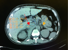
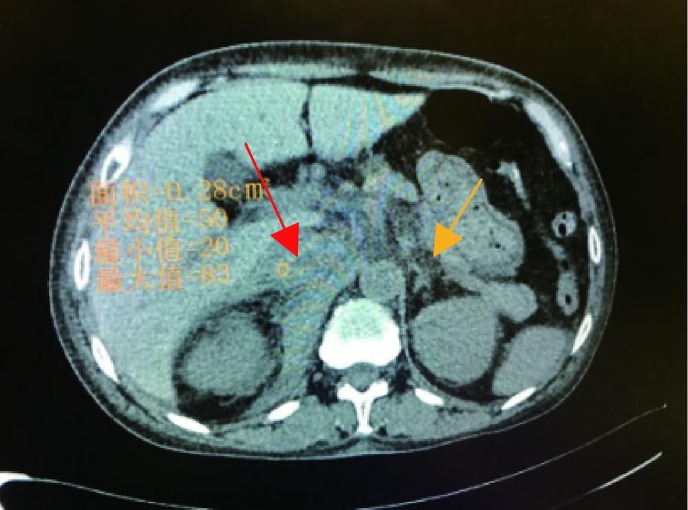
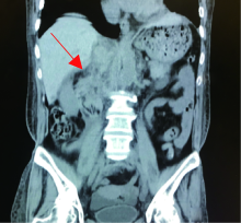
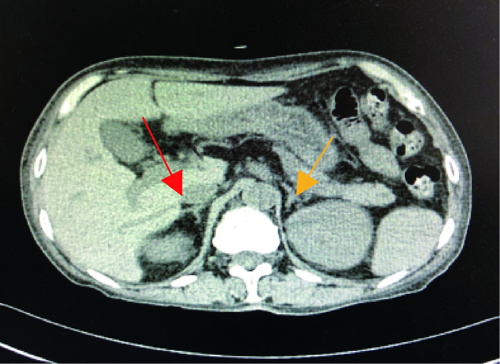
Adrenal hemorrhage in a patient with systemic lupus erythematosus

Yu-hua WANG(),Guo-hua ZHANG,Ling-ling ZHANG,Jun-li LUO,Lan GAO   

  1. Department of Rheumatology, Beijing Shijitan Hospital, Capital Medical University, Beijing 100038, China
  • Received:2019-09-11 Online:2019-12-18 Published:2019-12-19
  • Contact: Yu-hua WANG E-mail:wyhxxtt@163.com
  • Supported by:
    Supported by Capital Medical University Foundation-Clinical Research Cooperation Fund(13JL64)

Abstract:

A 58-year-old female was referred to our department with intermittent suffocation for 1.5 years, aggravated for a month. 1.5 years before she developed oral ulcer, raynaud phenomenon, proteinuria, bilateral pleural effusion, ANA and anti-dsDNA positive. This patient was diagnosed with systemic lupus erythematosus (SLE). After given hormones, hydroxychloroquine sulfate (HCQ), her symptom relieved soon. The patient stopped her pills 1 year ago. One month ago, she had chest tightness, increased urine foam, and suffered from oliguria. Her admission medical examination: blood pressure (BP) 130/80 mmHg, conjunctiva pale, and lower lung breath sounds reduced. There was no tenderness, rebound pain and abdominal muscle tension in the abdomen. Liver and spleen rib inferior, mobile dullness negative, and lower extremity edema. Blood routine tests were performed with hemoglobin (HGB) 57 g/L. Urine routine: BLD (3+). 24-hour urinary protein 3.2 g. serum albumin 20.5 g/L, C-reactive protein (CRP) 12.85 mg/L, erythrocyte sedimentation rate (ESR) 140 mm/h. Antinuclear antibody (ANA) (H)1 ∶10 000;, anti-dsDNA antibody 1 ∶3 200;, anti-Smith antibody, anti-U1-snRNP / Sm antibody were positive, blood complement 3(C3) 0.43 g/L, complement 4(C4) 0.07 g /L. Anticardiolipin antibody (ACL), anti-β2-GP1;, lupus anticoagulant (LA) were negative, HRCT suggested bilateral medial pleural cavity product liquid. Admission diagnosis: SLE lupus nephritis, anemia, pleural effusion, and hypoproteinemia. We treated her with methylprednisolone 1 000 mg×3 d;, late to 48 mg/d and cyclophosphamide 1.0 g, HCQ 0.2 g bid, gamma globulin 10 g×5 d. Day 2 of treatment;, this patient developed acute right upper quadrant pain, not accompanied by nausea, vomiting, blood stool and diarrhea. Antipyretic antispasmodic treatment was invalid, after the morning to ease their own abdominal pain. Day 4 of treatment, daytime blood HGB 77 g/L. Bilateral renal vascular ultrasound: bilateral renal artery blood flow velocity was reduced. The abdominal pain of the above symptoms recurred at night, BP was 120/80 mmHg, and no positive signs were found on abdominal examination. No abnormality was found in the vertical abdominal plain film. Blood routine examination: HGB 53 g/L, Plasma D dimer 2 515 μg/L;, amylase in hematuria was normal, the stool occult blood was negative. Abdominal computed tomography (CT): normal structure of right adrenal gland disappeared, irregular mass shadow could be seen in adrenal region, CT value was about 50 HU. Morphological density of left adrenal gland was not abnormal. The retroperitoneum descended along the inferior vena cava to the right iliac blood vessel and showed a bolus shadow. The density of some segments increased. The lesion involved the right renal periphery and reached the left side of abdominal aorta. Most lesions surrounded the inferior vena cava, the right renal vein and part of the small intestine. The boundary between the upper lesion and the vena cava was unclear. Iodine-containing contrast agent was taken orally. No sign of contrast agent overflowing was found in the abdominal cavity. Hematoma and exudative changes were considered in retroperitoneum. Conclusion of contrast-enhanced ultrasound of blood vessels: The retroperitoneal inferior vena cava (volume 3.5 cm×3.5 cm×1.5 cm) was hypoechoic and had no blood flow lesion. The adrenal gland had a high possibility of origin. Left renal vein thrombosis extended to inferior vena cava. According to the above data;, it was analyzed that the cause of retroperitoneal hematoma of the patient was left adrenal vein thrombosis caused by hypercoagulable state, which led to vascular rupture and hemorrhage caused by increased vascular pressure in adrenal gland. Therefore, on the basis of continuing to actively treat the primary disease, and on the basis of dynamic observation of no active hemorrhage for 3 days, the anticoagulant therapy was continued with 10 mg/d of apixaban. Clinical symptoms were gradually eased, HGB did not decrease. Two weeks later, the ultrasonic examination showed that the irregular cluster hypoechoic range behind the inferior vena cava was significantly smaller than that before (1.8 cm×1.2 cm×0.7 cm). Abdominal CT examination after 1 month showed that there was no abnormal morphological density of bilateral adrenal glands and basic absorption of retroperitoneal exudation. Adrenal hemorrhage is uncommon. SLE with adrenal hemorrhage is rarer. In SLE patients;, especially those complicated with APS, if abdominal pain accompanied by HGB decrease occurs, except after gastrointestinal hemorrhage, the possibility of adrenal hemorrhage should be warned.

Key words: Systemic lupus erythematosus (SLE), Adrenal hemorrhage, Antiphospholipid syndrome

CLC Number: 

  • R593

Figure 1

Abdominal CT (coronal section):retroperitoneal cavity along the inferior vena cava down to the right side of the iliac blood vessels can be seen next to the film group (2016-03-25)"

Figure 2

Abdominal CT (transverse section): normal structure of right adrenal gland disappears, irregular mass shadow can be seen in adrenal region, CT value is about 50 HU. Morphological density of left adrenal gland was not abnormal (2016-03-25)"

Figure 3

Abdominal CT (transverse section): the morphological density of bilateral adrenal glands is normal (2016-04-27)"

Figure 4

Abdominal CT (coronal section): retroperitoneal effusion was significantly higher than before absorption (2016-04-27)"

[1] Presotto F, Fornasini F, Betterle C , et al. Acute adrenal failure as the heralding symptom of primary antiphospholipid syndrome:report of a case and review of the literature[J]. Eur J Endocrinol, 2005,153(4):507-514.
[2] 张逸箫, 孔垂泽, 姜元军 . 肾上腺出血的诊断与治疗[J]. 国际外科学杂志, 2019,46(1):44-47.
[3] Ruiz-Irastorza G, Cuadrado MJ, Ruiz-Arruza I , et al. Evidence-based recommendations for the prevention and long-term management of thrombosis in antiphospholipid antibody-positive patients:report of a task force at the 13th international congress on antiphospholipid antibodies[J]. Lupus, 2011,20(2):206-218.
[4] 高辉, 姚海红, 安媛 , 等. 抗磷脂抗体综合征合并肾上腺血肿一例[J]. 中华风湿病学杂志, 2017,21(8):556-557.
[5] Aldaajani H, Albahrani S, Saleh K , et al. Bilateral adrenal hemorrhage in antiphospholipid syndrome. Anticoagulation for the treatment of hemorrhage[J]. Saudi Med J, 2018,39(8):829-833.
[6] Fukui S, Iwamoto N, Tsuji S , et al. Diffuse alveolar hemorrhage emerging one week after starting high-dose corticosteroid therapy for granulomatosis with polyangiitis (GPA) with systemic lupus erythematosus (SLE). Intern Med, 2015,54(20):2681-2686.
[7] Xu J, Zhou Q, Jiang N , et al. Bilateral adrenal hemorrhage revealed antiphospholipid syndrome in a male patient: benefit from comprehensive treatment[J]. Curr Med Res Opin, 2018,34(12):2165-2168.
[8] Abdulla MC, Alungal J, Hashim S . SLE presenting as multiple hemorrhagic complications[J]. Lupus, 2015,24(10):1103-1106.
[9] Heller T, Bergholtz M, Martin F , et al. Bilateral adrenal hemorrhage occurring two times in primary antiphospholipid syndrome (APS). Anticoagulation as treatment of hemorrhage[J]. Dtsch Med Wochenschr, 2002,127(33):1695-1698.
[10] Sanford Z, Nanjundappa A, Annie FH , et al. Adrenal hemorrhage in a patient anticoagulated with apixaban with antiphospholipid syndrome[J]. Cureus, 2019,11(7):e5108.
[1] Ji LI,Li ZHENG,Lian-jie SHI,Jing XU,Jian-long SHU,Xue-wu ZHANG. Increased serum soluble-endoglin level and its clinical significance in antiphospholipid syndrome [J]. Journal of Peking University(Health Sciences), 2018, 50(6): 1027-1032.
[2] Jie-yu GU,Cui LU,Hui SHI,Cheng-de YANG. Case series and clinical analysis of 14 cases of catastrophic antiphospholipid syndrome [J]. Journal of Peking University(Health Sciences), 2018, 50(6): 1033-1038.
[3] ZHENG Xiao-Juan, DENG Xiao-Li, LIU Xiang-Yuan. Pregnancy outcome in 54 patients with antiphospholipid syndrome:a retrospective clinical study [J]. Journal of Peking University(Health Sciences), 2014, 46(2): 323-328.
Viewed
Full text


Abstract

Cited

  Shared   
  Discussed   
[1] Author. English Title Test[J]. Journal of Peking University(Health Sciences), 2010, 42(1): 1 -10 .
[2] . [J]. Journal of Peking University(Health Sciences), 2009, 41(2): 188 -191 .
[3] . [J]. Journal of Peking University(Health Sciences), 2009, 41(3): 376 -379 .
[4] . [J]. Journal of Peking University(Health Sciences), 2009, 41(4): 459 -462 .
[5] . [J]. Journal of Peking University(Health Sciences), 2009, 41(6): 682 -686 .
[6] . [J]. Journal of Peking University(Health Sciences), 2010, 42(1): 82 -84 .
[7] . [J]. Journal of Peking University(Health Sciences), 2007, 39(3): 319 -322 .
[8] . [J]. Journal of Peking University(Health Sciences), 2007, 39(3): 333 -336 .
[9] . [J]. Journal of Peking University(Health Sciences), 2007, 39(3): 337 -340 .
[10] . [J]. Journal of Peking University(Health Sciences), 2007, 39(3): 225 -328 .