Journal of Peking University (Health Sciences) ›› 2021, Vol. 53 ›› Issue (1): 102-108. doi: 10.19723/j.issn.1671-167X.2021.01.016

Previous Articles     Next Articles

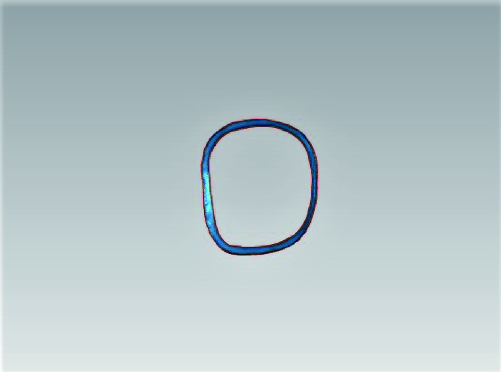
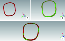
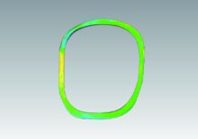
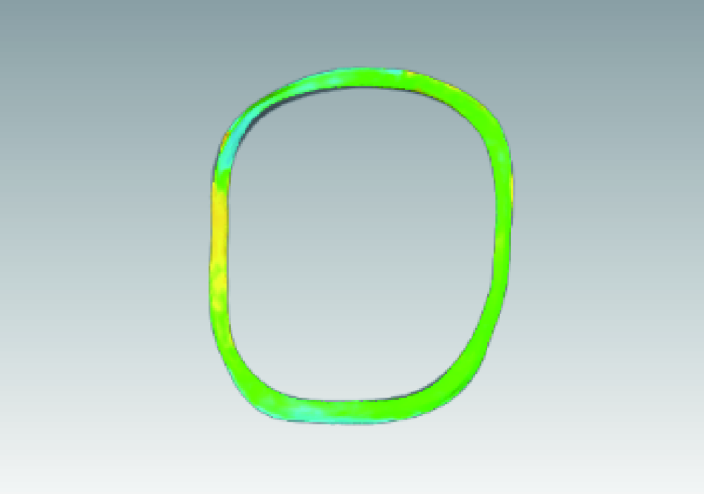
Influence of trueness for local finish lines of a full crown preparation on that of complete finish line

JIANG Nan,BAO Xu-dong(),YUE Lin   

  1. Department of Cariology and Endodontology, Peking University School and Hospital of Stomatology & National Clinical Research Center for Oral Diseases & National Engineering Laboratory for Digital and Material Technology of Stomatology & Beijing Key Laboratory of Digital Stomatology, Beijing 100081, China
  • Received:2019-07-25 Online:2021-02-18 Published:2021-02-07
  • Contact: Xu-dong BAO E-mail:baoxudong@vip.163.com

Abstract:

Objective: To investigate the factors affecting the trueness of finish lines of full crown preparations in digital impressions.Methods: A full crown preparation of the right maxillary molar was prepared on the standard resin dentition model, the trueness of the finish lines, the full crown preparation in the whole dentition and the isolated full crown preparation were measured respectively. Detection of scanning trueness of the finish lines of the full crown preparation in the whole dentition: (1) Using Imetric scanner to scan the full crown preparation, obtaining STL (Stereo lithographic) format data as the reference true value; (2) Using CEREC Omnicam oral scanner and 3Shape TRIOS oral scanner to scan the full crown preparation, obtaining all the STL format data, import Geomagic Studio 2013 software, extraction of images and data of the complete finish lines or local finish lines (mesial, distal, buccal, lingual) of the full crown preparation, then using the data to 3D Compare Analysis with the reference true value, outputting RMS (root mean square) values which could evaluate the scanning trueness, the lower RMS value was, the lower the trueness was. The detection of scanning trueness of the finish lines of the isolated full crown preparation: (1) the anterior and posterior adjacent teeth of the preparation were removed to establish the model of the isolated full crown preparation; (2) CEREC Omnicam oral scanner and 3Shape TRIOS oral scanner were used to scan the isolated preparations, and each group was repeated 6 times. Data collection and analysis were the same as the first part, that is, detection of scanning trueness of finish lines of the full crown preparation in the whole dentition.Results: In the whole dentition, (1) the RMS value [(44±7) μm] of CEREC Omnicam oral scanner scanning complete finish lines was greater than that of 3Shape TRIOS oral scanner [(35±6) μm](P<0.05). (2) the RMS values of the mesial [(45±9) μm], buccal [(38±3) μm] and lingual [(40±3) μm] finish lines in CEREC Omnicam oral scanner scanning were all lower than that in distal [(63±7) μm](P<0.05), and the RMS values in mesial were higher than that in buccal and lingual (P>0.05). The RMS values of 3Shape TRIOS oral scanner scanning mesial and distal finish lines were significantly higher than those of the buccal and lingual side (P>0.05), in which:(45±8) μm in mesial, (50±10) μm in the distal, (33±8) μm in the buccal and (33±6) μm in the lingual side. The RMS value of CEREC Omnicam oral scanner scanning distal finish line was greater than that of 3Shape TRIOS oral scanner (P<0.05). Without adjacent teeth, (1) the RMS values of the complete finish lines of CEREC Omnicam oral scanner and 3 Shape TRIOS oral scanner were significantly reduced (P<0.05), including CEREC Omnicam oral scanner (34±3) μm and 3 Shape TRIOS oral scanner (26±4) μm; (2) the RMS values of the buccal and lingual finish lines of CEREC Omnicam oral scanner and 3Shape TRIOS oral scanner showed no significant changes (P>0.05), among which, CEREC Omnicam oral scanner buccal (44±7) μm, lingual (43±3) μm, 3Shape TRIOS oral scanner buccal (29±5) μm, lingual (40±7) μm. The RMS values of CEREC Omnicam oral scanner and 3Shape TRIOS oral scanner were significantly reduced and there was no significant difference between them (P>0.05). CEREC Omnicam oral scanner (32±8) μm and 3Shape TRIOS oral scanner (32±6) μm. The RMS values of the distal finish lines of CEREC Omnicam oral scanner and 3 Shape TRIOS oral scanner also decreased significantly (P<0.05), and CEREC Omnicam oral scanner (38±3) μm decreased more significantly, similar to 3Shape TRIOS oral scanner (36±1) μm, P>0.05.Conclusion: When adjacent teeth exist in the full crown preparation, the mesial and distal parts of the finish line will be blocked by adjacent teeth, which will affect the trueness of the local finish line, and ultimately reduce the trueness of the complete finish line.

Key words: Intraoral scanners, Trueness, Preparation, Finish line

CLC Number: 

  • R782.1

Figure 1

An example of finish line of the full crown preparation"

Figure 2

Example of best fit alignment A, finish line of the full crown preparation obtained by Imetric scanner; B, finish line of the full crown preparation obtained by CEREC Omnicam oral scanner; C, finish line of the full crown preparation after best fit alignment."

Figure 3

Example of 3D compare analysis"

Figure 4

An example of local finish lines of the full crown preparation A, mesial finish line; B, distal finish line; C, buccal finish line; D, lingual finish line."

Table 1

Comparison of RMS values for different parts of finish lines(n=6)"

Site Scanner type t P
CEREC Omnicam oral scanner 3Shape TRIOS oral scanner
Mesial/μm 45±9 45±8 -0.36 0.74
Distal/μm 63±7 50±10 3.63 0.02
Buccal/μm 38±3 33±8 1.14 0.34
Lingual/μm 40±3 33±6 2.11 0.10
F 17.57 5.83
P <0.001 0.010

Table 2

Comparison of RMS values between finish lines of preparation in the dentition and isolated preparation(n=6)"

Preparation position CEREC Omnicam oral scanner 3Shape TRIOS oral scanner
Preparation in the dentition/μm 44±7 35±6
Isolated preparation/μm 34±3 26±4
t 2.37 3.49
P 0.04 0.02

Table 3

A comparison of RMS values for different parts of finish lines of the isolated preparation and preparation in the dentition(n=6)"

Site CEREC Omnicam oral scanner 3Shape TRIOS oral scanner
Preparation in the dentition Isolated preparation t P Preparation in the dentition Isolated preparation t P
Mesial/μm 45±9 32±8 2.92 0.04 45±8 32±6 3.30 <0.05
Distal/μm 63±7 38±3 6.61 0.01 50±10 36±1 3.50 0.04
Buccal/μm 38±3 44±7 -1.49 0.23 33±8 29±5 0.47 0.67
Lingual/μm 40±3 43±3 -2.60 0.08 33±6 40±7 -1.01 0.39
[1] Logozzo S, Zanetti EM, Franceschini G, et al. Recent advances in dental optics: Part I. 3D intraoral scanners for restorative dentistry[J]. Opt Laser Eng, 2014,54(3):203-221.
doi: 10.1016/j.optlaseng.2013.07.017
[2] 王勇. 口内数字印模技术[J]. 口腔医学, 2015,35(9):705-709, 743.
[3] 张馨月, 李虹, 赵一姣, 等. 两种结构光口内三维扫描仪获取单冠预备体数据质量的模型评价[J]. 中华口腔医学杂志, 2016,51(7):432-436.
[4] Mangano F, Gandolfi A, Luongo G, et al. Intraoral scanners in dentistry: a review of the current literature[J]. BMC Oral Health, 2017,17(1):1-11.
doi: 10.1186/s12903-016-0228-6 pmid: 27412290
[5] Lee JJ, Jeong ID, Park JY, et al. Accuracy of single-abutment digital cast obtained using intraoral and cast scanners[J]. J Prosthet Dent, 2016,117(2):253-259.
doi: 10.1016/j.prosdent.2016.07.021 pmid: 27666500
[6] Mou SH, Chai T, Wang JS, et al. Influence of different convergence angles and tooth preparation heights on the internal adaptation of Cerec crowns[J]. J Prosthet Dent, 2002,87(3):248-255.
doi: 10.1067/mpr.2002.122011 pmid: 11941350
[7] Nedelcu R, Olsson P, Nyström I, et al. Finish line distinctness and accuracy in 7 intraoral scanners versus conventional impression: an in vitro descriptive comparison[J]. BMC Oral Health, 2018,18(1):1-11.
doi: 10.1186/s12903-017-0444-8 pmid: 29301577
[8] Sim J, Jang Y, Kim W, et al. Comparing the accuracy (trueness and precision) of models of fixed dental prostheses fabricated by digital and conventional workflows[J]. J Prosthodont Res, 2019,63(1):25-30.
doi: 10.1016/j.jpor.2018.02.002 pmid: 29615324
[9] Re D, Cerutti F, Augusti G, et al. Comparison of marginal fit of Lava CAD/CAM crown-copings with two finish lines[J]. Int J Esthet Dent, 2014,9(3):426-435.
pmid: 25126621
[10] Schaefer O, Watts DC, Sigusch BW, et al. Marginal and internal fit of pressed lithium disilicate partial crowns in vitro: A three-dimensional analysis of accuracy and reproducibility[J]. Dental Materials, 2012,28(3):320-326.
doi: 10.1016/j.dental.2011.12.008
[11] 药学与临床研究编辑部. 如何正确运用组内相关系数进行一致性检验: 药物研究中的统计学(一)[J]. 药学与临床研究, 2018,26(1):7-8.
[12] 赵一姣, 王勇. 从工程技术角度谈口腔医学椅旁数字化技术[J]. 中华口腔医学杂志, 2018,53(4):230-235.
[1] LI Si-yu,DUAN Xue-fei,CAO Ye. Evaluation of the effect of using ultrasonic instruments to improve the shoulder of the preparations [J]. Journal of Peking University (Health Sciences), 2021, 53(1): 88-94.
[2] Ying ZHAN,Yi-tian DU,Zhen-zhen YANG,Chun-li ZHANG,Xian-rong QI. Preparation and characterization of paclitaxel microspheres in situ gel and its antitumor efficacy by local injection [J]. Journal of Peking University(Health Sciences), 2019, 51(3): 477-486.
[3] Yan-jun GE,Xiao-qiang LIU. Effects of loupes and microscope on laminate veneer preparation [J]. Journal of Peking University(Health Sciences), 2019, 51(1): 100-104.
[4] YANG Yin-jie, HOU Ben-xiang, HOU Xiao-mei. Effect of autoclave on surface microstructure and cyclic fatigue resistance of R-phase rotary instruments#br# [J]. Journal of Peking University(Health Sciences), 2018, 50(5): 882-886.
[5] CHEN Chen, ZHANG Wen-xin,QI Li-yuan,GAO Xue-jun, LIANG Yu-hong. Detection of root cracks after root canal preparation using rotary NiTi systems by optical coherence tomography (OCT) scan [J]. Journal of Peking University(Health Sciences), 2018, 50(3): 547-552.
[6] LI Hao, LIU Yu-hua, LUO Zhi-qiang. Effects of bioactive glass on reducing the hypersensitivity after full crown preparation [J]. Journal of Peking University(Health Sciences), 2017, 49(4): 709-713.
[7] TIAN Shi-yu, BAI Wei, LIANG Yu-hong. Impact of apical preparation diameter on fracture resistance of mandibular premolar roots [J]. Journal of Peking University(Health Sciences), 2017, 49(1): 92-095.
[8] SU Zheng, BAI Yu-hao, HOU Xiao-mei. Effects of different techniques on removal of vapor lock in the apical region of curved canals: a cone-beam computed tomography study [J]. Journal of Peking University(Health Sciences), 2017, 49(1): 76-080.
[9] SHI Nian-qiu, ZHANG Hong, ZHANG Yong, FENG Bo, LI Zheng-qiang, QI Xian-rong. Study on the properties of felodipine solid dispersions prepared by different technologies [J]. Journal of Peking University(Health Sciences), 2016, 48(6): 1067-1073.
[10] CHEN Xiao-bo, CHEN Chen, LIANG Yu-hong. Comparison of effectiveness and safety between Twisted File technique and ProTaper Universal rotary full sequence based on micro-computed tomography [J]. Journal of Peking University(Health Sciences), 2016, 48(1): 101-104.
[11] CHEN Chen, LIANG Yu-Hong, GAO Xue-Jun. Comparison of the incidences of apical root cracks after canal preparation with two nickel-titanium rotary systems: an in vitro study [J]. Journal of Peking University(Health Sciences), 2015, 47(1): 129-133.
[12] CAI Xue, NIE Jie, WANG Zu-Hua, TIAN Hong-Yan, ZHAO Ying, WANG Xiao-Yan. Effects of different cavosurface margins on color matching of the resin composite [J]. Journal of Peking University(Health Sciences), 2015, 47(1): 120-123.
[13] XIE Yao, ZHANG Sun, GE Li-Hong. Marginal microleakage of cavities prepared with Er:YAG laser on primary teeth  in vitro [J]. Journal of Peking University(Health Sciences), 2014, 46(3): 474-477.
Viewed
Full text


Abstract

Cited

  Shared   
  Discussed   
[1] Author. English Title Test[J]. Journal of Peking University(Health Sciences), 2010, 42(1): 1 -10 .
[2] . [J]. Journal of Peking University(Health Sciences), 2009, 41(4): 459 -462 .
[3] . [J]. Journal of Peking University(Health Sciences), 2010, 42(1): 82 -84 .
[4] . [J]. Journal of Peking University(Health Sciences), 2007, 39(3): 319 -322 .
[5] . [J]. Journal of Peking University(Health Sciences), 2007, 39(3): 337 -340 .
[6] . [J]. Journal of Peking University(Health Sciences), 2007, 39(4): 351 -354 .
[7] . [J]. Journal of Peking University(Health Sciences), 2007, 39(4): 377 -380 .
[8] . [J]. Journal of Peking University(Health Sciences), 2007, 39(4): 416 -419 .
[9] . [J]. Journal of Peking University(Health Sciences), 2007, 39(4): 420 -422 .
[10] . [J]. Journal of Peking University(Health Sciences), 2007, 39(4): 437 -439 .