Journal of Peking University (Health Sciences) ›› 2021, Vol. 53 ›› Issue (3): 594-597. doi: 10.19723/j.issn.1671-167X.2021.03.026
Previous Articles Next Articles
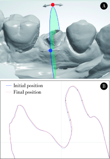
Three-dimensional movement of posterior teeth after losing the interproximal and occlusal contacts in adults
LIU Xiao-qiang1,YANG Yang1,ZHOU Jian-feng1,LIU Ming-yue2,Δ( ),TAN Jian-guo1,Δ()
- 1. Department of Prosthodontics, Peking University School and Hospital of Stomatology & National Clinical Research Center for Oral Diseases & National Engineering Laboratory for Digital and Material Technology of Stomatology & Beijing Key Laboratory of Digital Stomatology, Beijing 100081, China
2. First Clinical Division, Peking University School and Hospital of Stomatology & National Clinical Research Center for Oral Diseases & National Engineering Laboratory for Digital and Material Technology of Stomatology & Beijing Key Laboratory of Digital Stomatology, Beijing 100081, China
CLC Number:
- R783

| [1] | 周永胜. 口腔修复学[M]. 3版. 北京: 北京大学医学出版社, 2020: 119, 133. |
| [2] |
Love WD, Adams RL. Tooth movement into edentulous areas[J]. J Prosthet Dent, 1971,25(3):271-278.
pmid: 5276850 |
| [3] |
Compagnon D, Woda A. Supraeruption of the unopposed maxillary first molar[J]. J Prosthet Dent, 1991,66(1):29-34.
pmid: 1941670 |
| [4] |
Kiliaridis S, Lyka I, Friede H, et al. Vertical position, rotation, and tipping of molars without antagonists[J]. Int J Prosthodont, 2000,13(6):480-486.
pmid: 11203673 |
| [5] |
Christou P, Kiliaridis S. Three-dimensional changes in the position of unopposed molars in adults[J]. Eur J Orthod, 2007,29(6):543-549.
doi: 10.1093/ejo/cjm036 |
| [6] | Petridis HP, Tsiggos N, Michail A, et al. Three-dimensional positional changes of teeth adjacent to posterior edentulous spaces in relation to age at time of tooth loss and elapsed time[J]. Eur J Prosthodont Restor Dent, 2010,18(2):78-83. |
| [7] |
Lindskog-Stokland B, Hansen K, Tomasi C, et al. Changes in molar position associated with missing opposed and/or adjacent tooth: a 12-year study in women[J]. J Oral Rehabil, 2012,39(2):136-143.
doi: 10.1111/j.1365-2842.2011.02252.x pmid: 21902708 |
| [8] |
Garcia-Herraiz A, Silvestre FJ, Leiva-Garcia R, et al. Post-extraction mesio-distal gap reduction assessment by confocal laser scanning microscopy: a clinical 3-month follow-up study[J]. J Clin Periodontol, 2017,44(5):548-555.
doi: 10.1111/jcpe.2017.44.issue-5 |
| [9] |
Faggion CM Jr, Giannakopoulos NN, Listl S. How strong is the evidence for the need to restore posterior bounded edentulous spaces in adults? Grading the quality of evidence and the strength of recommendations[J]. J Dent, 2011,39(2):108-116.
doi: 10.1016/j.jdent.2010.11.002 |
| [10] |
Craddock HL, Youngson CC. A study of the incidence of overeruption and occlusal interferences in unopposed posterior teeth[J]. Br Dent J, 2004,196(6):341-348.
doi: 10.1038/sj.bdj.4811082 |
| [11] | Heij DG, Opdebeeck H, van Steenberghe D, et al. Facial development, continuous tooth eruption, and mesial drift as compromising factors for implant placement[J]. Int J Oral Maxillofac Implants, 2006,21(6):867-878. |
| [12] |
Firestone JM. Missing posterior teeth[J]. J Am Dent Assoc, 2001,132(1):14-18.
doi: 10.14219/jada.archive.2001.0004 |
| [13] |
Carlsson GE, Kiliaridis S. Tooth movement[J]. Br Dent J, 2005,198(7):420-421.
doi: 10.1038/sj.bdj.4812246 |
| [14] | 李德利, 谭建国. 一步一步做好美学临时修复[J]. 中华口腔医学杂志, 2021,56(2):226-230. |
| [15] |
Gragg KL, Shugars DA, Bader JD, et al. Movement of teeth adjacent to posterior bounded edentulous spaces[J]. J Dent Res, 2001,80(11):2021-2024.
pmid: 11759014 |
| [1] | Shishi BO,Chengzhi GAO. Tooth segmentation and identification on cone-beam computed tomography with convolutional neural network based on spatial embedding information [J]. Journal of Peking University (Health Sciences), 2024, 56(4): 735-740. |
| [2] | Xiuwen FEI,Si LIU,Bo WANG,Aimei DONG. Clinical characteristics and treatment in adults and children with histiocytic necroti-zing lymphadenitis [J]. Journal of Peking University (Health Sciences), 2024, 56(3): 533-540. |
| [3] | Yifan WU,Yingxiang YU,Lan XIE,Zhida ZHANG,Cuiqing CHANG. Characteristics of resting energy expenditure and evaluation of prediction formulas in young men with different body mass indexes [J]. Journal of Peking University (Health Sciences), 2024, 56(2): 247-252. |
| [4] | Xinyu XU,Ling WU,Fengqi SONG,Zili LI,Yi ZHANG,Xiaojing LIU. Mandibular condyle localization in orthognathic surgery based on mandibular movement trajectory and its preliminary accuracy verification [J]. Journal of Peking University (Health Sciences), 2024, 56(1): 57-65. |
| [5] | Hai-hong YAO,Fan YANG,Su-mei TANG,Xia ZHANG,Jing HE,Yuan JIA. Clinical characteristics and diagnostic indicators of macrophage activation syndrome in patients with systemic lupus erythematosus and adult-onset Still's disease [J]. Journal of Peking University (Health Sciences), 2023, 55(6): 966-974. |
| [6] | Zi-xuan XUE,Shi-ying TANG,Min QIU,Cheng LIU,Xiao-jun TIAN,Min LU,Jing-han DONG,Lu-lin MA,Shu-dong ZHANG. Clinicopathologic features and prognosis of young renal tumors with tumor thrombus [J]. Journal of Peking University (Health Sciences), 2023, 55(5): 802-811. |
| [7] | Yun-fei DAI,He LIU,Chu-fang PENG,Xi-jun JIANG. Retrospective evaluation of treatment outcomes in immature teeth treated with regenerative endodontic procedures with an over-36-month review [J]. Journal of Peking University (Health Sciences), 2023, 55(4): 729-735. |
| [8] | Ting WANG,Qiao-sheng LI,Hao-ran LIU,Wei-yan JIAN. Urban-rural differentials in the relationship between personality traits and changes in depressive symptoms [J]. Journal of Peking University (Health Sciences), 2023, 55(3): 385-391. |
| [9] | Si-yu WU,Ya-ning LI,Xiao ZHANG,Long-wei LV,Yun-song LIU,Hong-qiang YE,Yong-sheng ZHOU. Prediction, analysis and application of learning curve of tooth preparation for all ceramic crowns of maxillary central incisors [J]. Journal of Peking University (Health Sciences), 2023, 55(1): 108-113. |
| [10] | Xiao-xuan LIU,Shuo ZHANG,Yan MA,A-ping SUN,Ying-shuang ZHANG,Dong-sheng FAN. Diagnostic value of F wave changes in patients with Charcot-Marie-Tooth1A and chronic inflammatory demyelinating polyneuropathy [J]. Journal of Peking University (Health Sciences), 2023, 55(1): 160-166. |
| [11] | Jing WEN,Xiang-ying OUYANG,Xi-yan PEI,Shan-yong QIU,Jian-ru LIU,Wen-yi LIU,Cai-fang CAO. Multivariable analysis of tooth loss in subjects with severe periodontitis over 4-year natural progression [J]. Journal of Peking University (Health Sciences), 2023, 55(1): 70-77. |
| [12] | Wei YONG,Kun QIAN,Wen-hao ZHU,Xiao-yi ZHAO,Chang LIU,Jie PAN. X-ray evaluation of pulp calcification in adult permanent teeth after pulpotomy [J]. Journal of Peking University (Health Sciences), 2023, 55(1): 88-93. |
| [13] | Juan GAO,Hang-miao LV,Hui-min MA,Yi-jiao ZHAO,Xiao-tong LI. Evaluation of root resorption after surgical orthodontic treatment of skeletal Class Ⅲ malocclusion by three-dimensional volumetric measurement with cone-beam CT [J]. Journal of Peking University (Health Sciences), 2022, 54(4): 719-726. |
| [14] | Hai-long HE,Qing LI,Tao XU,Xiao-wei ZHANG. Treatment of adult-acquired buried penis with suprapubic liposuction combined with modified Devine operation [J]. Journal of Peking University (Health Sciences), 2022, 54(4): 741-745. |
| [15] | FENG Sha-wei,GUO Hui,WANG Yong,ZHAO Yi-jiao,LIU He. Initial establishment of digital reference standardized crown models of the primary teeth [J]. Journal of Peking University (Health Sciences), 2022, 54(2): 327-334. |
|
||