Journal of Peking University (Health Sciences) ›› 2022, Vol. 54 ›› Issue (5): 991-999. doi: 10.19723/j.issn.1671-167X.2022.05.028

Previous Articles     Next Articles

Evaluation of carotid atherosclerotic plaques by vascular plaque quantification (VPQ) technology of three-dimensional ultrasonography

Hai-ying XING1,2,Yu-hui CHEN3,Ke XU1,2,Dian-dian HUANG1,2,Qing PENG1,2,Ran LIU1,2,Wei SUN1,2,Yi-ning HUANG1,2,*()   

  1. 1. Department of Neurology, Peking University First Hospital, Beijing 100034, China
    2. Department of Neurology, Beijing Key Laboratory of Neurovascular Disease Discovery, Beijing, China
    3. Department of Neurology, Beijing Hospital, Beijing 100730, China
  • Received:2022-06-30 Online:2022-10-18 Published:2022-10-14
  • Contact: Yi-ning HUANG E-mail:ynhuang@bjmu.edu.cn

RICH HTML


Abstract:

Objective: To analyze the feasibility of using vascular plaque quantification (VPQ) to evaluate carotid atherosclerotic plaques and to observe the effect of statins on carotid atherosclerotic plaques. Methods: Patients with carotid plaques from January 2016 to September 2018 in Peking University First Hospital Neurology Department were recruited and underwent three-dimonsional ultrasound (3DUS). Their gray scale median (GSM) and other parameters of carotid plaques were measured with VPQ. The patients were divided into low GSM group (GSM < 40) and high GSM group (GSM≥40). The clinical characteristics and plaque characteristics of the patients in the two groups were compared to analyze the stability of plaques. According to whether taking statins or not, the patients were further divided into statin group and non-statin group, plaque GSM and other parameters of their carotid plaques were measured and the changes of carotid plaques at the end of 3 months and 2 years were observed. Results: A total of 120 patients were enrolled, including 79 males and 41 females, with an average age of (65.39±9.11) years. The patients were divided into low GSM group (31 cases) (GSM < 40) and high GSM group (89 cases) (GSM≥40). The stenosis of the lumen in the low GSM group was more severe (the area stenosis rate was 41.32%±21.37% vs. 29.79%±17.16%, P < 0.05). The nor-malized wall index (NWI) of plaque in low GSM group was significantly higher than that in high GSM group (0.61 ±0.14 vs. 0.52±0.12, P < 0.01). A total of 77 patients, including 51 males and 26 females, aged (64.96±9.58) years, were enrolled to observe the statin effects on carotid plaque. They were divided into statin group (n=56) and non-statin group (n=21) according to whether taking statins or not. At the baseline and 3-month follow-up, there were no significant differences in carotid plaque volume, area, degree of luminal stenosis and GSM between the two groups (P>0.05). At the end of the 2-year follow-up, GSM increased in the statin group [median 10.00 (2.00, 28.00)] but decreased in the non-statin group [median -7.00 (-11.00, 5.50)], with a statistically significant difference between the two groups (P < 0.01). There was no significant increase in carotid plaque volume in the statin group, while there was a slight increase in the non-statin group, but there was no significant difference between the two groups [median increase in plaque volume was 0.00 (-30.00, 40.00) mm3 in the statin group and 30.00 (10.00, 70.00) mm3 in the non-statin group, P>0.05]. Conclusion: The VPQ technology of 3DUS can be used to evaluate carotid atherosclerotic plaques. Patients with low GSM (GSM < 40) have more severe vascular stenosis and higher normalized wall index. VPQ technology can also be used to observe the effect of statins on carotid plaque, the GSM of plaques increase in patients who are taking moderate-intensity statin treatment for two years.

Key words: Three-dimensional ultrasonography, Vascular plaque quantification, Carotid plaque, Gray scale median, Statin

CLC Number: 

  • R543.4

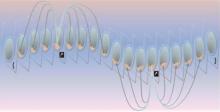
Figure 1

Determine begin frame, key frames and end frame in VPQ VPQ, vascular plaque quantification."

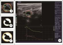
Figure 2

Parameters measured by vascular plaque quantification (VPQ) A, max plaque thickness (purple line); B, plaque area (white area) and lumen area (black area outlined by the blue line); C, total vessel area (Area outlined by the red line. Vessel wall area=Total vessel area-lumen area+plaque area); D, parameters measured by VPQ, including plaque volume, gray scale median (GSM), etc."

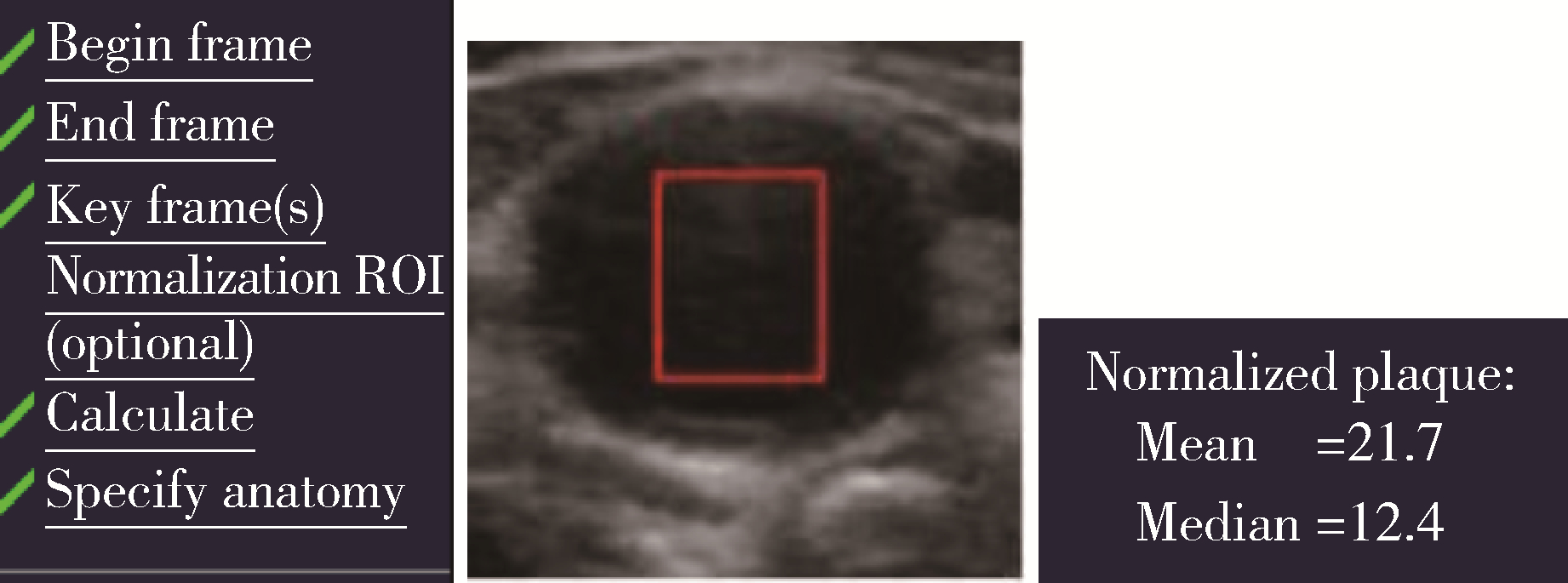
Figure 3

Normalization of gray scale median (GSM) in vascular plaque quantification (VPQ) ROI, region of interest."

Table 1

Baseline characteristics of low GSM group and high GSM group"

Items Total(n=120) Low GSM group(n=31) High GSM group(n=89) P
Age/years, $\bar x \pm s$ 65.39±9.11 66.1±9.29 65.15±9.09 0.62
Male, n (%) 79 (65.83) 18 (58.06) 61 (68.53) 0.44
Smoking,n (%) 50 (42.37) 10 (32.25) 40 (44.94) 0.25
Hypertension, n (%) 80 (67.80) 18 (58.06) 62 (69.66) 0.29
Diabetes mellitus, n (%) 42 (35.59) 13 (41.94) 29 (32.58) 0.31
ASCVD, n (%) 68 (61.82) 15 (48.38) 53 (62.92) 0.44
Stroke, n (%) 61 (51.70) 14 (45.16) 47 (52.81) 0.52
BMI/(kg/m2), $\bar x \pm s$ 24.64 ±3.31 24.76 ±3.05 24.59 ±3.41 0.80
SBP /mmHg, $\bar x \pm s$ 137.26 ±17.03 139.72 ±16.66 136.45 ±17.16 0.37
DBP /mmHg, $\bar x \pm s$ 74.06 ±12.74 72.45 ±13.47 74.59 ±12.52 0.45
Glucose/(mmol/L), $\bar x \pm s$ 6.13 ±1.73 6.00 ±1.20 6.16 ±1.86 0.61
TG/(mmol/L), $\bar x \pm s$ 1.38 ±0.73 1.41±0.69 1.37 ±0.75 0.79
LDL-C/(mmol/L), $\bar x \pm s$ 1.98 ±0.86 1.86 ±1.05 2.02±0.80 0.45
Antiplatelet, n (%) 99 (83.90) 25 (80.64) 74 (83.14) 0.92
Lipid lowering, n (%) 87 (72.50) 21 (67.74) 66 (74.16) 0.49

Table 2

Carotid plaque characteristics of low GSM group and high GSM group"

Items Total(n=120) Low GSM group(n=31) High GSM group(n=89) P
Plaque volume /mm3, M (P25, P75) 170.0
(100.0, 267.5)
180.0
(120.0, 270.0)
160.0
(95.0, 260.0)
0.29
Plaque area/mm2, $\bar x \pm s$ 15.97 ±11.37 18.02 ±13.13 15.26 ±10.69 0.30
Stenosis ratio/%, $\bar x \pm s$ 32.77 ±18.93 41.32 ±21.37 29.79 ±17.16 0.01
NWI, $\bar x \pm s$ 0.54 ±0.13 0.61 ±0.14 0.52 ±0.12 < 0.01

Table 3

Baseline, 3-month follow-up, and change from baseline carotid plaque volumes and areas"

Items Total(n=77) Statin group(n=56) Non-statin group (n=21) P*
Plaque volume /mm3, M (P25, P75)
  Baseline 170.00
(100.00, 230.00)
170.00
(107.50, 235.00)
140.00
(80.00, 245.00)
0.28
  3-month 160.00
(100.00, 260.00)
160.00
(100.00, 230.00)
160.00
(85.00, 275.00)
0.76
  Change 0.00
(-20.00, 20.00)
0.00
(-20.00, 12.50)
0.00
(-25.00, 40.00)
0.47
   P# 0.65 0.46 0.38
Plaque area/mm2
  Baseline, $\bar x \pm s$ 15.12±10.39 16.20±10.85 12.21±8.61 0.13
  3-month, $\bar x \pm s$ 14.35±10.57 14.44±9.93 14.10±12.37 0.90
  Change, M (P25, P75) -0.31 (-3.02, 2.89) -0.83 (-3.35, 1.81) 0.48 (-1.47, 4.10) 0.07
   P# 0.33 0.09 0.32
Stenosis ratio/%
  Baseline, $\bar x \pm s$ 31.69±18.75 33.63±18.52 26.52±18.81 0.14
  3-month, $\bar x \pm s$ 31.30±17.82 31.66±16.52 30.33±21.32 0.77
  Change, M (P25, P75) 0 (-3.50, 5.50) 0.00 (-7.00, 5.00) 2.00 (-1.00, 10.00) 0.10
   P# 0.79 0.52 0.09
NWI
  Baseline, $\bar x \pm s$ 0.54±0.14 0.55±0.14 0.51±0.13 0.25
  3-month, $\bar x \pm s$ 0.54±0.13 0.54±0.13 0.52±0.15 0.61
  Change, M (P25, P75) 0.00 (-0.04, 0.03) 0.00 (-0.05, 0.03) 0.00 (-0.03, 0.04) 0.53
   P# 0.68 0.93 0.31

Table 4

Baseline, 3-month follow-up, and change from baseline carotid plaque GSM"

Items Total(n=77) Statin group(n=56) Non-statin group(n=21) P*
Baseline, $\bar x \pm s$ 54.20±19.16 55.57±21.03 49.07±7.91 0.06
3-month, $\bar x \pm s$ 55.63±18.38 57.88±18.98 47.20±13.35 0.02
Change, M (P25, P75) 0.00
(-8.00, 11.00)
0.50
(-7.00, 13.00)
-3.00
(-9.00, 6.00)
0.23
P# 0.51 0.33 0.52

Table 5

Baseline, 2-year follow-up, and change from baseline carotid plaque volumes and areas"

Items Total(n=62) Statin group(n=47) Non-statin group(n=15) P*
Plaque volume /mm3, M (P25, P75)
  Baseline 180.00
(97.50, 312.50)
200.00
(90.00, 320.00)
140.00
(100.00, 280.00)
0.74
  2 years 185.00 (117.50, 302.50) 200.00
(120.00, 300.00)
160.00
(110.00, 360.00)
0.86
  Change 15.00
(-30.00, 50.00)
0.00
(-30.00, 40.00)
30.00
(10.00, 70.00)
0.09
   P# 0.11 0.56 0.02
Plaque area /mm2
  Baseline, $\bar x \pm s$ 15.97±12.63 15.41±10.19 17.72±8.69 0.65
  2 years, $\bar x \pm s$ 13.68±8.82 12.88±8.35 16.19±10.05 0.26
  Change, M (P25, P75) -0.44 (-7.10, 2.58) -0.81 (-8.47, 2.13) 1.46 (-6.17, 5.71) 0.14
   P# 0.13 0.07 0.89
Stenosis ratio/%
  Baseline, $\bar x \pm s$ 31.76±19.80 31.49±18.96 32.60±22.92 0.10
  2 years, $\bar x \pm s$ 31.40±17.25 30.79±17.07 33.33±18.29 0.64
  Change, M (P25, P75) -1.00 (-5.25, 8.00) -1.00 (-4.00, 8.00) -1.00 (-9.00, 9.00) 0.93
   P# 0.95 0.88 0.95
NWI
  Baseline, $\bar x \pm s$ 0.53±0.14 0.53±0.14 0.54±0.16 0.94
  2 years, $\bar x \pm s$ 0.54±0.12 0.53±0.12 0.54±0.13 0.94
  Change, M (P25, P75) -0.01 (-0.05, 0.06) -0.01
(-0.04, 0.06)
-0.02 (-0.06, 0.07) 0.96
   P# 0.87 0.87 0.93

Table 6

Baseline, 2-year follow-up, and change from baseline carotid plaque GSM"

Items Total(n=62) Statin group(n=47) Non-statin group (n=15) P*
Baseline, $\bar x \pm s$ 52.14±17.00 52.72±16.65 50.53±18.36 0.67
2 years, $\bar x \pm s$ 62.55±21.39 67.87±20.62 47.82±16.28 < 0.01
Change, M (P25, P75) 7.00
(-6.00, 21.75)
10.00
(2.00, 28.00)
-7.00
(-11.00, 5.50)
< 0.01
P# < 0.01 < 0.01 0.37
1 Lal BK , Hobson RW , Pappas PJ , et al. Pixel distribution analysis of B-mode ultrasound scan images predicts histologic features of atherosclerotic carotid plaques[J]. J Vasc Surg, 2002, 35 (6): 1210- 1217.
doi: 10.1067/mva.2002.122888
2 Herr JE , Hetu MF , Li TY , et al. Presence of calcium-like tissue composition in carotid plaque is indicative of significant coronary artery disease in high-risk patients[J]. J Am Soc Echocardiogr, 2019, 32 (5): 633- 642.
doi: 10.1016/j.echo.2019.01.001
3 Mujaj B , Bos D , Selwaness M , et al. Statin use is associated with carotid plaque composition: The Rotterdam study[J]. Int J Cardiol, 2018, 260, 213- 218.
doi: 10.1016/j.ijcard.2018.02.111
4 Vergallo R , Crea F . Atherosclerotic plaque healing[J]. N Engl J Med, 2020, 383 (9): 846- 857.
doi: 10.1056/NEJMra2000317
5 Touboul PJ , Hennerici MG , Meairs S , et al. Mannheim carotid intima-media thickness and plaque consensus (2004-2006-2011). An update on behalf of the advisory board of the 3rd, 4th and 5th watching the risk symposia, at the 13th, 15th and 20th European Stroke Conferences, Mannheim, Germany, 2004, Brussels, Belgium, 2006, and Hamburg, Germany, 2011[J]. Cerebrovasc Dis, 2012, 34 (4): 290- 296.
doi: 10.1159/000343145
6 中国成人血脂异常防治指南修订联合委员会. 中国成人血脂异常防治指南(2016年修订版)[J]. 中华心血管病杂志, 2016, 44 (10): 833- 853.
doi: 10.3760/cma.j.issn.0253-3758.2016.10.005
7 Geroulakos G , Ramaswami G , Nicolaides A , et al. Characterization of symptomatic and asymptomatic carotid plaques using high-resolution real-time ultrasonography[J]. Br J Surg, 1993, 80 (10): 1274- 1277.
8 Johri AM , Herr JE , Li TY , et al. Novel ultrasound methods to investigate carotid artery plaque vulnerability[J]. J Am Soc Echocardiogr, 2017, 30 (2): 139- 148.
doi: 10.1016/j.echo.2016.11.003
9 el-Barghouty N , Geroulakos G , Nicolaides A , et al. Computer-assisted carotid plaque characterisation[J]. Eur J Vasc Endovasc Surg, 1995, 9 (4): 389- 393.
doi: 10.1016/S1078-5884(05)80005-X
10 Spanos K , Tzorbatzoglou I , Lazari P , et al. Carotid artery plaque echomorphology and its association with histopathologic characteristics[J]. J Vasc Surg, 2018, 68 (6): 1772- 1780.
doi: 10.1016/j.jvs.2018.01.068
11 Mathiesen EB , Bonaa KH , Joakimsen O . Echolucent plaques are associated with high risk of ischemic cerebrovascular events in carotid stenosis: The tromso study[J]. Circulation, 2001, 103 (17): 2171- 2175.
doi: 10.1161/01.CIR.103.17.2171
12 Hashimoto H , Tagaya M , Niki H , et al. Computer-assisted analysis of heterogeneity on B-mode imaging predicts instability of asymptomatic carotid plaque[J]. Cerebrovasc Dis, 2009, 28 (4): 357- 364.
doi: 10.1159/000229554
13 Huibers A , de Borst GJ , Bulbulia R , et al. Plaque echolucency and the risk of ischaemic stroke in patients with asymptomatic carotid stenosis within the first asymptomatic carotid surgery trial (ACST-1)[J]. Eur J Vasc Endovasc Surg, 2016, 51 (5): 616- 621.
doi: 10.1016/j.ejvs.2015.11.013
14 Zhang M , Zhao Q , Gao Y , et al. Prediction for rupture risk of carotid artery plaques: A comparative study of 3D-GSM and CAS system[J]. Eur J Radiol, 2016, 85 (9): 1659- 1665.
doi: 10.1016/j.ejrad.2016.05.013
15 王秀玲, 张敏郁, 丁桂春, 等. 三维超声灰阶中位数定量评价颈动脉斑块的易损性[J]. 中华医学超声杂志: 电子版, 2017, 14 (1): 23- 28.
doi: 10.3877/cma.j.issn.1672-6448.2017.01.008
16 Urbak L , Sandholt BV , Graebe M , et al. Patients with unstable atherosclerosis have more echolucent carotid plaques compared with stable atherosclerotic patients: A 3-D ultrasound study[J]. Ultrasound Med Biol, 2020, 46 (9): 2164- 2172.
doi: 10.1016/j.ultrasmedbio.2020.04.002
17 Nicolaides AN , Kakkos SK , Kyriacou E , et al. Asymptomatic internal carotid artery stenosis and cerebrovascular risk stratification[J]. J Vasc Surg, 2010, 52 (6): 1486- 1496.
doi: 10.1016/j.jvs.2010.07.021
18 Ruiz-Ares G , Fuentes B , Martinez-Sanchez P , et al. A prediction model for unstable carotid atheromatous plaque in acute ischemic stroke patients: Proposal and internal validation[J]. Ultrasound Med Biol, 2014, 40 (9): 1958- 1965.
doi: 10.1016/j.ultrasmedbio.2014.04.015
19 Biasi GM , Froio A , Diethrich EB , et al. Carotid plaque echolucency increases the risk of stroke in carotid stenting: The imaging in carotid angioplasty and risk of stroke (ICAROS) study[J]. Circulation, 2004, 110 (6): 756- 762.
doi: 10.1161/01.CIR.0000138103.91187.E3
20 Ariyoshi K , Okuya S , Kunitsugu I , et al. Ultrasound analysis of gray-scale median value of carotid plaques is a useful reference index for cerebro-cardiovascular events in patients with type 2 diabetes[J]. J Diabetes Investig, 2015, 6 (1): 91- 97.
doi: 10.1111/jdi.12242
21 国家卫生健康委员会脑卒中防治专家委员会血管超声专业委员会, 中国超声医学工程学会浅表器官及外周血管超声专业委员会, 中国超声医学工程学会颅脑及颈部血管超声专业委员会, 中国超声医学工程学会颅脑及颈部血管超声专业委员会. 头颈部血管超声若干问题的专家共识(颈动脉部分)[J]. 中国脑血管病杂志, 2020, 17 (6): 346- 352.
doi: 10.3969/j.issn.1672-5921.2020.06.013
22 Kadoglou NP , Gerasimidis T , Moumtzouoglou A , et al. Intensive lipid-lowering therapy ameliorates novel calcification markers and GSM score in patients with carotid stenosis[J]. Eur J Vasc Endovasc Surg, 2008, 35 (6): 661- 668.
doi: 10.1016/j.ejvs.2007.12.011
23 Yamaguchi M , Sasaki M , Ohba H , et al. Quantitative assessment of changes in carotid plaques during cilostazol administration using three-dimensional ultrasonography and non-gated magnetic resonance plaque imaging[J]. Neuroradiology, 2012, 54 (9): 939- 945.
doi: 10.1007/s00234-012-1011-2
[1] Chao GONG,Qiu-ping LIU,Jia-min WANG,Xiao-fei LIU,Ming-lu ZHANG,Han YANG,Peng SHEN,Hong-bo LIN,Xun TANG,Pei GAO. Effectiveness of statin treatment strategies for primary prevention of cardiovascular diseases in a community-based Chinese population: A decision-analytic Markov model [J]. Journal of Peking University (Health Sciences), 2022, 54(3): 443-449.
[2] LIU Huan, WANG Hong-yu, LIU Jin-bo, ZHAO Hong-wei, ZHOU Ying-yan, LI Li-hong. Relationship between large artery elasticity function and coronary heart disease, lower extremity arterial disease in patients with carotid plaque [J]. Journal of Peking University(Health Sciences), 2017, 49(6): 1027-1033.
[3] JIA Li-jing, YI Liang, YANG Zhi-xu, WANG Shu-peng, LI Gang, ZHU Xi. Preventive effects of ulinastatin on acute respiratory distress syndrome [J]. Journal of Peking University(Health Sciences), 2016, 48(4): 672-679.
[4] ZHANG Jing, REN Jing-yi, CHEN Hong, HAN Guan-ping. Statins decreases expression of five inflammation-associated microRNAs in the plasma of patients with unstable angina [J]. Journal of Peking University(Health Sciences), 2015, 47(5): 761-768.
[5] 欧Meng-恩 , ZHANG Xiao, LIU Yun-Song, GE Yan-Jun, ZHOU Yong-Sheng. Ectopic osteogenesis of stromal cell-derived factor 1 combined with simvastatin loaded collagen scaffold in vivo [J]. Journal of Peking University(Health Sciences), 2015, 47(1): 47-51.
[6] XU Lu, WANG Chao, SHEN Wen-Wen, QI Rong. Effects of simvastatin nano-liposomes on osteogenic differentiation of bone marrow stromal cells [J]. Journal of Peking University(Health Sciences), 2014, 46(6): 883-888.
[7] GUAN Jing-Lin, JIANG Wen- , WANG Huai-Yu, ZENG Hui, ZHANG Su-Min, WANG Guang, MAO Jie-Ming. Multiple effect of simvastatin on vascular endothelium of hypercholesterolemia patients [J]. Journal of Peking University(Health Sciences), 2014, 46(5): 703-706.
[8] XIA Jing-Gang, QU Yang, Hu-Shao-Dong, XU Ji, YIN Chun-Lin, XU Dong. Cardioprotective effect and mechanism of intensive lipid modulation on patients with coronary artery disease undergoing noncardiac surgery [J]. Journal of Peking University(Health Sciences), 2014, 46(4): 601-605.
Viewed
Full text


Abstract

Cited

  Shared   
  Discussed   
No Suggested Reading articles found!