Journal of Peking University (Health Sciences) ›› 2023, Vol. 55 ›› Issue (4): 652-657. doi: 10.19723/j.issn.1671-167X.2023.04.014

Previous Articles     Next Articles

Prognosis of patients with spinal intramedullary cavernous hemangioma by different treatments

Bin CHEN,Chao WU,Bin LIU,Tao YU,Zhen-yu WANG*()   

  1. Department of Neurosurgery, Peking University Third Hospital, Beijing 100191, China
  • Received:2020-10-26 Online:2023-08-18 Published:2023-08-03
  • Contact: Zhen-yu WANG E-mail:wzyu502@126.com

RICH HTML


Abstract:

Objective: To investigate the improvement of spinal cord function in patients with spinal intramedullary cavernous hemangioma (SICH) treated with different methods at the last follow-up. Methods: A retrospective study of 30 patients with SICH in Peking University Third Hospital from January 2007 to December 2018 was conducted. Clinical data of 30 patients were collected including gender, age, clinical symptoms, and imaging manifestations were acquired from their clinical records. Spinal functions of the patients with SICH were evaluated by European myelopathy score (EMS). The functional status of the spinal cord before and after the last follow-up were analyzed. Results: Among the 30 patients, there were 14 male patients and 16 female patients (1 ∶ 1.14). The average age of the patients was (48.1±13.6) years (18-81 years). In the study, 3 cases were sensory disturbance; 2 cases manifested with only decreased muscle strength; 1 case showed simple pain; 1 case manifested with decreased muscle strength and pain; Sensory disturbance accompanied by decreased muscle strength occurred in 5 cases; 3 cases suffered from both sensory disturbance, decreased muscle strength and abnormal defecation; 3 cases suffered from sensory disturbance, decreased muscle strength and pain; 8 cases showed sensory disturbance and pain; 1 case had sensory disturbance, pain and abnormal defecation; 1 case had sensory disturbance, pain, decreased muscle strength and abnormal defecation; 2 cases were asymptomatic. There were 11 patients whose lesions were located in the cervical region, 2 patients located at the cervical-thoracic region, 15 patients located in the thoracic region, and 2 patients located in the lumbar spine. The average maximum diameter of hemangioma was (10.90±4.87) mm. Their magnetic resonance imaging (MRI) features were usually mixed signal and high signal on T2WI, and equal signal or mixed signal on T1WI. A total of 30 patients were followed up for (27.4±8.7) months, including 19 patients with surgical treatment and 11 patients with conservative treatment. The spinal cord function at the last follow up in surgical group was significantly improved. The difference was statistically significant (P < 0.05). Only one patient' s symptom in conservative treatment group improved. However, the symptoms of the two patients aggravated. Other patients remained stable. There was no significant difference in spinal cord function before and after treatment (P>0.05). Conclusion: The surgical treatment of SICH has obvious positive effect and good prognosis. The overall improvement rate of conservative treatment is relatively low with a risk of aggravation.

Key words: Spinal intramedullary cavernous hemangioma, Surgical treatment, Conservative treatment, European myelopathy score

CLC Number: 

  • R651.2

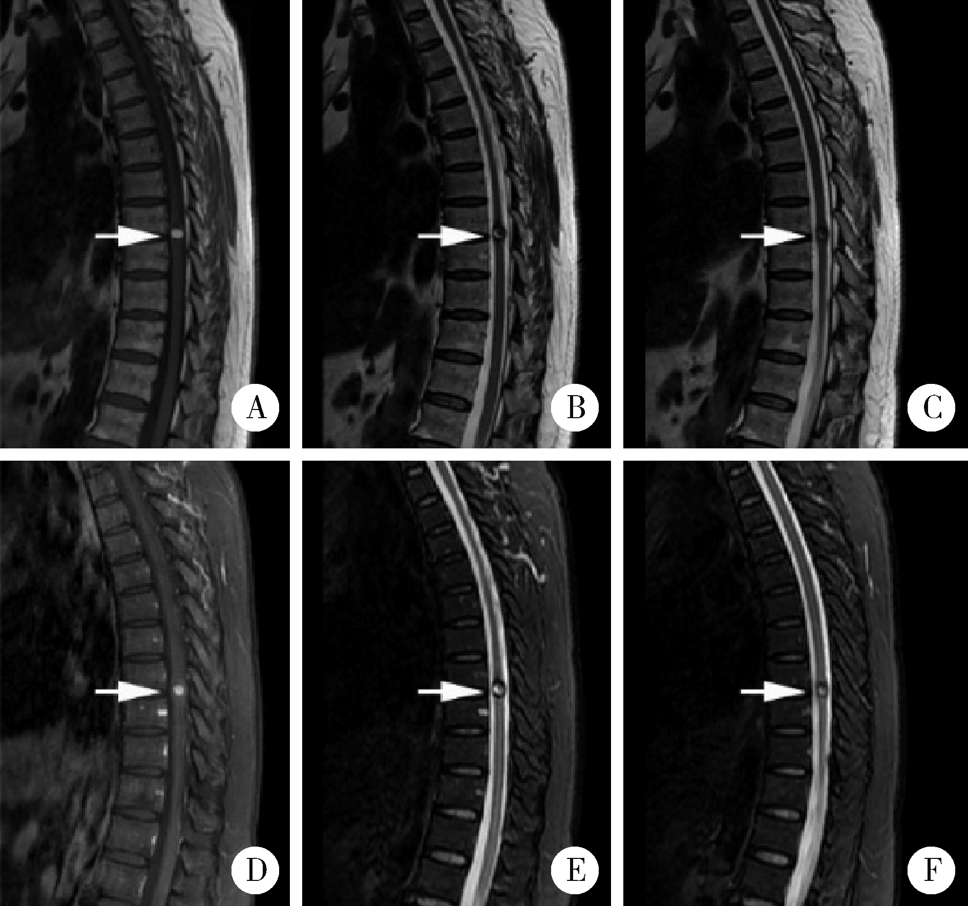
Figure 1

Typical image of cavernous hemangioma of thoracic spinal cord Preoperative magnetic resonance imaging of patient No.15. The patient is female with chronic onset symptoms, her symptoms include sensory disturbances and decreased muscle strength. A, B, C, magnetic resonance imaging showed that the lesion was located in the thoracic spinal canal, with high signal intensity on T1WI and mixed signal intensity on T2WI, enhanced magnetic resonance imaging showed that the lesions could be enhanced by enhanced scanning; D, E, F, the typical signs of "black ring". Arrows showed the spinal intramedullary cavernous hemangioma lesion before surgery."

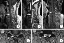
Figure 2

Typical image of intramedullary cavernous hemangioma of cervical spinal cord Preoperative magnetic resonance imaging of patient No.29. The patient is male with chronic onset symptoms, his symptoms were mainly mild sensory disturbances. A, B, lesions showed equal signals on T1WI and mixed signals on T2WI; C, lesion can be enhanced; D, E, typical signs of black rings can be seen. Arrows showed the spinal intramedullary caver-nous hemangioma lesion before surgery."

Figure 3

Comparison of magnetic resonance imaging before and after surgery for spinal intramedulary cavernous hemangioma A, preoperative magnetic resonance imaging of patient No.8. The patient is male with chronic onset symptoms, his symptoms include sensory disturbances, pain, and dysdefecation. Compared with the preoperative image, the lesion has been removed, and intramedullary mixed signal or high signal can be seen on T2WI; B, postoperative magnetic resonance imaging images of patient No.8. Arrow showed the spinal intramedullary cavernous hemangioma lesion before surgery."

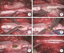
Figure 4

Microsurgical resection steps of spinal intramedulary cavernous hemangioma A, incision of dura mater; B, pull dura mater to both sides; C, expose the tumor; D, burn the tumor to inactivate it and extract the tumor; E, tumor was totally extracted; F, close the dura mater."

Figure 5

Pathological results of spinal intramedulary cavernous hemangioma after surgery A, under microscope, the vessel tube was composed of fibroblasts, and the lumen was lined with thin endothelial cells, lacking elastic fibers and muscle layers (HE ×100); B, there are many fibers between the lumens, no normal spinal cord tissue was found, a patch of hemorrhages can be seen under microscope (HE ×100)."

Table 1

General information of spinal intramedulary cavernous hemangioma patients"

No.GenderAge/yearsClinical presentationChronic/acuteLocationDiameter/mmTreatmentImproved/not improved
1Male50Downregulated strength of muscle, painChronicT77SurgeryImproved
2Male51Sensory disorder, painChronicC7-T113SurgeryNot Improved
3Female32Sensory disorder, painChronicL59SurgeryNot Improved
4Female45Sensory disorder, painChronicC6-713SurgeryImproved
5Male79Downregulated strength of muscleChronicT6-715SurgeryNot Improved
6Female30Sensory disorder, pain, downregulated strength of muscleChronicC27SurgeryImproved
7Male40Sensory disorder, downregulated strength of muscle,
defecation dysfunction
ChronicT35SurgeryImproved
8Male46Sensory disorder, pain, defecation dysfunctionChronicT118SurgeryImproved
9Male65Sensory disorder, pain, downregulated strength of muscle,
defecation dysfunction
AcuteC37SurgeryImproved
10Male37Sensory disorder, pain, downregulated strength of muscle,
defecation dysfunction
AcuteT9-1012SurgeryImproved
11Female40Sensory disorder, painChronicC6-T128SurgeryNot Improved
12Female55Sensory disorder, painChronicC26SurgeryImproved
13Female49Sensory disorder, pain, downregulated strength of muscle,
defecation dysfunction
AcuteT99SurgeryImproved
14Male34Sensory disorder, pain, downregulated strength of muscle, ChronicT37SurgeryImproved
15Female55Sensory disorder, downregulated strength of muscleChronicT8-911SurgeryImproved
16Female72Sensory disorderChronicT2-315SurgeryNot Improved
17Male81Sensory disorder, downregulated strength of muscleChronicT2-317SurgeryImproved
18Male52Sensory disorder, downregulated strength of muscle,
defecation dysfunction
ChronicT48SurgeryImproved
19Female18Sensory disorder, pain, downregulated strength of muscle,
defecation dysfunction
AcuteT47SurgeryImproved
20Male36Sensory disorder, pain, downregulated strength of muscle,
defecation dysfunction
AcuteT8-912ConservativeNot Improved
21Female50PainChronicC5-613ConservativeNot Improved
22Female43Sensory disorder, downregulated strength of muscleChronicC6-711ConservativeNot Improved
23Female31Sensory disorder, painChronicC77ConservativeNot Improved
24Female48Sensory disorder, painChronicL26ConservativeNot Improved
25Male31Sensory disorderChronicT1-324ConservativeNot Improved
26Female61Downregulated strength of muscleChronicC2-423ConservativeNot Improved
27Male58Sensory disorder, pain, downregulated strength of muscleChronicT11-1211ConservativeImproved
28Female52Sensory disorder, downregulated strength of muscleChronicC5-610ConservativeNot Improved
29Male47Sensory disorder, downregulated strength of muscleChronicC4-525ConservativeNot Improved
30Female54Sensory disorder, downregulated strength of muscleChronicC3-417ConservativeNot Improved
1 ZevgaridisD,MedeleR J,HamburgerC,et al.Cavernous haemangiomas of the spinal cord. A review of 117 cases[J].Acta Neurochir,1999,141(3):237-245.
doi: 10.1007/s007010050293
2 BadhiwalaJH,FarrokhyarF,AlhazzaniW,et al.Surgical outcomes and natural history of intramedullary spinal cord cavernous malformations: A single-center series and meta-analysis of indivi-dual patient data: Clinic article[J].J Neurosurg Spine,2014,21(4):662-676.
doi: 10.3171/2014.6.SPINE13949
3 DalitzK,VitzthumHE.Evaluation of five scoring systems for cervical spondylogenic myelopathy[J].Spine J,2019,19(2):e41-e46.
doi: 10.1016/j.spinee.2008.05.005
4 SchermanDB,RaoPJ,VarikattW,et al.Clinical presentation and surgical outcomes of an intramedullary C2 spinal cord cavernoma: A case report and review of the relevant literature[J].J Spine Surg,2016,2(2):139-142.
doi: 10.21037/jss.2016.04.01
5 PétillonP,WilmsG,RaftopoulosC,et al.Spinal intradural extramedullary cavernous hemangioma[J].Neuroradiology,2018,60(10):1085-1087.
doi: 10.1007/s00234-018-2073-6
6 NieQB,ChenZ,JianFZ,et al.Cavernous angioma of the cauda equina: A case report and systematic review of the literature[J].J Int Med Res,2012,40(5):2001-2008.
doi: 10.1177/030006051204000542
7 LiangJT,BaoYH,ZhangHQ,et al.Management and prognosis of symptomatic patients with intramedullary spinal cord cavernoma: Clinical article[J].J Neurosurg Spine,2011,15(4):447-456.
doi: 10.3171/2011.5.SPINE10735
8 ArdeshiriA,ÖzkanN,ChenB,et al.A retrospective and consecutive analysis of the epidemiology and management of spinal cavernomas over the last 20 years in a single center[J].Neurosurg Rev,2016,39(2):269-276.
doi: 10.1007/s10143-015-0674-7
9 BaldvinsdóttirB,ErlingsdóttirG,Kjartanssonó,et al.Extrame-dullar cavernous hemangioma with intra- and extradural growth and clinical symptoms of Brown-Séquard syndrome: Case report and review of the literature[J].World Neurosurg,2016,98(11):881. e5-881. e8.
10 OttenM,McCormickP.Natural history of spinal cavernous malformations[J].Handb Clin Neurol,2017,143,233-239.
11 MithaAP,TurnerJD,SpetzlerRF.Surgical approaches to intramedullary cavernous malformations of the spinal cord[J].Neurosurgery,2011,68(2):317-324.
12 ZhangL,YangW,JiaW,et al.Comparison of outcome between surgical and conservative management of symptomatic spinal cord cavernous malformations[J].Neurosurgery,2016,78(4):552-561.
doi: 10.1227/NEU.0000000000001075
13 ReitzM,BurkhardtT,VettorazziE,et al.Intramedullary spinal cavernoma: Clinical presentation, microsurgical approach, and long-term outcome in a cohort of 48 patients[J].Neurosurg Focus,2015,39(2):E19.
doi: 10.3171/2015.5.FOCUS15153
14 AoyamaT,HidaK,HoukinK.Intramedullary cavernous angiomas of the spinal cord: Clinical characteristics of 13 lesions[J].Neurol Med Chir (Tokyo),2011,51(8):561-566.
doi: 10.2176/nmc.51.561
15 李熊辉,王振宇,刘彬.脊髓髓内海绵状血管瘤的诊疗现状[J].中国脊柱脊髓杂志,2017,27(3):276-279.
16 AzadTD,VeeravaguA,LiA,et al.Long-term effectiveness of gross-total resection for symptomatic spinal cord cavernous malformations[J].Neurosurgery,2018,83(6):1201-1208.
doi: 10.1093/neuros/nyx610
17 董伟杰,刘馨蔓,苏月焦,等.脊髓髓内海绵状血管瘤的显微手术治疗[J].中华显微外科杂志,2018,41(2):105-108.
[1] Ying ZHOU,Ning ZHAO,Hongyuan HUANG,Qingxiang LI,Chuanbin GUO,Yuxing GUO. Application of double-layer soft tissue suture closure technique in the surgical treatment of patients with mandible medication-related osteonecrosis of the jaw of early and medium stages [J]. Journal of Peking University (Health Sciences), 2024, 56(1): 51-56.
[2] HONG Peng,TIAN Xiao-jun,ZHAO Xiao-yu,YANG Fei-long,LIU Zhuo,LU Min,ZHAO Lei,MA Lu-lin. Bilateral papillary renal cell carcinoma following kidney transplantation: A case report [J]. Journal of Peking University (Health Sciences), 2021, 53(4): 811-813.
[3] Jie YANG,Ran ZHANG,Yu-nan LIU,Dian-can WANG. Plunging ranula presenting as a giant retroauricular mass: A case report [J]. Journal of Peking University(Health Sciences), 2020, 52(1): 193-195.
[4] Rong YANG,Qing-xiang LI,Chi MAO,Xin PENG,Yang WANG,Yu-xing GUO,Chuan-bin GUO. Multimodal image fusion technology for diagnosis and treatment of the skull base-infratemporal tumors [J]. Journal of Peking University(Health Sciences), 2019, 51(1): 53-58.
Viewed
Full text


Abstract

Cited

  Shared   
  Discussed   
No Suggested Reading articles found!