Journal of Peking University (Health Sciences) ›› 2025, Vol. 57 ›› Issue (2): 384-387. doi: 10.19723/j.issn.1671-167X.2025.02.025
Previous Articles Next Articles
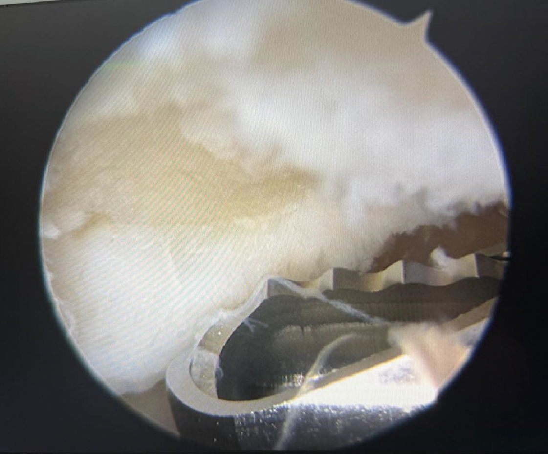
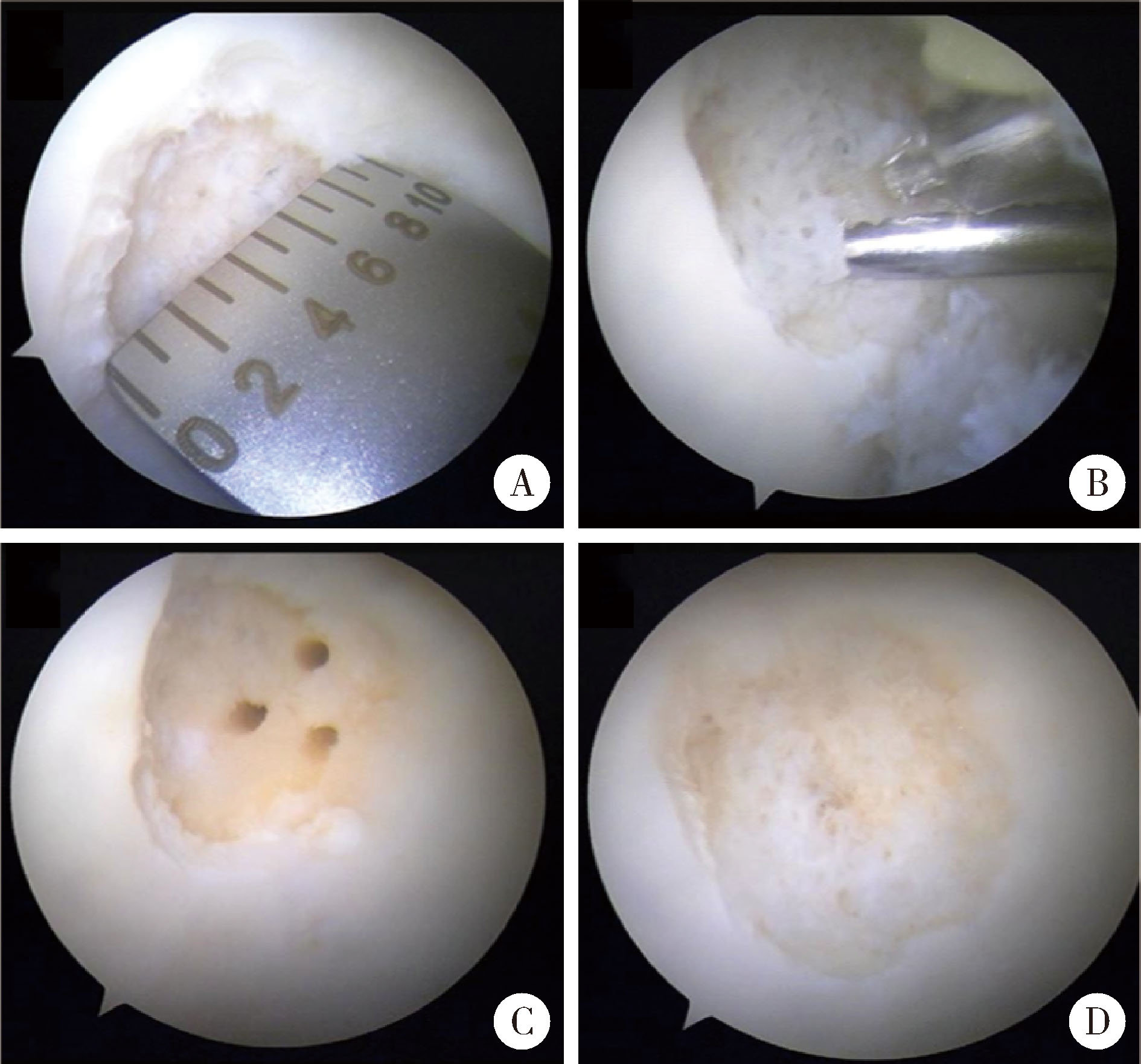
Arthroscopic tissue engineering scaffold repair for cartilage injuries
Zhenlong LIU, Zhenchen HOU, Xiaoqing HU, Shuang REN, Qinwei GUO, Yan XU, Xi GONG△( ), Yingfang AO△()
- Department of Sports Medicine, Peking University Third Hospital, Institute of Sports Medicine of Peking University, Beijing Key Laboratory of Sports Injuries, Beijing 100191, China
CLC Number:
- R687.4


| 1 | Schneider S , Kaiser R , Uterhark B , et al. Autologous surface repair: Autologous matrix-induced chondrogenesis and minced cartilage implantation[J]. JCJP, 2023, 3 (1): 100111. |
| 2 |
Fossum V , Hansen AK , Wilsgaard T , et al. Collagen-covered autologous chondrocyte implantation versus autologous matrix-induced chondrogenesis: A randomized trial comparing 2 methods for repair of cartilage defects of the knee[J]. Orthop J Sports Med, 2019, 7 (9): 2325967119868212.
doi: 10.1177/2325967119868212 |
| 3 |
Krych AJ , Saris DBF , Stuart MJ , et al. Cartilage Injury in the Knee: Assessment and treatment options[J]. J Am Acad Orthop Surg, 2020, 28 (22): 914- 922.
doi: 10.5435/JAAOS-D-20-00266 |
| 4 |
Makris EA , Gomoll AH , Malizos KN , et al. Repair and tissue engineering techniques for articular cartilage[J]. Nat Rev Rheumatol, 2015, 11 (1): 21- 34.
doi: 10.1038/nrrheum.2014.157 |
| 5 |
Kwon H , Brown WE , Lee CA , et al. Surgical and tissue engineering strategies for articular cartilage and meniscus repair[J]. Nat Rev Rheumatol, 2019, 15 (9): 550- 570.
doi: 10.1038/s41584-019-0255-1 |
| 6 |
Liu Z , Hu X , Man Z , et al. A novel rabbit model of early osteoarthritis exhibits gradual cartilage degeneration after medial collateral ligament transection outside the joint capsule[J]. Sci Rep, 2016, 6, 34423.
doi: 10.1038/srep34423 |
| 7 | JareckI J, Was'ko MK, Widuchowski W, et al. Knee cartilage lesion management-current trends in clinical practice[J/OL]. J Clin Med, 2023, 12(20)[2025-01-01]. https://doi.org/10.3390/jcm12206434. |
| 8 |
Kalairaj MS , Pradhan R , Saleem W , et al. Intra-articular injectable biomaterials for cartilage repair and regeneration[J]. Adv Healthc Mater, 2024, 13 (17): e2303794.
doi: 10.1002/adhm.202303794 |
| 9 |
Lories RJ , Luyten FP . The bone-cartilage unit in osteoarthritis[J]. Nat Rev Rheumatol, 2011, 7 (1): 43- 49.
doi: 10.1038/nrrheum.2010.197 |
| 10 |
Redondo ML , Beer AJ , Yanke AB . Cartilage restoration: Microfracture and osteochondral autograft transplantation[J]. J Knee Surg, 2018, 31 (3): 231- 238.
doi: 10.1055/s-0037-1618592 |
| 11 | Gikas PD , Bayliss L , Bentley G , et al. An overview of autologous chondrocyte implantation[J]. J Bone Joint Surg Br, 2009, 91 (8): 997- 1006. |
| 12 |
Migliorini F , Eschweiler J , Götze C , et al. Matrix-induced autologous chondrocyte implantation (mACI) versus autologous matrix-induced chondrogenesis (AMIC) for chondral defects of the knee: A systematic review[J]. Br Med Bull, 2022, 141 (1): 47- 59.
doi: 10.1093/bmb/ldac004 |
| 13 |
Bąkowski P , Grzywacz K , Prusińska A , et al. Autologous matrix-induced chondrogenesis (AMIC) for focal chondral lesions of the knee: A 2-year follow-up of clinical, proprioceptive, and isoki-netic evaluation[J]. J Funct Biomater, 2022, 13 (4): 277.
doi: 10.3390/jfb13040277 |
| 14 |
Vanlauwe J , Saris DB , Victor J , et al. Five-year outcome of characterized chondrocyte implantation versus microfracture for symptomatic cartilage defects of the knee: early treatment matters[J]. Am J Sports Med, 2011, 39 (12): 2566- 2574.
doi: 10.1177/0363546511422220 |
| 15 |
Benthien JP , Behrens P . The treatment of chondral and osteochondral defects of the knee with autologous matrix-induced chondrogenesis (AMIC): Method description and recent developments[J]. Knee Surg Sports Traumatol Arthrosc, 2011, 19 (8): 1316- 1319.
doi: 10.1007/s00167-010-1356-1 |
| 16 |
Skodacek D , Brandau S , Deutschle T , et al. Growth factors and scaffold composition influence properties of tissue engineered human septal cartilage implants in a murine model[J]. Int J Immunopathol Pharmacol, 2008, 21 (4): 807- 816.
doi: 10.1177/039463200802100405 |
| 17 |
Zhang X , Zheng Z , Liu P , et al. The synergistic effects of microfracture, perforated decalcified cortical bone matrix and adenovirus-bone morphogenetic protein-4 in cartilage defect repair[J]. Biomaterials, 2008, 29 (35): 4616- 4629.
doi: 10.1016/j.biomaterials.2008.07.051 |
| 18 |
Huang H , Zhang X , Hu X , et al. A functional biphasic biomaterial homing mesenchymal stem cells for in vivo cartilage regeneration[J]. Biomaterials, 2014, 35 (36): 9608- 9619.
doi: 10.1016/j.biomaterials.2014.08.020 |
| 19 | Liu Z, Hou Z, Pan T, et al. Tissue engineered cartilage repair using small-incision implantation of decalcified corticocancellous bone scaffold[J/OL]. Arthrosc Tech, 2024: 103346(2024-12-10)[2025-01-01]. https://doi.org/10.1016/j.eats.2024.103346. |
| 20 | Liu Z, Ye F, Ao Y, et al. Absorbable nail fixation of biologic membrane for treatment of cartilage defects by matrix-induced autologous chondrocyte implantation[J/OL]. Arthrosc Tech, 2024, 13(7): 102984[2025-01-01]. https://doi.org/10.1016/j.eats.2024.102984. |
| [1] | Zhen-xing SHAO,Qing-fa SONG,Yu-qing ZHAO,Guo-qing CUI. An arthroscopic “inlay” Bristow procedure with suture button fixation: Surgical technique and radiology evaluation [J]. Journal of Peking University (Health Sciences), 2021, 53(5): 896-901. |
| [2] | Jia-peng ZHENG,Qi XIAO,Hui-yun DENG,Qing-quan WU,Wen-liang ZHAI,Da-sheng LIN. Arthroscopic classification and management for the popliteal hiatus of the lateral meniscus tears [J]. Journal of Peking University (Health Sciences), 2021, 53(5): 891-895. |
| [3] | HAN Wei-hua,LUO Hai-yan,GUO Chuan-bin,NING Qi,MENG Juan-hong. Expression of cartilage oligomeric matrix protein in the synovial chondromatosis of the temporomandibular joint [J]. Journal of Peking University (Health Sciences), 2021, 53(1): 34-39. |
| [4] | Zhong-di LIU,Ting-min XU,Yu DANG,Dian-ying ZHANG,Zhong-guo FU. A mid-term clinical follow-up study on repair of the meniscus tears by a modified arthroscopic outside-in puncture suture technique [J]. Journal of Peking University (Health Sciences), 2020, 52(5): 870-874. |
| [5] | Dong JIANG,Yue-lin HU,Chen JIAO,Qin-wei GUO,Xing XIE,Lin-xin CHEN,Feng ZHAO,Yan-bin PI. Mid-to-long term outcomes and influence factors of postoperative concurrent chronic ankle instability and posterior ankle impingement [J]. Journal of Peking University(Health Sciences), 2019, 51(3): 505-509. |
| [6] | Cui-ping ZHANG,Pei-pei LIU,Qiang FU,Guan-ying GAO,Li-gang CUI,Yan XU,Jian-quan WANG. Application of ultrasound-guided hip joint drug injection in the postoperative rehabilitation of arthroscopie repair of acetabular labral tears [J]. Journal of Peking University(Health Sciences), 2019, 51(2): 265-267. |
| [7] | LIU Bo, CHEN Shan-lin, ZHU Jin, WANG Zhi-xin, YANG Chen, SHEN Jie, TIAN Guan-lei. Arthroscopic management of lesser arc perilunate injuries [J]. Journal of Peking University(Health Sciences), 2016, 48(2): 234-236. |
| [8] | PAN Li-ping, CAO Yong-ping, WEN Li-cheng, CHAI Wei-bing, DU Jun-bao, JIN Hong-fang, LIU Jia, YANG Xin, MENG Zhi-chao, LIU Heng, CUI Yun-peng, WANG Rui, WU Hao, ZHOU Xing-tong, LI Xiang. Hydrogen sulfide in cartilage and its inhibitory effect on matrix metalloproteinase 13 expression in chondrocytes induced by interlukin-1β [J]. Journal of Peking University(Health Sciences), 2016, 48(2): 194-201. |
| [9] | WU Guan, JIANG Chun-Yan, LU Yi, ZHU Yi-Ming, LI Feng-Long, LI Xu. Modified arthroscopic Latarjet procedure for the treatment of anterior shoulder instability [J]. Journal of Peking University(Health Sciences), 2015, 47(2): 321-325. |
| [10] | LI Feng-Long, JIANG Chun-Yan, LU Yi, ZHU Yi-Ming, LI Xu. Arthroscopic coracoclavicular ligament reconstruction versus open modified Weaver-Dunn procedure for acromioclavicular joint dislocations:comparison of curative effect [J]. Journal of Peking University(Health Sciences), 2015, 47(2): 253-257. |
|
||