Journal of Peking University (Health Sciences) ›› 2025, Vol. 57 ›› Issue (6): 1208-1212. doi: 10.19723/j.issn.1671-167X.2025.06.030

Previous Articles     Next Articles

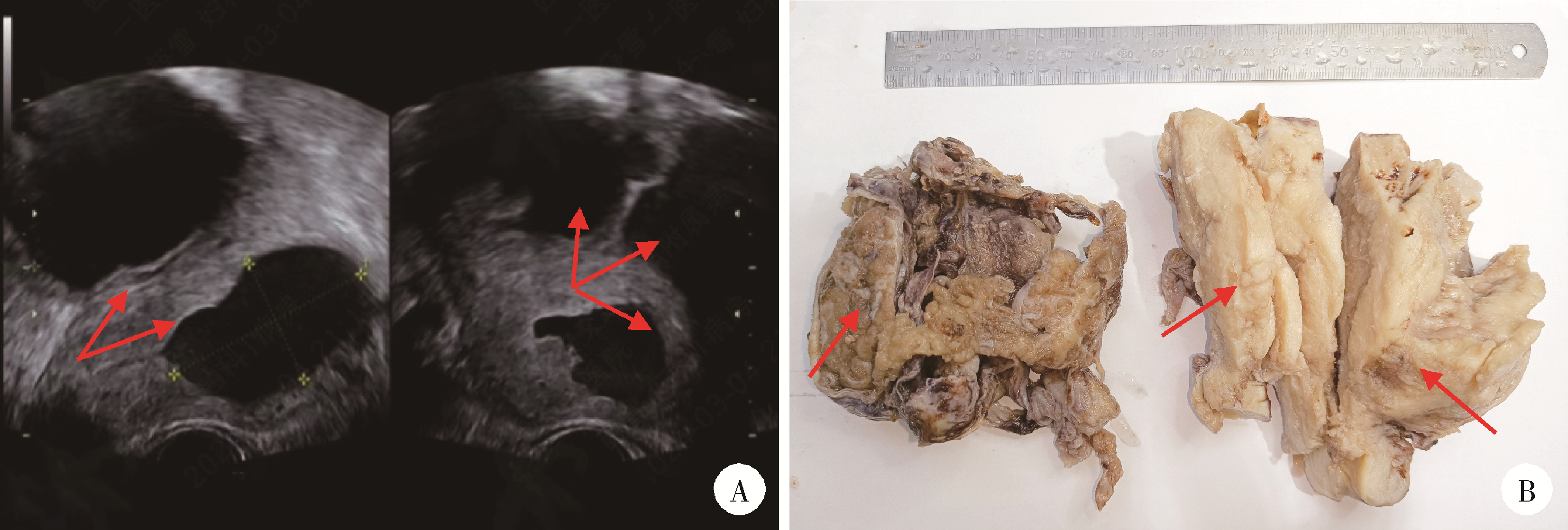
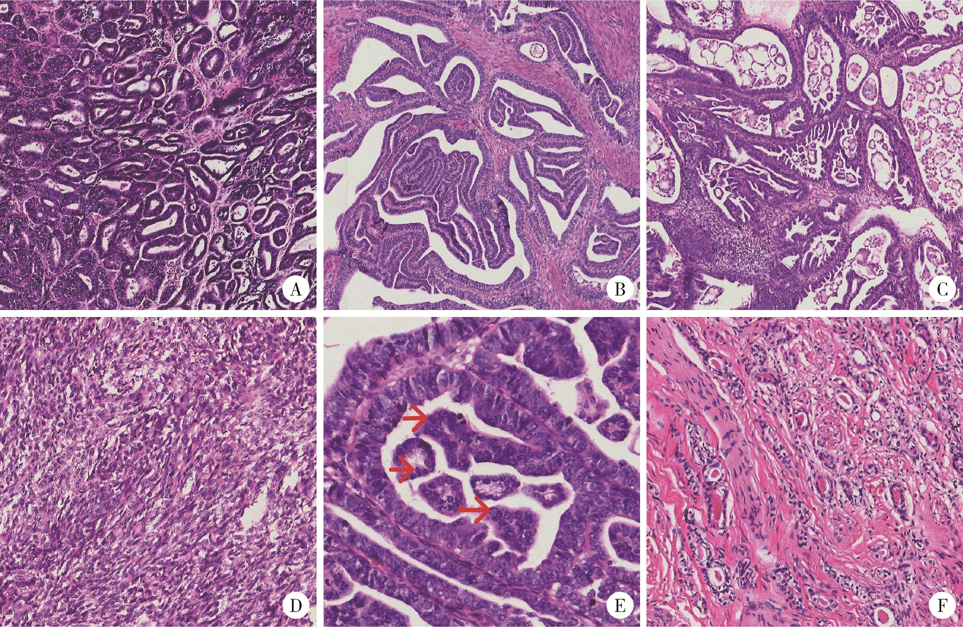
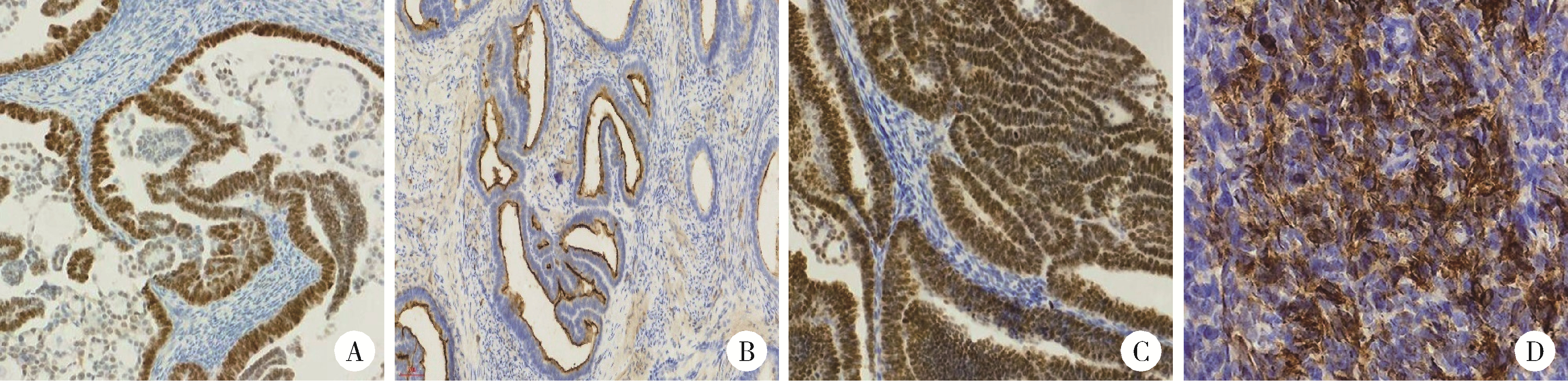
Clinicopathological analysis of mesonephric-like adenocarcinoma in the corpusuteri: A report of 3 cases

Xiaolin WANG1, Luyao LI1, Wen ZHANG2, Hongyan WANG1,*()   

  1. 1. Department of Pathology, the Frist Affiliated Hospital of Xi'an Jiaotong University, Xi'an 710061, China
    2. Department of Pathology, Northwest Women's and Children's Hospital, Xi'an 710061, China
  • Received:2023-11-29 Online:2025-11-07 Published:2025-11-07
  • Contact: Hongyan WANG

RICH HTML


Abstract:

Mesonephric-like adenocarcinoma (MLA) in the corpus uteri is a highly aggressive malignant tumor, which is easily confused with other types of endometrial cancer. When the tumor morphology and cellular characteristics are not consistent with the clinical biological behavior, attention should be paid to it. This study is to investigate the clinicopathologic features of MLA in the corpus uteri. Three cases of MLA in the corpus uteri diagnosed in the First Affiliated Hospital of Xi'an Jiaotong University from 2020 to 2023 were studied by clinical data, microscopic features and immunohistochemistry. The related literature was reviewed. The clinical manifestations of the three cases of MLA in the corpus uteri were nonspecific. One case was from our hospital and the other two cases were from other hospitals. The age range was 54-58 years. In the specimen description, there was a diffusely growing mass in the endometrium of the uterus, with an uneven surface and tough texture. The muscle wall was extensively invaded. At low magnification, the tumor cells were arranged in tubular, glandular, papillary, micropapillary and solid growth patterns. At high magnification, the cells lining the lumen were arranged in a single layer, cuboidal or columnar pattern, with mild to moderate atypia, with vesicular nuclei and nuclear furrows. Some of the lumens showed eosinophilic homogeneous pink staining without structural-like material. In the solid area, the cells were plat fusiform, arranged in bundles or whirlpools, with large nuclear atypia and frequent mitotic figures. A large number of intravascular cancer thrombus were observed in all the three cases. The tumor cells were positive for GATA3 and/or thyroid transcription factor-1 (TTF1), diffusely positive for pair box gene 2 (PAX2) and PAX8, and positive for CD10 in some luminal margins. Estrogen receptor (ER) was focal positive, and progesterone receptor (PR) was negative. KRAS mutation was detected in case 1. According to the 2023 updated International Federation of Gynecology and Obstetrics (FIGO) staging guidelines for endometrial cancer, all the three cases were in advanced stage. It is suggested that pathologists should make accurate diagnosis based on morphological manifestations, using a set of matched immunohistochemical markers and necessary molecular tests to avoid misdiagnosis and better guide clinical diagnosis and treatment.

Key words: Uterus, Mesonephric-like adenocarcinoma, Clinical pathology

CLC Number: 

  • R737.33

Figure 1

Imaging features and gross changes of case 1 A, the ultrasound image of case 1 showed mixed echogenic masses in the uterine cavity and bilateral ovaries with dark fluid areas (arrows indicate the fluid dark area of the mass); B, gross findings of case 1; The left side was the right ovary and the right side shows the whole uterus (arrows indicate the location of the mass)."

Figure 2

Microscopic features of the three patients (hematoxylin-eosin staining) A, the tumor cells were tubular or glandular (×200); B, the tumor cells were papillary (×200); C, the tumor cells were micropapillary (×200); D, the tumor cells were spindle-shaped (×200); E, vesicular nuclei and nuclear furrows (red arrows) are visible at high magnification (×400); F, luminal eosinophilic homogeneous pink staining without structured secretions (×200)."

Figure 3

Immunohistochemical staining characteristics of the three patients (EnVision) A, pair box gene 2 (PAX2) was diffusely positive in the nucleus (×200); B, the luminal margin was positive for CD10 (×200); C, thyroid transcription factor-1 (TTF1) was positive in the nucleus (×200); D, case 3 was positive for broad-spectrum CK in solid spindle cells (×200)."

Table 1

Surgical-pathological staging of the three patients"

Items Case 1 Case 2 Case 3
Depth of infiltration 2/3 myometrium Full myometrium and serosa 3/5 myometrium
Tumor emboli in blood vessels and lymphatic vessels >4 >4 >4
Limited spread Involving the cervix and both adnexa Involving both adnexa Involving the cervix
Pelvic lymph nodes Negative Negative Negative
Distant metastasis Negative Peritoneal metastasis Negative
1
Deolet E , Van Dorpe J , Van de Vijver K . Mesonephric-like adenocarcinoma of the endometrium: Diagnostic advances to spot this wolf in sheep's clothing. A review of the literature[J]. J Clin Med, 2021, 10 (4): 698.

doi: 10.3390/jcm10040698
2
Berek JS , Matias-Guiu X , Creutzberg C , et al. FIGO staging of endometrial cancer: 2023[J]. Int J Gynaecol Obstet, 2023, 162 (2): 383- 394.

doi: 10.1002/ijgo.14923
3
McFarland M , Quick CM , McCluggage WG . Hormone receptor-negative, thyroid transcription factor 1-positive uterine and ovarian adenocarcinomas: Report of a series of mesonephric-like adenocarcinomas[J]. Histopathology, 2016, 68 (7): 1013- 1020.

doi: 10.1111/his.12895
4
Pors J , Segura S , Chiu DS , et al. Clinicopathologic characteristics of mesonephric adenocarcinomas and mesonephric-like adenocarcinomas in the gynecologic tract: A multi-institutional study[J]. Am J Surg Pathol, 2021, 45 (4): 498- 506.

doi: 10.1097/PAS.0000000000001612
5
Kim HG , Kim H , Yeo MK , et al. Mesonephric-like adenocarcinoma of the uterine corpus: Comprehensive analyses of clinicopathological, molecular, and prognostic characteristics with retrospective review of 237 endometrial carcinoma cases[J]. Cancer Genomics Proteomics, 2022, 19 (4): 526- 539.

doi: 10.21873/cgp.20338
6
Pors J , Cheng A , Leo JM , et al. A comparison of GATA3, TTF1, CD10, and calretinin in identifying mesonephric and mesonephric-like carcinomas of the gynecologic tract[J]. Am J Surg Pathol, 2018, 42 (12): 1596- 1606.

doi: 10.1097/PAS.0000000000001142
7
Euscher ED , Bassett R , Duose DY , et al. Mesonephric-like carcinoma of the endometrium: A subset of endometrial carcinoma with an aggressive behavior[J]. Am J Surg Pathol, 2020, 44 (4): 429- 443.

doi: 10.1097/PAS.0000000000001401
8
Kim H , Kim HS . Mesonephric-like adenocarcinoma of the uterine corpus: Comparison between mismatch repair protein immuno-staining and microsatellite instability testing[J]. Anticancer Res, 2023, 43 (4): 1785- 1795.

doi: 10.21873/anticanres.16332
9
Mirkovic J , McFarland M , Garcia E , et al. Targeted genomic profiling reveals recurrent KRAS mutations in mesonephric-like adenocarcinomas of the female genital tract[J]. Am J Surg Pathol, 2018, 42 (2): 227- 233.

doi: 10.1097/PAS.0000000000000958
10
Howitt BE , Nucci MR . Mesonephric proliferations of the female genital tract[J]. Pathology, 2018, 50 (2): 141- 150.

doi: 10.1016/j.pathol.2017.11.084
[1] Guangyan YU, Xin PENG, Min GAO, Peng YE, Na GE, Mengqi JIA, Bingyu LI, Zunan TANG, Leihao HU, Wenbo ZHANG. Research progress in diagnosis and treatment of salivary gland tumors [J]. Journal of Peking University (Health Sciences), 2025, 57(1): 1-6.
[2] Dan LUO, Haijian HUANG, Xin CHEN, Xiaoyan CHEN. Primary uterine hepatoid adenocarcinoma: Clinicopathological analysis of 2 cases and literature review [J]. Journal of Peking University (Health Sciences), 2024, 56(6): 1126-1131.
[3] Bo-han NING,Qing-xia ZHANG,Hui YANG,Ying DONG. Endometrioid adenocarcinoma with proliferated stromal cells, hyalinization and cord-like formations: A case report [J]. Journal of Peking University (Health Sciences), 2023, 55(2): 366-369.
[4] Xue-mei HA,Yong-zheng YAO,Li-hua SUN,Chun-yang XIN,Yan XIONG. Solid placental transmogrification of the lung: A case report and literature review [J]. Journal of Peking University (Health Sciences), 2023, 55(2): 357-361.
[5] Wei-hua HOU,Shu-jie SONG,Zhong-yue SHI,Mu-lan JIN. Clinicopathological features of Helicobacter pylori-negative early gastric cancer [J]. Journal of Peking University (Health Sciences), 2023, 55(2): 292-298.
[6] Qi SHEN,Yi-xiao LIU,Qun HE. Mucinous tubular and spindle cell carcinoma of kidney: Clinicopathology and prognosis [J]. Journal of Peking University (Health Sciences), 2023, 55(2): 276-282.
[7] Qian SU,Xin PENG,Chuan-xiang ZHOU,Guang-yan YU. Clinicopathological characteristics and prognosis of non-Hodgkin lymphoma in oral and maxillofacial regions: An analysis of 369 cases [J]. Journal of Peking University (Health Sciences), 2023, 55(1): 13-21.
[8] Er-shu BO,Peng HONG,Yu ZHANG,Shao-hui DENG,Li-yuan GE,Min LU,Nan LI,Lu-lin MA,Shu-dong ZHANG. Clinicopathological features and prognostic analysis of papillary renal cell carcinoma [J]. Journal of Peking University (Health Sciences), 2022, 54(4): 615-620.
[9] Qian SU,Xin PENG,Chuan-xiang ZHOU,Guang-yan YU. Clinicopathological features and possible prognostic factors in parotid lymphomas [J]. Journal of Peking University(Health Sciences), 2019, 51(1): 35-42.
[10] HUANG Zi-xiong, DU Yi-qing, ZHANG Xiao-peng, LIU Shi-jun, XU Tao. Clinical and pathological analysis of renal cell carcinoma bone metastasis [J]. Journal of Peking University(Health Sciences), 2018, 50(5): 811-815.
[11] MU Tian, WANG Yan, LIU Guo-li, WANG Jian-liuMU Tian, WANG Yan, LIU Guo-li, WANG Jian-liu. Application of seven prediction models of vaginal birth after cesarean in a Chinese hospital [J]. Journal of Peking University(Health Sciences), 2016, 48(5): 795-800.
Viewed
Full text


Abstract

Cited

  Shared   
  Discussed   
No Suggested Reading articles found!