miR-488-5p promotes osteogenic and neurogenic differentiation of rat bone marrow mesenchymal stem cells and enhances neuralized bone regeneration

  • Liting ZENG 1 ,
  • Kaiyuan CHENG 1, 2 ,
  • Zhongning LIU 1 ,
  • Jian LI 1 ,
  • Jingwen YANG , 1, * ,
  • Ting JIANG , 1, *
Expand
  • 1. Department of Prosthodontics, Peking University School and Hospital of Stomatology & National Center for Stomatology & National Clinical Research Center for Oral Diseases & National Engineering Research Center of Oral Biomaterials and Digital Medical Devices, Beijing 100081, China
  • 2. Department Ⅰ of Prosthodontics, Tianjin Stomatological Hospital, School of Medicine, Nankai University, Tianjin Key Laboratory of Oral and Maxillofacial Function Reconstruction, Tianjin 300041, China
YANG Jingwen, e-mail,
JIANG Ting, e-mail,

Received date: 2025-10-10

  Online published: 2025-12-10

Supported by

the National Natural Science Foundation of China(82170928)

the National Natural Science Foundation of China(82201022)

Copyright

All rights reserved. Unauthorized reproduction is prohibited.

Abstract

Objective: To investigate the role of microRNA miR-488-5p, which showed increased expression after the disconnection of the inferior alveolar nerve, in promoting the osteogenic and neurogenic differentiation of rat bone marrow mesenchymal stem cells (rBMSCs), as well as its effect on promoting the neuralized tissue engineered bone regeneration. Methods: rBMSCs were subjected to in vitro neural or osteogenic differentiation induction cultures. The expression levels of miR-488-5p at different time points (days 0, 2, 4, and 7) were detected by quantitative real-time polymerase chain reaction (qRT-PCR). miR-488-5p overexpression or low expression in rBMSCs was achieved by transfection with miR-488-5p mimics or inhibitors. Four groups, the miR-488-5p mimics, the miR-488-5p inhibitor, and their respective negative controls (NC), were established to investigate the effects of miR-488-5p on the neural differentiation and osteogenic differentiation of rBMSCs.A 5 mm diameter, full-thickness circular critical bone defect was created in the rat calvaria. The rats were treated with light-cured gelatin methacryloyl (GelMA) seeded with rBMSCs. The rats were divided into four groups: ①BLANK group: GelMA; ②BMSCs group: GelMA + rBMSCs; ③NC-BMSCs group: GelMA + rBMSCs transfected with miR-488-5p mimics NC; and ④miR-488-5p-BMSCs group: GelMA + rBMSCs transfected with miR-488-5p mimics. Specimens were obtained 4 and 8 weeks after surgery, and micro-CT was performed to measure and analyze bone mineral density (BMD), bone volume/total volume (BV/TV), bone surface area/total volume (BS/TV) and trabecular number (Tb.N). The effects of neuralized tissue engineering bone formation in the defect area were assessed using Hematoxylin-Eosin (HE) staining, Masson staining, and tissue immunofluorescence staining of the nerve-specific protein soluble protein-100 (S100). Results: As rBMSCs progressed toward neural or osteogenic differentiation, miR-488-5p expression increased significantly from day 0 to day 7. Regarding neural differentiation, the mimics group showed increased expression of neural-related genes and proteins compared with the mimics NC group, while the opposite result was observed in the inhibitor group. As for osteogenic differentiation, the mimics group showed increased expression of osteogenic genes and proteins, more intense alkaline phosphatase (ALP) and alizarin red staining (ARS) staining, and enhanced ALP activity compared with the mimics NC group, while the opposite result was observed in the inhibitor group. 4 and 8 weeks after critical calvarial defect construction in rats, the BLANK group had the least amount of new bone formation, while the BMSCs group and the NC-BMSCs group had similar and intermediate amounts of new bone formation. The miR-488-5p-BMSCs group had the most new bone formation. At 4 weeks, the BMD [(0.63±0.05) g/cm3 vs. (0.51±0.03) g/cm3], BV/TV (33.17%±6.43% vs. 18.11%±1.52%), BS/TV [(3.43±0.69) /mm vs. (2.46±0.20) /mm], and Tb.N [(0.92±0.21) /mm vs.(0.59±0.07) /mm] in the miR-488-5p-BMSCs group were significantly higher than those in the NC-BMSCs group. At 8 weeks, the BMD [(0.80±0.04) g/cm3 vs. (0.68±0.04) g/cm3], BV/TV (56.69%±6.22% vs. 42.36%±3.86%), and the number of S100-labeled nerve cells around the new bone (46.33±4.04 vs. 26.00±3.61) in the miR-488-5p-BMSCs group was also significantly higher than that in the NC-BMSCs group. Conclusion: miR-488-5p promoted the osteogenic and neurogenic differentiation of rBMSCs and promoted the formation of neuralized tissue-engineered bone in rat calvarial defects.

Cite this article

Liting ZENG , Kaiyuan CHENG , Zhongning LIU , Jian LI , Jingwen YANG , Ting JIANG . miR-488-5p promotes osteogenic and neurogenic differentiation of rat bone marrow mesenchymal stem cells and enhances neuralized bone regeneration[J]. Journal of Peking University(Health Sciences), 2026 , 58(1) : 10 -21 . DOI: 10.19723/j.issn.1671-167X.2026.01.002

骨组织是一个高度动态代谢且神经支配丰富的器官,其稳态维持和再生过程与神经系统密切相关。神经系统不仅为骨骼提供感觉输入,还在骨折愈合、骨代谢以及骨组织工程中发挥关键作用[1-3]。神经在骨再生中的重要性已被广泛认可,当前的神经化骨组织工程研究手段主要通过在骨支架中加入神经成分,如神经细胞[4-5]、神经营养因子[6-7]、具有神经诱导功能的生物材料等[8-9],从而促进骨骼再生。如何能有效实现神经和骨组织协同再生是神经化骨组织工程需要解决的关键问题之一[1]。微小RNA(microRNA,miRNA)作为内源性调控分子,在成骨和神经生成过程中均具有重要调控作用,可通过靶向关键的成骨转录因子和信号通路来调节骨髓间充质干细胞的成骨分化和骨重塑过程[10-12],也可参与调控神经干细胞的分化、轴突导向、突触形成以及神经损伤后的修复过程[13]。目前,大多数研究主要关注miRNA在单一谱系(成骨或成神经)中的作用,系统性地寻找和验证一个内源性的能够在神经损伤后被激活,并同时具有促进成骨和成神经双重功能的miRNA的研究仍非常匮乏。鉴于miRNA具有广泛靶向性和精细调控能力,深入探究能同时调控神经和骨再生的miRNA,将为神经化骨组织工程提供新的分子靶点和治疗策略。三叉神经是颅面部重要的感觉神经,其分支下牙槽神经的损伤、离断,不仅会引起感觉异常和疼痛[14-15],还会影响骨组织的愈合效果和骨密度[16]。本课题组前期研究发现,离断大鼠下颌骨下牙槽神经后,其拔牙窝愈合骨质变差、骨小梁稀疏、骨密度降低、下颌皮质骨骨沉积减慢[17]。在离断下牙槽神经1周后取其上端三叉神经节进行miRNA高通量测序,发现miR-488-5p在神经离断组中高表达且差异较大,预实验进一步显示miR-488-5p可以促进大鼠骨髓间充质干细胞(rat bone marrow mesenchymal stem cells,rBMSCs)神经相关标志基因如巢蛋白(nestin,Nes)、可溶性蛋白100(soluble protein-100,S100)、Ⅲ类β-微管蛋白(class Ⅲ β-tubulin,Tubb3)的mRNA表达,提示miR-488-5p可能具有促进成神经的功能,但对成骨的作用尚未可知。因此,本研究假设miR-488-5p可能同时具有促进rBMSCs神经向分化及成骨分化的双重功能,计划通过体外研究验证miR-488-5p对rBMSCs成神经分化和成骨分化的作用,通过体内研究促进rBMSCs的miR-488-5p表达对骨缺损区骨和神经再生的影响,为神经化骨组织工程领域的研究探索新的方法。

1 材料与方法

1.1 主要材料及试剂

最低必需培养基α(minimum essential medium α,α-MEM)、改良Eagle/F12培养基(Dulbecco ’ s modified Eagle’ s medium/F12,DMEM/F12)、胎牛血清(fetal bovine serum,FBS)、青-链霉素双抗、胰蛋白酶、B27购自美国Gibco公司;成神经诱导液组分表皮生长因子(epidermal growth factor,EGF)和碱性成纤维细胞生长因子(basic fibroblast growth factor,bFGF)购自美国Peprotech公司;地塞米松、β-甘油磷酸钠、抗坏血酸购自美国Sigma-Aldrich公司;LipofectamineTM 2000转染试剂购自美国Invitrogen公司;miR-488-5p模拟物、miR-488-5p抑制剂及各自阴性对照(negative control,NC)由苏州吉玛基因公司设计合成;逆转录试剂盒、SYBR Green试剂盒及实时荧光定量聚合酶链反应(quantitative real-time polymerase chain reaction,qRT-PCR)引物购自湖南艾克瑞生物工程有限公司;动物RNA抽提试剂盒、二喹啉甲酸(bicinchoninic acid,BCA)蛋白浓度测定试剂盒、BCIP/NBT碱性磷酸酶(alkaline phosphatase,ALP)显色试剂盒购自上海碧云天生物技术有限公司;ALP定量检测试剂盒购自南京建成生物制品公司;茜素红染色液、Triton X-100溶液购自北京索莱宝科技有限公司;十六烷基吡啶粉末购自上海麦克林生化科技有限公司;兔源抗大鼠βⅢ-Tubulin抗体(A17913)、兔源抗大鼠Gap43抗体(A19055)、兔源抗大鼠Runx2抗体(A2851)、兔源抗大鼠Osx抗体(A18699)购自武汉爱博泰克生物科技有限公司;小鼠源抗大鼠S100抗体(15146-1-AP)购自武汉三鹰生物技术有限公司;小鼠源抗大鼠β-actin抗体购自北京中杉金桥生物技术有限公司;甲基丙烯酰化明胶(gelatin methacryloyl,GelMA)购自苏州永沁泉智能设备有限公司。
本研究开始前已经北京大学生物医学伦理委员会实验动物福利伦理分委员会审查批准(LA2019314),所应用的SD(Sprague Dawley)大鼠购自北京大学医学部实验动物中心。

1.2 原代rBMSCs的提取与培养

将4周龄SD大鼠断颈法处死,75%(体积分数)乙醇溶液浸泡消毒5 min后,无菌条件下分离股骨及胫骨,无菌镊子剪开胫骨、股骨干骺端,用5 mL注射器抽取含有10%(体积分数)FBS、1%(体积分数)青-链霉素双抗的α-MEM培养基,反复冲洗骨髓腔至骨壁透亮。收集冲洗出来的骨髓,以1 000 r/min离心5 min,弃掉上清液,加入组分为α-MEM培养基、10%FBS、1%青-链霉素双抗的细胞增殖培养基(proliferation medium,PM)重悬,接种于10 cm培养皿中,置于37 ℃恒温、5%(体积分数)CO2孵箱内培养,避免移动,视情况每3天换液,待细胞长满后传代,之后每2~3天使用PM进行换液,细胞培养至第2~4代用于实验。

1.3 rBMSCs成神经诱导

神经诱导培养基(neurogenic medium,NM)分为预诱导液及正式诱导液。预诱导液的配制:DMEM/F12基础培养基+ 1%青-链霉素双抗+20 μg/L EGF+20 μg/L bFGF+2%(体积分数)B27。将预诱导液加入接种了细胞的孔板中,每2~3天换液,预诱导7 d后加入成神经正式诱导液(DMEM/F12基础培养基+1%青-链霉素双抗+10%FBS +10 μg/L EGF+10 μg/L bFGF+2%B27),每2~3天换液,成神经诱导5 d后收集细胞。

1.4 rBMSCs成骨诱导

配制成骨诱导培养基(osteogenic medium,OM):α-MEM基础培养基+1%青-链霉素双抗+10% FBS +100 nmol/L地塞米松+10 mmol/L β-甘油磷酸钠+50 μg/mL抗坏血酸。在接种了细胞的孔板中加入OM,每2~3天换液,成骨诱导至第7天、第21天收集细胞。

1.5 miRNA转染

将rBMSCs以一定密度接种至孔板内,使第2天转染时细胞汇合度达到约70%。按照LipofectamineTM 2000试剂盒说明书进行转染,以六孔板为例,转染前使用不含青-链霉素的10%FBS的α-MEM培养基换液,每孔2 mL培养基,分别使用120 μL无血清培养基以200 pmol/孔的终浓度稀释miR-488-5p模拟物、模拟物阴性对照、miR-488-5p抑制剂及抑制剂阴性对照,混匀,随后加入12 μL LipofectamineTM 2000转染试剂,吹打,室温孵育10 min后将混合液加入六孔板内,将孔板置于细胞孵育箱中继续培养,转染6~8 h后换液。

1.6 RNA提取、逆转录与qRT-PCR

收集细胞后使用动物RNA抽提试剂盒提取RNA,通过分光光度法进行定量,使用逆转录试剂盒合成cDNA。使用SYBR Green试剂盒进行qRT-PCR,分别以GapdhU6为内参基因,检测成神经相关标志物基因(包括NesS100Tubb3)、微管相关蛋白2(microtubule-associated protein 2,Map2)、神经/胶质细胞抗原2(neural/glial antigen 2,Ng2)、神经生长因子(nerve growth factor,Ngf)及成骨相关标志物基因包括Runt相关转录因子2(runt-related transcription factor 2,Runx2)、碱性磷酸酶(alkaline phosphatase,Alp)、Ⅰ型胶原α1链(collagen type Ⅰ alpha 1 chain,Col1a1)、骨桥蛋白(osteopontin,Opn)、骨形态发生蛋白2(bone morphogenetic protein 2,Bmp2)、骨钙素(osteocalcin,Ocn)的mRNA和miR-488-5p的表达情况,实验结果以2-ΔΔCt相对表达法进行分析。引物序列见表 1
表1 qRT-PCR引物序列

Table 1 Primer sequences used for qRT-PCR

Gene Forward primer (5′ to 3′) Reverse primer (5′ to 3′)
U6 CGCTTCGGCAGCACATATAC CGAATTTGCGTGTCATCCTT
miR-488-5p GGCACCCAGATAATGGCAC AGTGCAGGGTCCGAGGTATT
Gapdh AGGTCGGTGTGAACGGATTT GAACTTGCCGTGGGTAGAGT
*Map2 ACAGCAACAAGTGGTGAATCAG GGAGGATGGAGGAAGGTCTTG
*Ng2 TTCTCACACAGAGGAGCCC CACTCAAGCTCTGGCTGCT
*Tubb3 CAGATGCTGGCCATTCAGAGTAAG TGTTGCCGATGAAGGTGGAC
*Ngf TGCCAAGGACGCAGCTTTC TGAAGTTTAGTCCAGTGGGCTTCAG
*Nes TGAACAAGAGACCCAACAAACAC TTCCAAGAGGCTTCGGTAACT
*S100 CTGTCAAGAACCTGCTCCGA AGTGGGCATGGAACACATTGA
#Runx2 GCCTTCAAGGTTGTAGCCCT TGAACCTGGCCACTTGGTTT
#Alp CATGGTGAGTGACACGGACA CCATGACGTGGGGGATGTAG
#Ocn CCGTTTAGGGCATGTGTTGC TTTCGAGGCAGAGAGAGGGA
#Bmp2 CGGGAACAAATGCAGGAAGC AAGGACATTCCCCATGGCAG
#Col1a1 CCCCAGCCGCAAAGAGTCTA CAGCTGACTTCAGGGATGTCTTC
#Opn CCAGCCAAGGACCAACTACA AGTGTTTGCTGTAATGCGCC

* neurogenic-related genes; # osteogenic-related genes; qRT-PCR, quantitative real-time polymerase chain reaction; Map2, microtubule-associated protein 2; Ng2, neural/glial antigen 2; Tubb3, Class Ⅲ β-tubulin; Ngf,nerve growth factor;Nes, nestin; S100, soluble protein-100; Runx2, runt-related transcription factor 2; Alp, alkaline phosphatase; Ocn, osteocalcin; Bmp2, bone morphogenetic protein 2; Col1a1, collagen type Ⅰ alpha 1 chain; Opn, osteopontin.

1.7 蛋白免疫印迹实验

收集细胞并使用裂解液提取总蛋白,使用BCA蛋白浓度测定试剂盒测定总蛋白浓度,在蛋白样品中加入上样缓冲液,煮沸,进行电泳、转印,使用5%(质量分数)脱脂奶粉封闭1 h,按说明书推荐比例稀释一抗,4 ℃孵育过夜,再按说明书推荐比例稀释二抗,室温孵育1 h,之后显色、曝光。

1.8 细胞免疫荧光实验

接种的细胞使用4%(体积分数)多聚甲醛固定30 min、0.1%(体积分数)的TritonX-100溶液室温透化细胞20 min、5%(体积分数)牛血清白蛋白封闭30 min。按照说明书推荐比例稀释一抗,4 ℃孵育过夜。避光条件下使用荧光二抗孵育1 h、DAPI染细胞核约20 min,后续利用荧光显微镜避光条件下采集图像,使用ImageJ软件对细胞荧光强度进行定量分析。

1.9 ALP染色与定量实验

在对rBMSCs成骨诱导7 d后,用4%多聚甲醛固定细胞20 min,使用BCIP/NBT碱性磷酸酶显色试剂盒进行ALP染色。向孔板中加入1%TritonX-100溶液裂解细胞并收集至EP管中,使用BCA蛋白浓度测定试剂盒和ALP定量检测试剂盒对ALP活性进行定量分析。

1.10 茜素红矿化结节染色(alizarin red staining,ARS)与半定量实验

在对rBMSCs成骨诱导21 d后,用4%多聚甲醛固定细胞20 min,使用茜素红染液染色5 min及以上,观察显色效果,当达到最佳显色效果时,吸净染液,蒸馏水冲洗去净非特异性着色。配制10 mmol/L十六烷基吡啶溶液,在染色后的各孔板中加入十六烷基吡啶溶液,室温下摇床轻晃溶解1 h,依次吸取100 μL溶解液至96孔板中,使用全波长酶标仪测量样品在562 nm波长下的光密度。

1.11 大鼠颅骨缺损模型构建与实验分组

将32只6周龄雄性SD大鼠随机分为4组:①BLANK组:单纯GelMA;②BMSCs组:GelMA+未转染rBMSCs;③NC-BMSCs组:GelMA+转染miR-488-5p模拟物NC的rBMSCs;④miR-488-5p-BMSCs组:GelMA+转染miR-488-5p模拟物的rBMSCs。每组8只大鼠(各组又分为术后4周组及术后8周组,每个时间点组各4只大鼠)。依照实验分组,于术前按照GelMA说明书配制GelMA溶液,使用细菌过滤器除菌,备用。按照第1.5小节中的方法提前制备不同组的细胞:来源于同一代的未进行转染操作的rBMSCs、转染miR-488-5p模拟物组NC的rBMSCs及转染miR-488-5p模拟物组的rBMSCs,培养48 h待转染效果稳定后,胰蛋白酶消化细胞,1 000 r/min离心5 min,弃上清液,以5×10⁶个细胞/mL的浓度重悬于GelMA光固化水凝胶溶液中,用于后续动物实验。
使用1%(体积分数)戊巴比妥钠溶液按照50 mg/kg体重腹腔注射麻醉,在颅顶骨区备皮,碘伏和酒精消毒,铺巾,使用无菌手术刀沿头盖骨正中线纵向切开皮肤形成约3 cm的切口,钝性分离皮下组织及骨膜达骨面,充分暴露术区,用直径5 mm环钻于远离颅缝处左右对称制造圆形全层骨缺损,注意不损伤深层脑组织和血管,随后在骨缺损处注射20 μL不负载或负载不同组细胞的GelMA光固化水凝胶,使用光强为10 mW/cm2的405 nm紫外灯下照射10 s,检查水凝胶固化和在位,无明显移动。逐层缝合关闭创口,肌肉注射8万单位青霉素预防术后感染。

1.12 显微计算机断层扫描术(micro computed tomography,micro-CT)与骨参数分析

分别于手术后4周、8周,采用二氧化碳过量麻醉法处死各组大鼠,取出颅骨术区标本,使用10%(体积分数)中性甲醛溶液固定48 h后,进行micro-CT扫描,使用三维可视化软件对获得的micro-CT图像进行三维重建,使用CTan、CTVol等软件进行定量分析,测量骨矿物质密度(bone mineral density,BMD)、骨体积分数(bone volume/total volume,BV/TV)、骨表面积密度(bone surface area/total volume,BS/TV)和骨小梁数量(trabecular number,Tb.N)。

1.13 组织形态学分析

将进行micro-CT扫描后的颅骨标本进行脱钙、包埋、切片后,进行苏木精-伊红(Hematoxylin-Eosin,HE)染色、Masson染色及神经特异性蛋白S100的组织免疫荧光染色,在显微镜下观察和拍照,后续使用ImageJ软件标记出Masson染色切片中缺损两端的新生骨组织并计算新生骨面积,使用ImageJ软件对S100免疫荧光染色标记的阳性细胞数量进行定量分析。

1.14 统计学分析

采用GraphPad Prism 9.0软件,计量资料使用Shapiro-Wilk检验数据均服从正态分布,以均数±标准差表示,对于两组样本比较采用双侧独立样本t检验,多组比较采用单因素方差分析结合Tukey事后检验进行组间比较,该检验已对多重性进行校正,结果的P值均为经Turkey校正后的P值,P<0.05认为差异有统计学意义。

2 结果

2.1 miR-488-5p在rBMSCs成神经分化过程中表达上调
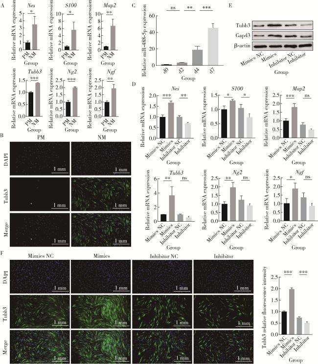
对rBMSCs进行神经向分化诱导12 d后,检测成神经诱导是否成功,qRT-PCR结果显示,NM组的神经相关标志物基因Map2NesNg2NgfS100Tubb3的表达水平显著高于未进行诱导的PM组(图 1A);免疫荧光染色结果进一步显示,NM组Tubb3蛋白荧光强度明显高于PM组(图 1B),表明神经向分化诱导成功。
图1 miR-488-5p促进rBMSCs神经向分化

Figure 1 miR-488-5p promotes neurogenic differentiation of rBMSCs

A, mRNA expression levels of neurogenic-related genes Nes, S100, Map2, Ng2, Tubb3, Ngf on day 12 of neurogenic differentiation in the NM and PM groups as detected by qRT-PCR; B, immunofluorescence detection of Tubb3 in the NM and PM groups; C, miR-488-5p expression levels on days 0, 2, 4 and 7 of neurogenic differentiation as detected by qRT-PCR; D, mRNA expression levels of neurogenic-related genes Nes, S100, Map2, Ng2, Tubb3, Ngf on day 12 of neurogenic differentiation as detected by qRT-PCR; E, protein expression levels of Tubb3 and Gap43 as detected by Western blot; F, immunofluorescence detection of Tubb3 and quantification of Tubb3 fluorescence intensity. ns, not significant. * P < 0.05, * * P < 0.01, * * * P < 0.001. PM, proliferation medium; NM, neurogenic medium; Mimics NC, negative control for miR-488-5p mimics; Inhibitor NC, negative control for miR-488-5p inhibitor; Nes, nestin; S100, soluble protein-100; Map2, microtubule-associated protein 2; Ng2, neural/glial antigen 2; Tubb3, class Ⅲ β-tubulin; Ngf, nerve growth factor; Gap43, growth associated protein 43;DAPI, 4', 6-diamidino-2-phenylindole; rBMSCs, rat bone marrow mesenchymal stem cells; qRT-PCR, quantitative real-time polymerase chain reaction.

在确认rBMSCs神经向分化模型建立有效的基础上,通过qRT-PCR检测miR-488-5p在神经诱导过程第0、2、4、7天的表达变化,发现随着神经向分化进程的推进,miR-488-5p的表达水平显著上升(F=109.3,P < 0.001,图 1C)。

2.2 miR-488-5p促进rBMSCs成神经分化

第2.1小节的结果提示miR-488-5p可能参与rBMSCs成神经分化的正向调控,为了进一步验证,通过向rBMSCs中分别转染miR-488-5p模拟物、miR-488-5p抑制剂以及各自的阴性对照,以过表达或抑制miR-488-5p。经神经向诱导12 d后,检测各组成神经相关基因及蛋白的表达情况,发现相比于模拟物阴性对照组,miR-488-5p模拟物组的成神经相关标志物基因包括Nes(P < 0.001)、S100 (P < 0.05)、Map2 (P < 0.001)、Tubb3 (P < 0.01)、Ng2 (P < 0.01)及Ngf(P < 0.05)的表达均显著上调(图 1D),Tubb3、神经生长相关蛋白43(growth associated protein 43,Gap43)表达增加(图 1E),细胞免疫荧光实验结果进一步证实了模拟物组的Tubb3蛋白表达显著增强(P < 0.001,图 1F);相反,抑制剂组的Nes(P < 0.01)、S100 (P < 0.05)基因表达水平显著低于其对照组,Tubb3、Gap43蛋白表达减少,Tubb3蛋白荧光强度更弱(P < 0.001)。

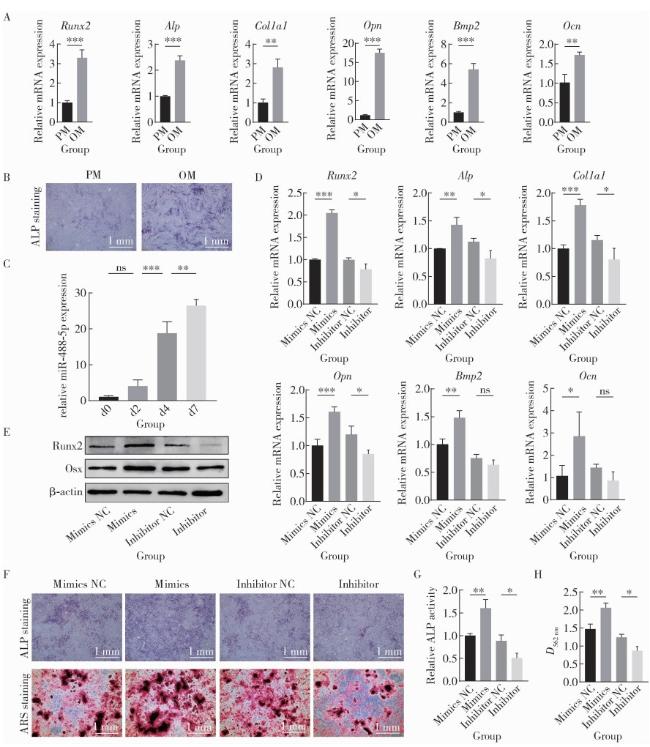
2.3 miR-488-5p在rBMSCs成骨分化过程中表达上调

对rBMSCs进行成骨分化诱导7 d后,检测成骨诱导是否成功,qRT-PCR结果显示(图 2),OM组的成骨相关标志物基因Runx2ALPCol1a1OpnBmp2Ocn的表达水平均显著高于PM组(图 2A);ALP染色结果显示,OM组较PM组染色更深(图 2B),表明成骨向分化诱导成功。
图2 miR-488-5p促进rBMSCs成骨向分化

Figure 2 miR-488-5p promotes osteogenic differentiation of rBMSCs

A, mRNA expression levels of osteogenic-related genes Runx2, Alp, Col1a1, Opn, Bmp2, Ocn on day 7 of osteogenic differentiation in the OM and PM groups as detected by qRT-PCR; B, ALP staining on day 7 of osteogenic differentiation in the OM and PM groups; C, miR-488-5p expression levels on days 0, 2, 4 and 7 of osteogenic differentiation as detected by qRT-PCR; D, mRNA expression levels of osteogenic-related genes Runx2, Alp, Col1a1, Opn, Bmp2, Ocn on day 7 of osteogenic differentiation as detected by qRT-PCR; E, protein expression levels of Runx2 and Osx as detected by Western blot; F, ALP staining on day 7 and ARS staining on day 21 of osteogenic differentiation; G, ALP activity on day 7 of osteogenic differentiation; H, quantification of mineralization by measuring ARS absorbance at 562 nm. ns, not significant. * P < 0.05, * * P < 0.01, * * * P < 0.001. PM, proliferation medium; OM, osteogenic medium; d, day; Mimics NC, negative control for miR-488-5p mimics; Inhibitor NC, negative control for miR-488-5p inhibitor; Runx2, Runt-related transcription factor 2; ALP, alkaline phosphatase; Ocn, Osteocalcin; Bmp2, bone morphogenetic protein 2; Col1a1, collagen type Ⅰ alpha 1 chain; Opn, osteopontin; ARS, alizarin red S; rBMSCs, rat bone marrow mesenchymal stem cells; qRT-PCR, quantitative real-time polymerase chain reaction.

在确认rBMSCs成骨分化模型建立有效的基础上,通过qRT-PCR检测miR-488-5p在成骨诱导过程第0、2、4、7天的表达水平变化,发现随着成骨分化进展,miR-488-5p的表达水平显著上升(F=89.37,P < 0.001,图 2C)。

2.4 miR-488-5p促进rBMSCs成骨分化

第2.3小节的结果提示miR-488-5p可能参与rBMSCs成骨分化的正向调控。在成骨诱导第7天,与模拟物阴性对照组相比,miR-488-5p模拟物组的成骨相关标志物基因Runx2 (P < 0.001)、Alp(P < 0.01)、Col1a1 (P < 0.001)、Opn(P < 0.001)、Bmp2 (P < 0.01)及Ocn(P < 0.05)表达显著升高(图 2D),Runx2、Osx蛋白表达水平也显著升高(图 2E)。此外,模拟物组的ALP染色更深(图 2F),ALP活性更强(P < 0.01,图 2G),在成骨诱导第21天,茜素红染色显示模拟物组形成了更多的矿化结节(图 2F2HP < 0.01)。相反,抑制剂组的Runx2 (P < 0.05)、Alp(P < 0.05)、Col1a1 (P < 0.05)、Opn(P < 0.05)基因表达水平及Runx2、Osx蛋白表达水平显著低于对照组,ALP染色更浅,ALP活性更弱(P < 0.05),形成的矿化结节也更少(P < 0.05)。

2.5 过表达miR-488-5p促进大鼠临界尺寸颅骨缺损神经化组织工程骨形成
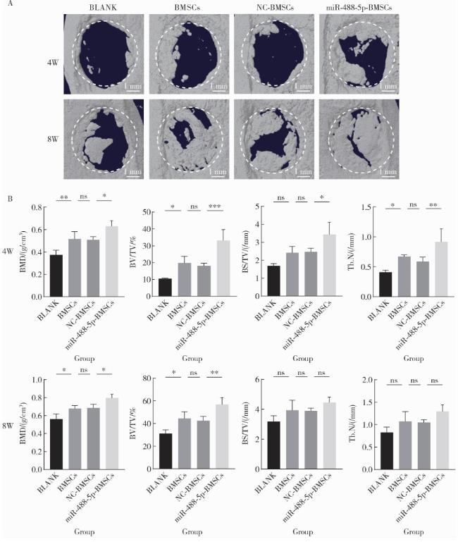
通过构建大鼠临界颅骨缺损模型进一步探究过表达miR-488-5p能否在体内促进骨缺损区域神经化的骨组织再生,利用商品化的GelMA水凝胶作为支架以辅助rBMSCs在骨缺损位置发挥作用。手术后4周和8周收集标本,micro-CT扫描结果(图 3A)显示,BLANK组骨缺损范围最大;BMSCs组和NC-BMSCs组缺损范围相近,且较BLANK组明显缩小,表明rBMSCs的转染操作未产生明显细胞毒性,且与未负载细胞的GelMA组相比,使用GelMA负载BMSCs的治疗方法促进了骨缺损愈合;miR-488-5p-BMSCs组的缺损范围最小,至术后8周,绝大部分骨缺损区已经被新生骨组织充填,表明miR-488-5p可以有效促进骨生成。对micro-CT扫描后的图像进行骨参数量化分析(图 3B),进一步证实了上述结果,术后4周,BMSCs组的BMD(P < 0.01)、BV/TV(P < 0.05)及Tb.N(P < 0.05)显著高于BLANK组,与NC-BMSCs组比较差异无统计学意义(P > 0.05),miR-488-5p-BMSCs组的BMD(P < 0.05)、BV/TV(P < 0.001)、BS/TV(P < 0.05)及Tb.N(P < 0.01)显著高于NC-BMSCs组;术后8周,BMSCs组的BMD、BV/TV显著高于BLANK组(P < 0.05),与NC-BMSCs组比较差异无统计学意义(P>0.05),miR-488-5p-BMSCs组的BMD(P < 0.05)及BV/TV(P < 0.01)显著高于NC-BMSCs组。
图3 大鼠颅骨缺损术后4周和8周micro-CT影像学分析

Figure 3 micro-CT imaging analysis of rat calvarial defects at 4 and 8 weeks after operation

A, 3D reconstructed images after micro-CT scanning showing bone defect areas at both 4 and 8 weeks after operation; B, quantitative comparisons of BMD, BV/TV, BS/TV, Tb.N at both 4 and 8 weeks after operation. ns, not significant. * P < 0.05, * * P < 0.01, * * * P < 0.001. W, weeks after operation. BLANK, the control group treated with GelMA only; BMSCs, the group treated with GelMA and normal rBMSCs; NC-BMSCs, the group treated with GelMA and rBMSCs transfected with negative control (NC) for miR-488-5p mimics; miR-488-5p-BMSCs, the group treated with GelMA and rBMSCs transfected with miR-488-5p mimics; BMD, bone mineral density; BV/TV, bone volume/total volume; BS/TV, bone surface area/total volume; Tb.N, trabecular number; 3D, 3-dimensional; BMSCs, bone marrow mesenchymal stem cells.

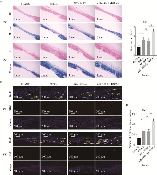
HE和Masson染色的结果显示,颅骨缺损术后4周(图 4A),BLANK组仅缺损边缘可见极少量新生骨质,缺损中央主要为纤维结缔组织,BMSCs组及NC-BMSCs组有少量新骨形成,而miR-488-5p-BMSCs组已有大量新生游离骨;术后8周,所有组均较同组术后4周结果表现出更多骨愈合,BLANK组新生骨面积最小,BMSCs组新生骨面积显著大于BLANK组[(1.65±0.31) mm2 vs. (0.87±0.19) mm2P < 0.05,图 4B],与NC-BMSCs组比较,差异无统计学意义[(1.65±0.31) mm2 vs. (1.51±0.30) mm2P>0.05],而miR-488-5p-BMSCs组的新生骨面积显著大于NC-BMSCs组[(2.53±0.26) mm2 vs. (1.51±0.30) mm2P < 0.01],且骨缺损区域大部分已被相对完整的新生骨覆盖。
图4 大鼠颅骨缺损术后4周和8周组织形态学分析

Figure 4 Histomorphological analysis of rat calvarial defects at 4 and 8 weeks after operation

A, HE staining and Masson staining; B, quantitative measurement of new bone area in Masson staining at 8 weeks after operation; C, immunofluorescence detection of S100 protein; D, analysis of the number of S100-positive cells in immunofluorescence staining at 8 weeks after operation. HE, Hematoxylin-Eosin; NB, new bone; S100, soluble protein-100; DAPI, 4', 6-diamidino-2-phenvlindole; W, weeks after operation; BLANK, the control group treated with GelMA only; BMSCs, the group treated with GelMA and normal rBMSCs; NC-BMSCs, the group treated with GelMA and rBMSCs transfected with negative control (NC) for miR-488-5p mimics; miR-488-5p-BMSCs, the group treated with GelMA and rBMSCs transfected with miR-488-5p mimics; BMSCs, bone marrow mesenchymal stem cells. ns, not significant; * P < 0.05, * * P < 0.01.

神经标志物蛋白S100可用于标记神经胶质细胞,其组织免疫荧光染色结果(图 4C)显示,在术后8周BLANK组被标记的神经细胞数量最少,BMSCs组的神经细胞数量显著多于BLANK组(26.67±7.57 vs. 12.67±2.52,P < 0.05,图 4D),且与NC-BMSCs组比较,差异无统计学意义(26.67±7.57 vs. 26.00±3.61,P>0.05),miR-488-5p-BMSCs组被标记的神经细胞数量显著多于NC-BMSCs组(46.33±4.04 vs. 26.00±3.61,P < 0.01)。以上结果表明,miR-488-5p有效促进了大鼠临界颅骨缺损区域神经化组织工程骨的形成。

3 讨论

骨代谢与神经调控并不是互相独立的过程,而是在骨微环境中相互作用[18]。本研究结果表明,miR-488-5p对rBMSCs的成骨分化和成神经分化均有正向调控作用,验证了最初的假设。促进rBMSCs中miR-488-5p的表达,可促进其在体外成骨和成神经分化,以及体内的神经化组织工程骨形成。
本研究在离断大鼠下牙槽神经后的miRNA测序中发现miR-488-5p表达上调,提示miR-488-5p可能参与神经损伤的修复和再生过程,但是其上调是参与修复的主动升高或是继发于损伤的被动反应尚不明确。进一步研究发现,miR-488-5p在rBMSCs成神经和成骨分化的过程中表达也显著上调,从第0天至第7天其表达量增加了20~40倍,但以往未见关于miR-488-5p在神经和骨方面的研究报道,miR-488-5p的上调也可能是主动性地促进rBMSCs成骨或成神经,亦或仅仅是伴随现象。
为了验证miR-488-5p是否具有促进rBMSCs成神经及成骨的作用,本研究体外实验结果表明,过表达miR-488-5p能主动增强rBMSCs的成骨与成神经分化能力;反之,抑制其表达则削弱了这种分化潜能。这证实了miR-488-5p同时具有促进rBMSCs成骨和成神经分化的双重功能,其表达上调是功能性的,而非伴随现象。体内研究表明,在大鼠颅骨缺损模型中,在缺损局部植入过表达miR-488-5p的rBMSCs,能显著促进缺损区域的新生骨与新生神经的形成,最终促进骨愈合。这一结果表明,在体内环境中miR-488-5p的上调具有积极的保护性与修复性意义。此外,本研究也证实了miR-488-5p在神经损伤后的表达上调是修复神经损伤的主动表现。
miRNA通过靶向不同基因,在多种细胞和生理过程中发挥关键调控作用,如miR-222通过靶向Pten(phosphatase and tensin homolog)促进坐骨神经离断后成年背根神经节神经突的生长[19],又通过靶向NLK(nemo-like kinase)促进BMSCs神经向分化[20]。已有研究证实Tet3 (tet methylcytosine dioxygenase 3)是miR-488-5p的靶基因之一[21]Tet3可抑制Wnt信号通路[22],有研究发现miR-26b-5p通过靶向Tet3促进BMSCs的成骨向分化[23],这提示Tet3可能是miR-488-5p调控BMSCs成骨或成神经分化的潜在靶点之一,课题组今后可进一步研究与验证。与此同时,深入研究miR-488-5p在神经损伤修复过程中的调控机制,包括其在神经损伤后的表达时空变化,是否在某种发挥修复功能的靶细胞中特异性地表达上调等,对于完整揭示miR-488-5p的生物学功能及其发挥功能的具体机制也至关重要。
BMSCs具有分化为骨细胞、软骨细胞、脂肪细胞、神经细胞和肌细胞等多种类型细胞的分化潜能,作为骨组织工程的种子细胞已被广泛研究和运用[24-25],也是神经化骨组织工程的理想种子细胞。本研究采用GelMA作为支架组织负载BMSCs作为治疗骨缺损的手段。GelMA水凝胶因其生物相容性好,可降解性和仿细胞外基质特性等优点,已广泛用于骨组织工程[26-28]。使用GelMA负载BMSCs的骨组织工程手段已被证实可以有效促进骨再生[29-30],本研究将其运用于神经化骨组织工程中,利用转染了miR-488-5p的BMSCs作为骨缺损局部骨和神经再生早期的种子细胞,希望能同时诱导rBMSCs发生神经分化和成骨分化,研究结果显示有效促进了大鼠颅骨缺损处的神经再生和骨再生,实现了较好的神经化组织工程骨效果。鉴于体内骨愈合过程的复杂性,其中的具体机制尚待深入研究。
在口腔领域,种植体植入颌骨形成骨结合后,可恢复一定程度的精细感觉,称为“骨感知”[31],通过促进种植体周围神经再生对于改善种植体骨感知和种植体长期稳定尤为必要。本课题组未来研究可探索如何将miR-488-5p运用于种植体表面骨整合,以及其对种植体周围骨再生、神经再生以及骨感知的影响,为临床治疗提供新思路。
综上所述,本研究发现并验证了miR-488-5p同时具有促进rBMSCs成骨及成神经的作用,并证实其可促进骨缺损区域神经化骨再生,为神经化骨组织工程领域的研究提供了新的干预靶点和研究思路,具有治疗牙槽骨缺损、颌面部创伤、大型骨缺损,乃至伴有神经损伤的骨科疾病的潜在应用价值,但有关分子机制及应用手段还需进一步研究。

利益冲突  所有作者均声明不存在利益冲突。

作者贡献声明  曾立婷:设计、实施实验,收集、整理数据,撰写论文;程凯远:提供关键研究基础和初步数据;刘中宁:提供研究思路及指导实验;李健:指导数据处理及审核数据;杨静文:总体把关和修改论文,经费支持;姜婷:总体把关和审定论文,设计研究方案,经费支持。所有作者均参与论文修改,并对最终文稿进行审读和确认。

1
Sun W, Ye B, Chen S, et al. Neuro-bone tissue engineering: Emerging mechanisms, potential strategies, and current challenges[J]. Bone Res, 2023, 11(1): 65.

DOI

2
Liu S, Liu S, Li S, et al. Nerves within bone and their application in tissue engineering of bone regeneration[J]. Front Neurol, 2022, 13, 1085560.

3
Zhao X, Yao M, Wang Y, et al. Neuroregulation during bone formation and regeneration: mechanisms and strategies[J]. ACS Appl Mater Interfaces, 2025, 17(5): 7223- 7250.

DOI

4
Jones RE, Salhotra A, Robertson KS, et al. Skeletal stem cell-Schwann cell circuitry in mandibular repair[J]. Cell Rep, 2019, 28(11): 2757. e5- 2766. e5.

5
Zhang X, Jiang X, Jiang S, et al. Schwann cells promote prevascularization and osteogenesis of tissue-engineered bone via bone marrow mesenchymal stem cell-derived endothelial cells[J]. Stem Cell Res Ther, 2021, 12, 382.

DOI

6
Fitzpatrick V, Moldes ZM, Deck A, et al. Functionalized 3D-printed silk-hydroxyapatite scaffolds for enhanced bone regeneration with innervation and vascularization[J]. Biomaterials, 2021, 276, 120995.

DOI

7
Zhang Z, Wang F, Huang X, et al. Engineered sensory nerve guides self-adaptive bone healing via NGF-TrkA signaling pathway[J]. Adv Sci (Weinh), 2023, 10(10): 2206155.

DOI

8
Heng BC, Bai Y, Li X, et al. Electroactive biomaterials for facilitating bone defect repair under pathological conditions[J]. Adv Sci (Weinh), 2022, 10(2): 2204502.

9
Deng L, Hou M, Lv N, et al. Melatonin-encapsuled silk fibroin electrospun nanofibers promote vascularized bone regeneration through regulation of osteogenesis-angiogenesis coupling[J]. Mater Today Bio, 2024, 25, 100985.

DOI

10
Zhao W, Zhang S, Wang B, et al. Runx2 and microRNA regulation in bone and cartilage diseases[J]. Ann N Y Acad Sci, 2016, 1383(1): 80- 87.

DOI

11
Fang S, Deng Y, Gu P, et al. microRNAs regulate bone development and regeneration[J]. Int J Mol Sci, 2015, 16(4): 8227- 8253.

DOI

12
Liu J, Dang L, Wu X, et al. microRNA-mediated regulation of bone remodeling: A brief review[J]. JBMR Plus, 2019, 3(9): 10213.

DOI

13
Nguyen LH, Diao HJ, Chew SY. MicroRNAs and their potential therapeutic applications in neural tissue engineering[J]. Adv Drug Deliv Rev, 2015, 88, 53- 66.

DOI

14
Li YK, Zhang YY, Lin J, et al. Metabotropic glutamate receptor 5-mediated inhibition of inward-rectifying K+ channel 4.1 contributes to orofacial ectopic mechanical allodynia following inferior alveolar nerve transection in male mice[J]. J Neurosci Res, 2023, 101(7): 1170- 1187.

DOI

15
Lee J, Ohara K, Shinoda M, et al. Involvement of satellite cell activation via nitric oxide signaling in ectopic orofacial hypersensitivity[J]. Int J Mol Sci, 2020, 21(4): 1252.

DOI

16
Xu Y, Xia M, Chen T, et al. Inferior alveolar nerve transection disturbs innate immune responses and bone healing after tooth extraction[J]. Ann N Y Acad Sci, 2019, 1448(1): 52- 64.

DOI

17
程凯远, 张美子, 刘中宁, 等. 下牙槽神经离断对下颌骨骨质和拔牙创愈合的影响研究[J]. 口腔颌面修复学杂志, 2025, 26(2): 105- 115.

18
Wan QQ, Qin WP, Ma YX, et al. Crosstalk between bone and nerves within bone[J]. Adv Sci (Weinh), 2021, 8(7): 2003390.

DOI

19
Zhou S, Shen D, Wang Y, et al. microRNA-222 Targeting PTEN promotes neurite outgrowth from adult dorsal root ganglion neurons following sciatic nerve transection[J]. PLoS One, 2012, 7(9): 44768.

DOI

20
Lei L, Liu Z, Yuan P, et al. Injectable colloidal hydrogel with mesoporous silica nanoparticles for sustained co-release of micro-RNA-222 and aspirin to achieve innervated bone regeneration in rat mandibular defects[J]. J Mater Chem B, 2019, 7(16): 2722- 2735.

DOI

21
Qiu J, Wu S, Wang P, et al. miR-488-5p mitigates hepatic stellate cell activation and hepatic fibrosis via suppressing TET3 expression[J]. Hepatol Int, 2022, 17(2): 463- 475.

22
Li X, Yue X, Pastor WA, et al. Tet proteins influence the balance between neuroectodermal and mesodermal fate choice by inhibiting Wnt signaling[J]. Proc Natl Acad Sci USA, 2016, 113(51): E8267- E8276.

23
Ye W, Zhang C, Fan Z. MiR-26b-5p/TET3 regulates the osteogenic differentiation of human bone mesenchymal stem cells and bone reconstruction in female rats with calvarial defects[J]. Mol Biol Rep, 2024, 51(1): 632.

DOI

24
Leo AJ, Grande DA. Mesenchymal stem cells in tissue engineering[J]. Cells Tissues Organs, 2006, 183(3): 112- 122.

DOI

25
Liu Y, Peng L, Li L, et al. 3D-bioprinted BMSC-laden biomimetic multiphasic scaffolds for efficient repair of osteochondral defects in an osteoarthritic rat model[J]. Biomaterials, 2021, 279, 121216.

DOI

26
Yu H, Luo X, Li Y, et al. Advanced hybrid strategies of GelMA composite hydrogels in bone defect repair[J]. Polymers (Basel), 2024, 16(21): 3039.

DOI

27
Zhou B, Jiang X, Zhou X, et al. GelMA-based bioactive hydrogel scaffolds with multiple bone defect repair functions: Therapeutic strategies and recent advances[J]. Biomater Res, 2023, 27(1): 86.

DOI

28
Mamidi N, Ijadi F, Norahan MH. Leveraging the recent advancements in GelMA scaffolds for bone tissue engineering: An assessment of challenges and opportunities[J]. Biomacromolecules, 2024, 25(4): 2075- 2113.

DOI

29
丁梦, 李强, 李筱叶, 等. 光固化水凝胶负载骨髓间充质干细胞修复大鼠颅骨缺损[J]. 口腔疾病防治, 2024, 32(5): 330- 340.

30
Ai Y, Dai F, Li W, et al. Photo-crosslinked bioactive BG/BMSCs@GelMA hydrogels for bone-defect repairs[J]. Mater Today Bio, 2023, 23, 100882.

DOI

31
Sekido D, Otsuka T, Shimazaki T, et al. Comparison of cerebral cortex activation induced by tactile stimulation between natural teeth and implants[J]. J Clin Exp Dent, 2020, 12(11): 1021- 1026.

Outlines

/