Microbial communities in extraradicular infections of post-treatment apical periodontitis without or with sinus tracts

  • Rentao TANG ,
  • Liuchang YANG ,
  • Jie NIE , * ,
  • Xiaoyan WANG
Expand
  • Department of Cariology and Endodontology, Peking University School and Hospital of Stomatology & National Center for Stomatology & National Clinical Research Center for Oral Diseases & National Engineering Research Center of Oral Biomaterials and Digital Medical Devices, Beijing 100081, China
NIE Jie, e-mail,

Received date: 2025-10-10

  Online published: 2025-12-09

Supported by

the Foundation of Peking University School and Hospital of Stomatology(PKUSS20180101)

Copyright

All rights reserved. Unauthorized reproduction is prohibited.

Abstract

Objective: To compare the composition and structure of extraradicular bacterial communities in periapical lesions of teeth with post-treatment apical periodontitis (PoAP) between cases with and without sinus tracts. Methods: Patients requiring apical surgery were recruited from the Department of Cariology and Endodontology, Peking University School and Hospital of Stomatology. Periapical lesion samples were collected from teeth with sinus tracts and without sinus tracts during apical microsurgery. The remaining lesion samples were submitted for pathological examination. Following the extraction of the bacterial DNA, the V3-V4 region of the bacterial 16S rRNA gene was subjected to high-throughput sequencing using Illumina NovaSeq 6000. Bioinformatics analysis was performed using QIIME2 software. α diversity indices (Shannon, Simpson, Chao1) were calculated, and intergroup differences were analyzed using Wilcoxon test. β diversity was assessed by principal coordinate analysis based on Weighted Unifrac test, and community composition differences were analyzed with permutational multivariate analysis of variance (PERMANOVA). Differences in relative abundance between the groups were compared using the Wilcoxon test, followed by Benjamini-Hochberg false discovery rate (FDR) correction to convert P value to q value. Results: A total of 66 subjects were enrolled, including 21 male and 45 female patients, with a mean age of (33.91±9.16) years. Three subjects each had two teeth enrolled. In total, 69 teeth were included. These teeth included 47 cases without sinus tracts and 22 cases with sinus tracts. No significant differences were observed between the two groups in the distribution of obturation quality or pathological types (P>0.05). No statistically significant differences were found in α diversity indices between the two groups (P>0.05). However, β diversity analysis revealed a significant difference between the two groups (P=0.008). At the phylum level, the relative abundance of Bacteroidota was significantly higher in the group with sinus tracts (16.98% vs. 9.22%, q < 0.01), while the relative abundance of Pseudomonadota was significantly lower in the group with sinus tracts (30.70% vs. 42.19%, q < 0.05). At the genus level, Porphyromonas, Tannerella, Segatella, Phocaeicola, and Hoylesella were significantly enriched in the group with sinus tracts (q < 0.05), whereas Bacillus was more abundant in the group without sinus tracts (q < 0.05). Conclusion: The extraradicular bacterial community structure significantly differs between PoAP with and without sinus tracts. Porphyromonas, Tannerella, Segatella, Phocaeicola, and Hoylesella may be associated with the formation and persistence of sinus tracts.

Cite this article

Rentao TANG , Liuchang YANG , Jie NIE , Xiaoyan WANG . Microbial communities in extraradicular infections of post-treatment apical periodontitis without or with sinus tracts[J]. Journal of Peking University(Health Sciences), 2026 , 58(1) : 43 -49 . DOI: 10.19723/j.issn.1671-167X.2026.01.006

根管治疗后慢性根尖周炎(post-treatment apical periodontitis,PoAP)是根管系统内及/或根管外微生物持续感染所导致的,是根管治疗失败的主要原因之一[1]。根管治疗的目标是彻底清除根管内的感染源,但由于根管解剖系统的复杂性、根管内生物膜的抵抗特性造成细菌残留,部分患牙根尖周组织仍持续受到细菌及其毒性产物的刺激,导致病变持续不愈[2]。尤其是根管外感染,即定植于根尖孔外根面牙骨质或根尖周病变组织中的细菌生物膜,被认为是导致感染持续和根管治疗失败的主要原因[3]。由于根管外独特的微环境,导致其菌群结构与致病机制可能与根管内菌群存在差异[4-5]
窦道是慢性根尖周炎的临床表现之一,其发生率可达18.1%[6],窦道的存在被视为影响根尖周病变愈合的重要不良因素[7]。此外,伴有窦道的根尖周炎通常提示根尖周骨质破坏更严重。因此,探究与窦道形成和持续存在相关的微生物学特征,不仅有助于阐明其发病机制,还能为临床诊疗策略的优化提供理论依据。
研究表明,在原发性慢性根尖周炎中无窦型和有窦型患牙根管内菌群存在显著差异[8-9]。早期通过细菌培养法的研究提示根管内产黑色素拟杆菌(Prevotella melaninogenica)可能与窦道形成相关[10]。采用棋盘式DNA-DNA杂交、基因芯片、变性梯度凝胶电泳等分子生物学技术进一步提示牙龈卟啉单胞菌(Porphyromonas gingivalis)、牙髓卟啉单胞菌(Porphyromonas endodontalis)、具核梭杆菌(Fusobacterium nucleatum)、Dialister invisus等与窦道存在具有相关性[9, 11-12]。然而,这些技术常局限于对已知微生物的靶向检测,难以揭示复杂的菌群结构。16S rRNA高通量测序技术不依赖既往微生物培养结果,能够更完整地解析菌群组成结构,有助于发现潜在关键致病菌,相关研究表明根管内的卟啉单胞菌属(Porphyromonas)、放线菌属(Actinomyces)、假单胞菌属(Pseudomonas)、脱硫弧菌属(Desulfovibrio)、Pseudoramibacter可能与窦道的形成密切相关[8]。值得注意的是,根管内与根管外的感染菌群结构存在显著差异[4-5],而窦道与根管外环境连通,探究根管外菌群与窦道的关系有助于进一步理解细菌在窦道发生发展过程中的作用。
本研究旨在利用16S rRNA高通量测序技术,分析无窦型与有窦型PoAP患牙根尖周病变组织中的菌群结构,并比较二者在菌群多样性、菌群组成及物种上的差异,探寻与根尖周病损持续存在及窦道形成相关的细菌,为阐明PoAP的病因和精准诊疗提供微生物学依据。

1 资料与方法

1.1 受试者纳入及排除标准

本试验获得北京大学口腔医学院生物医学伦理委员会批准(PKUSSIRB-202387060),研究对象包括患者和健康人, 均签署知情同意书。选择2023年9月至2025年1月于北京大学口腔医院牙体牙髓科诊断为PoAP需行显微根尖手术治疗的患者。对患者进行常规口腔检查及影像学检查,根据是否存在窦道将患牙分为无窦型组和有窦型组。
纳入标准:(1)18~60岁;(2)患有慢性根尖周炎需行显微根尖手术的单根前牙或前磨牙;(3)患牙(或经牙周基础治疗后)牙周探诊深度(probing depth,PD)≤ 3 mm,牙龈缘无明显红肿,不松动;(4)锥形束计算机断层扫描(cone-beam computed tomography,CBCT)显示根尖周病损,最大径范围5~10 mm,且有皮质骨缺损。排除标准:(1)急性感染期;(2)术前或术中检查发现患牙有根裂或其他不能保留患牙的情况;(3)患有严重的或未经控制的系统性疾病;(4)有既往因肿瘤、外伤、正颌手术原因导致的颌面部手术史。

1.2 样本采集及DNA提取

样本采集方法参照既往研究[13],简述如下:使用0.4%(质量分数)阿替卡因肾上腺素局部麻醉后,口内使用0.12%(质量分数)氯己定含漱消毒,常规消毒铺巾,手术显微镜下做沟内切口及垂直切口,翻全厚黏骨膜瓣,必要时使用裂钻去骨,暴露根尖病变组织。用0.9%(质量分数)生理盐水冲洗后,使用无菌刮匙刮取根尖周病变组织样本,并将其立即放入含有1 mL磷酸盐缓冲液(phosphate-buffered saline,PBS)的无菌1.5 mL离心管中,随即置于冰盒中并转移至-80 ℃冰箱保存,用于16S rRNA高通量测序。经搔刮取样后的剩余根尖病变组织取样进行病理分析。所有样本采集后常规完成显微根尖手术。
用于16S rRNA高通量测序的样本解冻后,在4 ℃条件下13 000×g离心20 min,弃上清,使用FastDNA SPIN DNA提取试剂盒(MP生物医疗公司,美国)提取基因组DNA后,使用Qubit荧光定量仪(Thermo Fisher Scientific公司,美国)检测基因组DNA浓度,通过琼脂糖凝胶电泳和实时荧光定量聚合酶链式反应(quantitative real-time polymerase chain reaction,qPCR)进行质量控制,确保符合测序所需的DNA浓度与质量。

1.3 16S rRNA测序及生物信息学分析

委托上海昊为泰生物科技有限公司进行细菌16S rRNA高通量测序。采用覆盖V3~V4高变区的上游引物(5′-CCTACGGGNGGCWGCAG-3′)和下游引物(5′-GACTACHVGGGTATCTAATCC-3′)进行聚合酶链式反应(polymerase chain reaction,PCR)扩增,进行3次重复扩增实验。将所有PCR产物混合后构建测序文库,通过琼脂糖凝胶电泳进行质控,使用Illumina NovaSeq 6000平台(Illumina公司,美国)进行扩增子测序。
使用QIIME2软件[14]cutadapt插件对原始测序数据去除adapter序列及引物,并使用DADA2插件进行过滤、降噪、去重复、去嵌合体,完成质量控制,形成扩增序列变体(amplicon sequence variants,ASVs)。参考Silva数据库,使用classify-sklearn插件对ASVs序列进行物种注释(置信度:0.7)。使用FastTree插件构建进化树。此外,利用QIIME2软件计算α多样性并绘制稀释曲线。基于Weighted Unifrac距离进行主坐标分析,评估样本的β多样性,并用置换多元方差分析(permutational multi-variate analysis of variance,PERMANOVA)比较两组菌群结构差异。

1.4 统计学分析

采用SPSS 27.0软件和R语言3.5.1软件对数据进行统计分析及作图。计量资料使用Shapiro-wilk进行正态性检验,对符合正态分布数据采用均数±标准差表示;不符合正态分布的数据采用中位数和四分位数表示,比较采用Wilcoxon秩和检验。对计数数据采用Fisher ’ s精确检验。采用Wilcoxon秩和检验比较两组间的物种相对丰度差异,并通过Benjamini-Hochberg法进行错误发现率(false discovery rate,FDR)校正,将P值校正为q值,校正后q < 0.05视为差异具有统计学意义。均采用双侧检验,显著性水平α设为0.05。

2 结果

2.1 样本纳入情况

纳入66例符合标准的受试者,平均年龄(33.91± 9.16)岁,男性21例,女性45例。66例受试者中3例各有2颗患牙被纳入,共纳入69颗患牙,其中无窦型组47颗,有窦型组22颗,所有患牙冠方封闭严密。在根充质量上,40颗患牙为恰填(无窦型组30颗,有窦型组10颗),17颗患牙为欠填(无窦型组11颗,有窦型组6颗),12颗患牙为超填(无窦型组6颗,有窦组6颗),两组在根充质量分布上的差异无统计学意义(P>0.05)。所有患牙均为根管治疗后3个月以上,但根尖周病变仍未愈合或无愈合趋势,根管治疗后临床症状未缓解,符合根尖手术适应证。
根尖手术后病理结果:除2颗患牙因送检样本过少,缺乏有效术后病理结果(无窦型组和有窦型组分别1颗)外,其余67颗均可见根尖周组织慢性炎症,其中13颗可见囊肿样结构(无窦型组8颗,有窦型组5颗),其余为肉芽肿样结构(无窦型组38颗,有窦型组16颗),两组在病理表现分布上的差异无统计学意义(P>0.05)。

2.2 菌群多样性分析

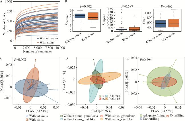
69个根尖周病变组织样本共获得543 0742条高质量序列,分属于51个门、135个纲、312个目、548个科、1 416个属、946个种。所有样本的覆盖度均达到0.99以上。通过ASVs数目构建稀释曲线(图 1A),可见所有样本曲线均趋向平缓,表明测序深度足够。无窦型与有窦型两组样本的α多样性指数(如Shannon、Simpson、Chao1)差异无统计学意义(P>0.05,图 1B)。采用基于Weighted Unifrac距离的主坐标分析,以评估两组样本在ASVs水平上的β多样性,可同时兼顾进化关系及物种丰度差异,结果可见两组样本呈分离趋势,通过PERMANOVA检验显示无窦型与有窦型两组样本间的菌群结构差异有统计学意义(P < 0.01,图 1C)。根据病理表现进一步分层分析组间差异(图 1D),无窦型及有窦型组内不同病理表现的菌群结构差异均无统计学意义(P>0.05)。同时,不同根管充填质量的PoAP根管外菌群结构差异也无统计学意义(P>0.05,图 1E)。
图1 菌群的多样性分析

Figure 1 Analysis of microbial community diversity

A, rarefaction curves based on ASVs; B, α diversity index (Shannon, Simpson, Chao1) of bacterial communities in the periapical lesions of PoAP without and with sinus tracts, Wilcoxon test was used to compare differences, Boxplots were used to present the median and quartiles; C, β diversity of bacterial communities in the periapical lesions of PoAP without and with sinus tracts; D, β diversity of bacterial communities in PoAP with different pathological manifestations; E, β diversity of bacterial communities in PoAP with different obturation qualities (adequate filling, underfilling, over-filling). ASVs, amplicon sequence variants; PoAP, post-treatment apical periodontitis; PCoA, principal coordinates analysis.

2.3 菌群组成比较

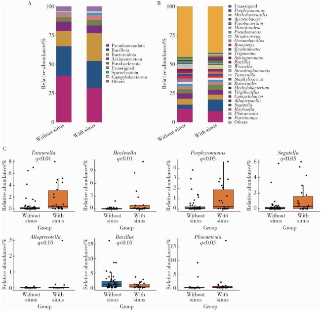
在门水平上(图 2A),PoAP的根管外菌群中的优势菌主要为假单胞菌门(Pseudomonadota)、厚壁菌门(Bacillota)、拟杆菌门(Bacteroidota)、放线菌门(Actinomycetota)、梭杆菌门(Fusobacteriota)、螺旋体门(Spirochaetota),这些菌门在无窦型组及有窦型组中的平均相对丰度均大于1%,其总相对丰度合计达到90%。在这些优势菌中,两组存在明显差异,拟杆菌门的相对丰度在有窦型组中显著高于无窦型组(16.98% vs. 9.22%, q < 0.01),假单胞菌门的相对丰度在有窦型组显著低于无窦型组(30.70% vs. 42.19%, q < 0.05)。
图2 菌群物种组成及差异

Figure 2 Comparison of microbial composition

A, component of bacterial communities at phylum level in the periapical lesions of PoAP without and with sinus tracts; B, component of bacterial communities at genus level in the periapical lesions of PoAP without and with sinus tracts; C, comparison of bacterial communities at genus level in the periapical lesions of PoAP without and with sinus tracts. Wilcoxon test was used to compare differences followed by Benjamini-Hochberg FDR correction to convert P value to q value. Boxplots were used to present the median and quartiles. PoAP, post-treatment apical periodontitis; FDR, false discovery rate.

在属水平上(图 2B),PoAP的根管外菌群的优势菌中主要为Methyloversatilis、不动杆菌属(Acinetobacter)、卟啉单胞菌属、梭杆菌属(Fusobacterium)、假单胞菌属、Roseateles、赤杆菌属(Erythrobacter)、链球菌属(Streptococcus)、鞘氨醇单胞菌属(Sphingomonas)和拟杆菌属(Bacteroides),这些细菌在无窦型组及有窦型组中的平均相对丰度均大于1%。在菌群差异比较中(图 2C),卟啉单胞菌属、坦纳菌属(Tannerella)、SegatellaPhocaeicolaHoylesella的相对丰度在无窦型组中显著小于有窦型组(q < 0.05),而芽孢杆菌属(Bacillus)的相对丰度在无窦型组中显著大于有窦型组(q < 0.05)。

3 讨论

本研究利用16S rRNA高通量测序技术系统分析了较大样本量的PoAP患牙根尖周病变组织中的菌群特征。α多样性反映了菌群的生物多样性水平(如物种丰富度等),通常在疾病状态下或是某种细菌占主导的情况下,α多样性会显著降低。本研究中无窦型与有窦型患牙的根管外菌群比较中,α多样性指数差异无统计学意义,这表明PoAP根管外感染的生物多样性与窦道是否存在无关。β多样性则反映不同菌群之间的组成及结构差异(丰度及发育关系等)。本研究结果表明,无窦型和有窦型PoAP根管外菌群结构存在显著差异,一方面可能是具有特定菌群结构的根管外感染易于形成窦道,另一方面,窦道可将根尖周病变与口腔环境相连通,可能改变局部微环境,从而进一步导致特定菌的富集、非优势菌的丰度下降。同时,本研究中不同个体间的菌群差异同样较大,这与既往研究[4, 8]结果一致。在Sun等[4]的研究中,比较了5例有窦型与5例无窦型PoAP的根管内外菌群差异,其研究结果表明,根管内外菌群中的卟啉单胞菌属、EubacteriumPhocaeicolaTreponema与窦道存在显著相关。本研究中,卟啉单胞菌属和Phocaeicola在有窦型PoAP的根管外菌群中也表现出更高的相对丰度。
本研究结果显示,有窦型PoAP中的拟杆菌门相对丰度显著高于无窦型,提示拟杆菌门丰度升高可能会造成更严重的骨破坏,导致窦道的形成。拟杆菌门是一类革兰氏阴性细菌,在根尖周炎根管内及根管外菌群中均为优势菌[15-17],既往研究[18]表明,其下属的Bacteroidaceae G.1.sp.oral taxon 272是有窦型慢性根尖周炎根管内的核心菌群。该菌门的成员具有蛋白水解能力和脂多糖介导的促炎活性,能够降解宿主组织蛋白、破坏细胞外基质,并激活宿主免疫反应[19],可能是参与根尖周病损形成的关键菌群,其具体作用机制需要进一步研究。
本研究中,卟啉单胞菌属、坦纳菌属在有窦型PoAP中相对丰度更高,这与既往根管内菌群的研究[4, 11]结果一致,二者均为革兰氏阴性菌,是根管内感染的主要优势菌之一[20-21]。同时,其下属的牙龈卟啉单胞菌和福塞坦菌(Tannerella forsythia)是牙周炎感染中的“红色复合体”,可促进炎症反应、加速骨吸收导致牙周组织破坏[22]。卟啉单胞菌属、坦纳菌属在有窦型PoAP中相对丰度升高,一方面可能是其加速了牙周组织破坏,从而形成窦道;另一方面可能是由于窦道的形成,使口腔中牙周致病菌沿窦道进入根尖周病变区,进而促进根尖周病变的骨破坏。
本研究还发现革兰氏阴性菌SegatellaPho-caeicolaHoylesella也与有窦型PoAP显著相关。既往研究通过分子生物学技术也能够在根管内及根管外检出这些菌属[4, 16, 23],但目前这些细菌在根尖周炎致病过程中发挥的作用尚不清楚,有待进一步研究。
芽孢杆菌属在无窦型PoAP中具有更高的丰度,提示芽孢杆菌属可能与窦道的形成负相关,但其在根尖周炎发展过程中的致病作用尚不清楚。有研究表明其下属的枯草芽孢杆菌(Bacillus subtilis)具有拮抗牙龈卟啉单胞菌的作用[24],并能改善患者的牙周炎状况[25]。但这一拮抗作用在复杂的根尖周炎菌群中是否成立尚需进一步研究。
此外,虽然根充质量和病理类型可能影响根尖病变的愈合[26],但本研究发现二者与菌群结构均无显著相关性;相反,窦道这一可以改变根尖周微环境的因素与菌群结构密切相关,提示局部微环境的变化对根尖周菌群具有更关键的影响。
综上,本研究通过16s rRNA高通量测序技术揭示了无窦型与有窦型PoAP根管外菌群的组成和结构存在显著差异,提示卟啉单胞菌属、坦纳菌属、SegatellaPhocaeicolaHoylesella等可能是导致根尖周病损持续存在和窦道形成的关键菌,而芽孢杆菌属与无窦型PoAP相关。由于有窦型PoAP其根管外感染中革兰氏阴性菌显著富集,提示临床上对于有窦型PoAP的治疗,可以采用针对革兰氏阴性菌的局部抗菌剂,或针对特定的优势菌开发靶向制剂,以提高临床疗效。
本研究的局限性为观察性研究,难以控制患者的全身及治疗史等混杂因素,未来尚待多因素模型进一步检验,并验证潜在致病菌与PoAP及窦道形成的因果关系。

利益冲突  所有作者均声明不存在利益冲突。

作者贡献声明  唐仁韬:设计研究方案,实施手术,收集样本,分析数据,撰写论文;杨流畅:收集样本,分析、整理数据,撰写论文;聂杰:设计研究方案,分析数据,撰写、审定论文;王晓燕:设计研究方案,审定论文。所有作者均参与论文修改,并对最终文稿进行审读和确认。

1
Nair PNR, Henry S, Cano V, et al. Microbial status of apical root canal system of human mandibular first molars with primary apical periodontitis after "one-visit" endodontic treatment[J]. Oral Surg Oral Med Oral Pathol Oral Radiol Endod, 2005, 99(2): 231- 252.

DOI

2
Nair PNR. On the causes of persistent apical periodontitis: A review[J]. Int Endod J, 2006, 39(4): 249- 281.

DOI

3
Bronzato JD, Davidian MES, de Castro M, et al. Bacteria and virulence factors in periapical lesions associated with teeth following primary and secondary root canal treatment[J]. Int Endod J, 2021, 54(5): 660- 671.

DOI

4
Sun X, Yang Z, Nie Y, et al. Microbial communities in the extraradicular and intraradicular infections associated with persistent apical periodontitis[J]. Front Cell Infect Microbiol, 2022, 11, 798367.

DOI

5
Zhang JL, Yun J, Yue L, et al. Distinctive microbiota distribution from healthy oral to post-treatment apical periodontitis[J]. Front Cell Infect Microbiol, 2022, 12, 980157.

DOI

6
Gupta R, Hasselgren G. Prevalence of odontogenic sinus tracts in patients referred for endodontic therapy[J]. J Endod, 2003, 29(12): 798- 800.

DOI

7
Ng YL, Mann V, Gulabivala K. A prospective study of the factors affecting outcomes of nonsurgical root canal treatment: Part 1: Periapical health[J]. Int Endod J, 2011, 44(7): 583- 609.

DOI

8
Wang W, Qu Y, Chen H, et al. The microbial co-infection interaction network in apical periodontitis with sinus tracts[J]. J Dent, 2025, 153, 105496.

DOI

9
漆正楠, 尹君, 唐子圣, 等. 有窦型与无窦型根尖周炎微生物群落比较[J]. 牙体牙髓牙周病学杂志, 2016, 26(1): 1- 6.

10
Griffee MB, Patterson SS, Miller CH, et al. The relationship of Bacteroides melaninogenicus to symptoms associated with pulpal necrosis[J]. Oral Surg Oral Med Oral Pathol, 1980, 50(5): 457- 461.

DOI

11
Sassone LM, Fidel R, Faveri M, et al. Microbiological evaluation of primary endodontic infections in teeth with and without sinus tract[J]. Int Endod J, 2008, 41(6): 508- 515.

DOI

12
Qi Z, Cao H, Jiang H, et al. Combinations of bacterial species associated with symptomatic endodontic infections in a Chinese population[J]. Int Endod J, 2016, 49(1): 17- 25.

DOI

13
Zhang C, Yang Z, Hou B. Diverse bacterial profile in extraradicular biofilms and periradicular lesions associated with persistent apical periodontitis[J]. Int Endod J, 2021, 54(9): 1425- 1433.

DOI

14
Bolyen E, Rideout JR, Dillon MR, et al. Reproducible, interactive, scalable and extensible microbiome data science using QIIME 2[J]. Nat Biotechnol, 2019, 37(8): 852- 857.

DOI

15
Alquria TA, Acharya A, Kabir B, et al. Clinical investigation of bacteriome in primary endodontic infections with apical periodontitis using high-throughput sequencing analysis[J]. J Endod, 2024, 50(10): 1393- 1402.

DOI

16
Buonavoglia A, Zamparini F, Lanave G, et al. Endodontic microbial communities in apical periodontitis[J]. J Endod, 2023, 49(2): 178- 189.

DOI

17
李昱志, 苏旭, 陈晓涛, 等. 基于16S rDNA测序的慢性牙髓炎及根尖周炎感染根管内菌群多样性研究[J]. 安徽医科大学学报, 2024, 59(9): 1669- 1674.

18
Mominkhan D, Brito LCN, Yakubu AR, et al. Exploring relationships within the microbiome of root canal infections and the influence of associated clinical parameters[J]. Int Endod J, 2025, 58(11): 1751- 1763.

DOI

19
Lyu P, Song Y, Bi R, et al. Protective actions in apical periodontitis: The regenerative bioactivities led by mesenchymal stem cells[J]. Biomolecules, 2022, 12(12): 1737.

DOI

20
Barbosa-Ribeiro M, Arruda-Vasconcelos R, Louzada LM, et al. Microbiological analysis of endodontically treated teeth with apical periodontitis before and after endodontic retreatment[J]. Clin Oral Investig, 2021, 25(4): 2017- 2027.

DOI

21
Ordinola-Zapata R, Costalonga M, Dietz M, et al. The root canal microbiome diversity and function: A whole-metagenome shotgun analysis[J]. Int Endod J, 2024, 57(7): 872- 884.

DOI

22
Darveau RP. Periodontitis: A polymicrobial disruption of host homeostasis[J]. Nat Rev Microbiol, 2010, 8(7): 481- 490.

DOI

23
Tak EJ, Park OJ, Lee JS, et al. Microbiota associated with caries and apical periodontitis: A next-generation sequencing study[J]. Int Endod J, 2025, 58(6): 890- 901.

DOI

24
Zhao W, Ji L, Li J, et al. Mesaconate from bacillus subtilis R0179 supernatant attenuates periodontitis by inhibiting porphyromonas gingivalis in mice[J]. J Periodontal Res, 2025, 60(6): 617- 627.

DOI

25
Tsubura S, Mizunuma H, Ishikawa S, et al. The effect of Bacillus subtilis mouth rinsing in patients with periodontitis[J]. Eur J Clin Microbiol Infect Dis, 2009, 28(11): 1353- 1356.

DOI

26
Turan Gökduman C, Çanakçi BC, Arili Öztürk E, et al. Factors affecting long-term success and survival in non-surgical root canal retreatment: A retrospective cohort study[J]. Clin Oral Investig, 2025, 29(11): 491.

DOI

Outlines

/