Value of direct immunofluorescence in the diagnosis of oral mucosal pemphigus vulgaris: A retrospective study based on multi-index combined analysis

  • Yanting CHI 1 ,
  • Hongjie JIANG 1, 2 ,
  • Yan CHEN 1 ,
  • Zhixiu XU , 1, * ,
  • Binbin LI , 1, *
Expand
  • 1. Department of Oral Pathology, Peking University School and Hospital of Stomatology & National Center for Stomatology & National Clinical Research Center for Oral Diseases & National Engineering Research Center of Oral Biomaterials and Digital Medical Devices & Beijing Key Laboratory of Digital Stomatology, Beijing 100081, China
  • 2. The Affiliated Stomatological Hospital of Chongqing Medical University & Chongqing Key Laboratory of Oral Diseases & Municipal Key Laboratory of Oral Biomedical Engineering of Higher Education & Chongqing Municipal Health Commission Key Laboratory of Oral Biomedical Engineering, Chongqing 401147, China
XU Zhixiu, e-mail,
LI Binbin, e-mail,

Received date: 2025-09-19

  Online published: 2026-01-06

Copyright

All rights reserved. Unauthorized reproduction is prohibited.

Abstract

Objective: To clarify the diagnostic efficacy of various diagnostic methods through multi-index combined analysis, and to explore the core diagnostic value of direct immunofluorescence (DIF) in oral mucosal pemphigus vulgaris (PV). Methods: A total of 53 patients with confirmed oral mucosal PV were included, retrospectively. Their data of DIF, histopathology, serum enzyme-linked immunosorbent assay (ELISA) and clinical diagnosis were systematically analyzed, and diagnostic efficacy of each method was evaluated using indicators, such as sensitivity, specificity, and area under curve (AUC) of the receiver operating characteristic (ROC) curve. Results: The results showed that among the 53 patients, middle-aged and elderly females were predominant, the buccal mucosa was the most common involved site, and most patients had a history of blistering. The positive rate of DIF was 96.23%, mainly manifested as a reticular fluorescent deposition between epithelial spinous cells, demonstrating the highest diagnostic value with a sensitivity of 96.23%, specificity of 100.00%, and AUC of 0.981. Histopathology ranked second, with a sensitivity of 94.34%, a specificity of 100.00%, and an AUC value of 0.972. The ELISA test had a sensitivity of 82.61%, a specificity of 82.35%, and an AUC value of 0.825. Although the sensitivity of clinical diagnosis was acceptable, its specificity was relatively low. Additionally, DIF exhibited complementarity with histopathology, ELISA, and clinical diagnosis, and combined testing could improve diagnostic accuracy. Conclusion: DIF is the "gold standard" for the diagnosis of oral mucosal PV. A comprehensive diagnostic workflow of "clinical manifestation-DIF-histopathology-ELISA" is proposed. This integrated diagnostic system not only significantly improves the accuracy of oral mucosal PV diagnosis but also aligns with the core principles of precision medicine, providing a basis for indivi-dualized treatment.

Cite this article

Yanting CHI , Hongjie JIANG , Yan CHEN , Zhixiu XU , Binbin LI . Value of direct immunofluorescence in the diagnosis of oral mucosal pemphigus vulgaris: A retrospective study based on multi-index combined analysis[J]. Journal of Peking University(Health Sciences), 2026 , 58(1) : 68 -73 . DOI: 10.19723/j.issn.1671-167X.2026.01.009

寻常型天疱疮(pemphigus vulgaris,PV)是一种慢性自身免疫性大疱性疾病,也是最常见和最严重的天疱疮亚型,主要影响40~60岁人群[1-2]。PV的发病机制是角质形成细胞表面存在主要针对桥粒芯糖蛋白(Dsg1、Dsg3)的自身抗体,后者破坏上皮层桥粒的黏附功能,导致棘层松解、水疱形成[3]。PV水疱的疱壁往往较薄,易破溃。口腔黏膜作为PV常见受累部位,其临床表现常缺乏特异性,几乎看不见完整水疱,主要表现为水疱破溃后遗留的不规则糜烂或溃疡,给诊断造成了一定的挑战,且容易与黏膜类天疱疮、扁平苔藓等口腔黏膜疾病相混淆。
PV的口腔黏膜病变通常早于皮肤病变,早期诊断对于其精准治疗和预后十分重要, 可降低严重并发症和死亡的发生率。目前,PV诊断方法主要包括直接免疫荧光(direct immunofluorescence,DIF)、组织病理学、血清学酶联免疫吸附试验(enzyme-linked immunosorbnent assay,ELISA)检测及临床表现[如尼氏征(Nikolsky sign)阳性等]。但各指标在诊断中的权重及相关性尚未取得共识,影响了PV诊断标准的同质性。有学者认为,DIF应作为自身免疫性大疱性疾病诊断的金标准[4-5],但也有学者认为,应综合以上指标联合分析[6-7]
基于以上情况,本研究回顾性总结口腔黏膜PV患者的临床资料,系统评估DIF的表现特征和诊断价值,并与临床表现、组织病理学特征、血清ELISA检测进行比较,明确各诊断方法的价值及相互关系,旨在为临床实践提供可靠的诊断依据。

1 资料与方法

1.1 临床资料

本研究纳入2023—2024年就诊于北京大学口腔医院,临床印象为天疱疮的患者。纳入标准:(1)临床表现为尼氏征阳性的口腔黏膜糜烂或溃疡;(2)有完整的组织病理学检查、DIF检测、临床症状记录。排除标准:(1)患有其他系统性疾病;(2)合并其他严重自身免疫性疾病;(3)具有头颈部放疗史。临床资料的收集包括患者性别、年龄、病损部位、病程及活组织检查前用药情况。本研究经北京大学口腔医院生物医学伦理委员会审核批准(批准号:PKUSSIRB-2025114171)。

1.2 活体组织取材

选择口腔黏膜邻近水疱或糜烂边缘3~10 mm内黏膜,取材深度包含上皮全层及固有层浅层[8-9]。切取组织并立即冰冻制片,进行DIF检测,荧光显微镜下观察;冰冻剩余组织用甲醛溶液固定,行常规组织病理学苏木精-伊红(hematoxylin-eosin,HE)染色,光镜下观察。

1.3 DIF检测

5 μm厚度冰冻切片经丙酮固定10 min后,分别滴加山羊抗人免疫球蛋白G(immunoglobulin G,IgG)、山羊抗人IgM、山羊抗人IgA、兔抗人补体C3及兔抗人纤维蛋白原(均为上海基因科技产品,1 ∶ 50稀释,货号分别为:GF020229、GF020329、GF020429、GF020129、GF011129),37 ℃湿盒孵育30 min,磷酸盐缓冲液洗片,封片。结果判读标准:上皮细胞间见翠绿色网状荧光,或松解的天疱疮细胞(Tzanck细胞)的细胞膜周围见翠绿色荧光环,则判读为阳性。

1.4 ELISA检测

收集患者病历资料中记录的ELISA检测结果,包括血清中抗Dsg1和抗Dsg3抗体的含量。ELISA检测采用欧蒙医学诊断(中国)有限公司抗Dsg1抗体、抗Dsg3抗体检测试剂盒(货号:EA_1495-4801_G、EA_1496-4801_G),并严格按试剂盒说明书进行操作。结果判读标准:(1)抗Dsg1抗体水平>20 U/mL为阳性,14~20 U/mL为不确定, < 14 U/mL为阴性;(2)抗Dsg3抗体水平>20 U/mL为阳性,7~20 U/mL为不确定, < 7 U/mL为阴性。

1.5 统计学方法

采用SPSS 24.0统计软件进行数据分析。计量资料若呈正态分布则以${\bar x}$±s表示,若呈偏态分布则以M(P25, P75)表示;计数资料以n(%)表示。通过四格表法计算各项诊断指标的灵敏度、特异度、阳性预测值、阴性预测值、漏诊率及误诊率。采用受试者工作特征(receiver operating characteristic,ROC)曲线评估DIF、组织病理学、ELISA与临床诊断对口腔黏膜PV的诊断效能,计算曲线下面积(area under curve,AUC)并进行组间比较。对于配对分类资料诊断一致性的比较,采用McNemar检验。P < 0.05为差异具有统计学意义。

2 结果

2.1 临床资料

本研究共收集临床表现印象为口腔黏膜PV的患者73例,依据诊断标准[8, 10],最终有53例(72.6%)患者被纳入研究,其中包括女性31例(58.49%),男性22例(41.51%)。患者发病年龄以30~60岁为主,占全部病例的62.26%;病变部位以颊部黏膜最为常见(58.49%),其次为牙龈(26.42%)、舌部(9.43%)及唇部(5.66%);30例患者(56.60%)伴有口腔黏膜起疱史,14例(26.42%)伴有皮肤疱性症状。在病程分布上,患者的病程为0.5~108个月,中位病程为5(2,12)个月,病程在1年内的患者占81.13%,病程1~4年的患者占9.43%,此外,还有9.43%的患者病程超过4年。38例(71.70%)确诊患者有药物治疗史,但仅42.11%(16/38)用药后缓解。

2.2 组织病理特征

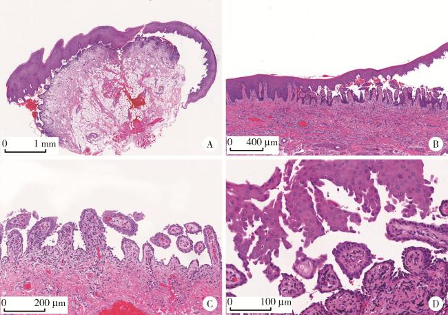
53例患者中,组织病理学检查显示50例(94.34%)呈现典型的病理改变,表现为棘层松解、上皮内疱形成。疱主要位于基底层和棘层之间,基底细胞附着于基底膜,呈“墓碑样”排列; 松解的棘层细胞单个或成簇的游离于疱液中,即经典的天疱疮细胞; 部分病例因为疱壁脱落,虽无法观察到完整棘层细胞,但仍可观察到基底细胞位于基底膜; 固有层内可见程度不等的慢性炎细胞浸润(图 1)。其余3例患者病理表现不典型,镜下均表现为黏膜轻度慢性炎,未见确切的上皮内疱的形成。
图1 口腔黏膜寻常型天疱疮的组织病理学特征(HE染色)

Figure 1 Histopathological features of oral mucosal pemphigus vulgaris (HE staining)

A, intraepithelial blister formation (×20); B, acantholysis (×40); C, blister roof detachment, with epithelial basal cells attached to the surface of connective tissue papillae showing a villous appearance (×100); D, acantholysis with scattered Tzanck cells: swollen round cells with hyperchromatic nuclei, often surrounded by a perinuclear cytoplasmic halo (×200). HE, hematoxylin-eosin.

2.3 DIF检测结果

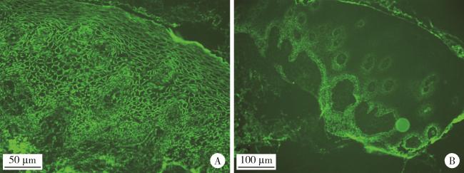
53例患者中,DIF检测显示51例阳性,阳性率为96.23%,主要表现为上皮棘细胞间特征性网状荧光沉积,呈翠绿色,部分病例可见松解的天疱疮细胞膜周围翠绿色荧光环(图 2)。5个检测指标中IgG阳性率最高,达92.45%(49例),C3阳性率为52.83%(28例),纤维蛋白原阳性率为20.75%(11例),而IgA和IgM阳性率较低,分别为5.66%(3例)和1.89%(1例);其中Ig G和C3联合检测的阳性率可达96.23%,与5个指标联合检测的阳性率一致。另外,2例DIF检测阴性患者的临床特征和血清学结果均符合PV诊断,但病理学特征不典型(表 1)。
图2 口腔黏膜寻常型天疱疮的直接免疫荧光表现

Figure 2 Direct immunofluorescence of oral mucosal pemphigus vulgaris

A, a reticular pattern of IgG deposition is observed in the intercellular spaces of the epithelial stratum spinosum (excitation wavelength 488 nm, emission wavelength 520-530 nm, ×400); B, a reticular pattern of C3 deposition is observed in the intercellular spaces of the epithelial stratum spinosum (excitation wavelength 488 nm, emission wavelength 520-530 nm, ×200).

表1 2例直接免疫荧光结果阴性患者的诊断及治疗情况

Table 1 Diagnosis and treatment status of 2 patients with negative direct immunofluorescence results

Patient Diagnosis Treatment
Clinical manifestation Histopathology Antibody level by ELISA/(U/mL) Treated with pemphigus protocol Used glucocorticoids Treatment outcome
No.1 Typical Atypical Anti-Dsg3: 93.70 (>20) Anti-Dsg1: 0 (< 20) Yes Yes Marked
No.2 Typical Atypical Anti-Dsg3: 58.44 (>20) Anti-Dsg1: 11.33 (< 20) Yes No Mild

ELISA, enzyme-linked immunosorbnent assay.

2.4 ELISA检测结果

53例确诊PV的患者中,有46例进行了血清学抗Dsg1和抗Dsg3抗体检测,其中38例(82.61%) 表达为阳性,包括抗Dsg1抗体阳性18例(39.13%)、抗Dsg3抗体阳性36例(78.26%)、抗Dsg1和抗Dsg3抗体同时阳性14例(30.43%)。

2.5 DIF的诊断效能评估

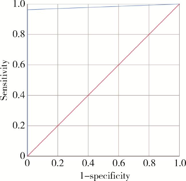
综合使用5个抗体指标的DIF在口腔黏膜天疱疮诊断中的灵敏度为96.23%,特异度为100.00%,其中灵敏度较高的是IgG和C3,特异度较高的是IgM和IgA(表 2)。DIF的ROC曲线下面积为0.981(P < 0.05,图 3)。
表2 直接免疫荧光各抗体指标的诊断效能

Table 2 Diagnostic performance of various direct immunofluorescence antibody indicators

Detection indicator Sensitivity Specificity Positive predictive value Negative predictive value Missed diagnosis rate False positive rate
IgG 92.45% 55.00% 84.48% 73.33% 7.55% 45.00%
C3 52.83% 60.00% 77.78% 32.43% 47.17% 40.00%
Fibrinogen 20.75% 75.00% 68.75% 26.32% 79.25% 25.00%
IgA 5.66% 85.00% 50.00% 25.37% 94.34% 15.00%
IgM 1.89% 95.00% 50.00% 26.76% 98.11% 5.00%
DIF 96.23% 100.00% 100.00% 90.91% 3.77% 0.00%

Ig, immunoglobulin; C3, complement 3; DIF, direct immunofluorescence.

图3 直接免疫荧光诊断口腔黏膜寻常型天疱疮的受试者工作特征曲线

Figure 3 Receiver operating characteristic curve of direct immunofluorescence for the diagnosis of oral mucosal pemphigus vulgaris

2.6 不同检测方法诊断效能的比较

表 3所示,不同检测方法中DIF的诊断价值最高,灵敏度为96.23%,特异度为100.00%,AUC值达0.981;组织病理学次之,灵敏度为94.34%,特异度为100.00%,AUC值为0.972;ELISA检测的灵敏度为82.61%,特异度为82.35%,AUC值为0.825;此外,虽然临床诊断的灵敏度可达86.79%,但其特异度比较低,仅为15.00%,AUC值也只有0.509。McNemar检验结果显示,DIF与组织病理学(P>0.999)、临床诊断(P=0.180)、ELISA检测(P=0.109)之间的灵敏度差异均无统计学意义。
表3 口腔黏膜天疱疮中不同检测方法诊断效能的比较

Table 3 Comparison of diagnostic performance of different detection methods in oral mucosal pemphigus vulgaris

Detection method Sensitivity Specificity AUC
DIF 96.23% 100.00% 0.981
Histopathology 94.34% 100.00% 0.972
ELISA 82.61% 82.35% 0.825
Clinical diagnosis 86.79% 15.00% 0.509

DIF, direct immunofluorescence; ELISA, enzyme-linked immunosorbnent assay; AUC, area under curve.

3 讨论

3.1 DIF在口腔黏膜PV中的特征和诊断价值

本研究通过回顾性分析53例口腔黏膜PV患者的临床资料、组织学特征、DIF特征及ELISA结果,系统评估了DIF在口腔黏膜PV诊断中的核心价值,并与其他三种诊断方法进行了比较。本研究数据显示,应用DIF诊断口腔黏膜PV的灵敏度为96.23%,特异度为100.00%,AUC值为0.981,表明DIF对于诊断口腔黏膜PV具有极高的效能,主要表现为上皮棘细胞间特征性网状荧光沉积,这是由于自身免疫复合物在上皮细胞间沉积,从而使得DIF具有高特异性。此外,DIF的假阴性率仅为3.77%,这可能与活组织检查部位的选择或操作技术有关。因此,建议临床上行活组织检查时要选择病变周围3~10 mm看似正常的黏膜,以避免因上皮脱落而导致的假阴性结果。若临床高度怀疑PV,而DIF检测为阴性,必要时可重复进行活组织检查和DIF检测,以明确诊断,尽早行针对性的治疗,改善患者预后[9]。DIF诊断口腔黏膜PV的灵敏度未达100.00%,可能受以下因素影响[7, 11-13]:(1)活组织检查部位选择和操作技术规范:由于抗体主要沉积于病损周围正常黏膜,若取材部位为活动性糜烂区、水疱中央或陈旧性皮损处,易导致假阴性结果;此外,小口径活组织检查也易造成漏检。(2)样本处理与技术因素:组织样本保存或运输不当(误用甲醛溶液,未使用Michel溶液或生理盐水,室温或冷藏保存超时等)均可致抗原降解;同时,检测试剂过期,染色流程操作不规范等技术环节问题也会降低检测灵敏度。(3)临床治疗影响:治疗可降低抗体水平,从而影响DIF检测结果。因此,在临床应用中需严格规范活组织检查操作流程,优化样本处理标准,并结合患者治疗史进行综合评估,以尽可能降低DIF假阴性风险,提高其灵敏度。
在DIF的5项免疫指标中,IgG阳性率最高(92.45%),是口腔黏膜PV中最主要的自身抗体亚类,表明其是PV最核心的诊断指标。此外,补体C3阳性率为52.83%,提示补体原位激活参与此疾病的发生[14]。同时,本研究还发现IgG与C3之间存在很强的相关性,IgG和C3的联合使用与5个指标的组合使用在诊断口腔黏膜PV时的阳性率(96.23%)是一致的。因此,可以优先考虑联合检测IgG和C3,这和先前的研究结果也具有一致性[15]。纤维蛋白原在基底膜区呈线性沉积,阳性率为20.75%。值得注意的是,本研究中IgA(5.66%)和IgM(1.89%)阳性率较低,提示PV是由IgG自身抗体介导的,若DIF检测仅显示出IgM或IgA沉积,则需与其他类型的自身免疫性大疱性疾病相鉴别。临床上,口腔黏膜出现糜烂或水疱的疾病种类繁多,如黏膜类天疱疮、扁平苔藓、多形性红斑及线状IgA病等。这些疾病的DIF检测的表现与口腔黏膜PV显著不同,黏膜类天疱疮主要表现为基底膜区线状IgG、C3或IgA沉积,扁平苔藓可见基底膜区线状IgG和C3沉积及表皮层内胶样小体,而线状IgA病则表现为基底膜区线状IgA沉积[5, 15-16]。本研究中口腔黏膜PV的DIF特征是上皮棘层细胞间网状IgG或C3沉积,可与其他自身免疫性大疱性疾病相鉴别。

3.2 DIF与其他诊断方法的综合分析

本研究中,组织病理诊断的灵敏度为94.34%,与DIF检测结果(96.23%)接近,但由于其诊断主要依赖于典型的棘层松解和上皮内疱形成,而口腔黏膜病损又常以薄壁水疱为特征,容易破裂,表现为非特异性的糜烂,从而导致组织病理学检查出现假阴性[17]。本研究发现,部分组织病理学特征不典型的病例可以通过DIF检测明确诊断,这表明在口腔黏膜PV的诊断中DIF与组织病理学具有互补性,组织病理学侧重于形态学观察,而DIF聚焦于免疫学检测。联合应用两种方法可通过形态-免疫学双重证据链,提高诊断的准确性,特别是对于早期或不典型的口腔黏膜PV患者,可以避免漏诊或误诊。
本研究中血清学ELISA检测的整体灵敏度为82.61%,低于DIF(96.23%)和组织病理学诊断(94.34%)。进一步分析显示,抗Dsg3抗体阳性率为78.26%,显著高于抗Dsg1抗体阳性率(39.13%),这一差异与Dsg1和Dsg3在皮肤及黏膜组织中的特异性分布差异密切相关,并可直接影响其临床表现。Dsg1主要集中在表皮浅层,而Dsg3则集中分布于基底层和副基底层。口腔黏膜PV患者体内主要产生抗Dsg3自身抗体,致使抗Dsg3抗体在口腔黏膜PV中占主导地位,而抗Dsg1抗体的表达较少[3, 18]。尽管血清学ELISA检测的敏感性相对有限,但其具有无创、可重复检测的优势,使其在天疱疮的临床管理中具有不可替代的价值,可为疾病动态监测、病情评估和预后判断提供重要的依据[19-21]。此外,对于DIF检测阴性或因客观条件无法开展DIF检测的病例,ELISA检测可作为重要的辅助诊断方法。本研究中2例DIF阴性的确诊患者,通过典型的临床症状和血清学阳性结果进行了明确诊断,这也充分体现了多指标联合诊断的重要性。
本研究结果显示,临床诊断虽然具有较高的灵敏度(86.79%),但其特异度仅为15.00%,这是由于口腔黏膜PV的临床症状缺乏特异性,水疱、糜烂、溃疡等病损表现与黏膜类天疱疮、扁平苔藓、多形性红斑等黏膜疾病存在明显的重叠,容易混淆而导致误诊[16]。因此,单纯依靠临床症状诊断口腔黏膜PV存在一定的局限性,临床上需结合DIF、组织病理学及ELISA等实验室检查进行综合诊断。
此外,作为单中心的回顾性研究,本研究也具有一定的局限性:样本量较小,且缺乏长期随访和预后数据。未来应通过多中心协作扩大样本量,结合其他转录组学、蛋白组学等分子生物学技术,深入探究DIF荧光模式与疾病临床亚型、严重程度、活动度及治疗反应的内在关联,为口腔黏膜PV的精准诊疗提供更加全面的理论依据。
综上,DIF在口腔黏膜PV诊断中具有极高的灵敏度和特异度,可作为该自身免疫性疾病诊断的核心指标。在临床实践中,建议采用多模态诊断策略,以典型临床表现作为初筛依据,DIF检测作为核心确诊手段,联合组织病理学提供形态学证据,并辅以ELISA检测进行抗体定量分析,从而构建“临床表现-DIF-组织病理学-ELISA”口腔黏膜PV综合诊断流程。这一综合诊断体系不仅能显著提高口腔黏膜PV诊断的准确性,更能为患者个体化治疗方案的制定提供理论基础。

利益冲突  所有作者均声明不存在利益冲突。

作者贡献声明  池彦廷: 收集分析整理数据, 实验,统计分析, 撰写文章; 蒋鸿杰: 收集数据; 陈艳: 分析数据; 徐志秀:实验支持;李斌斌: 设计课题, 指导文章撰写。所有作者均参与论文修改,并对最终文稿进行审读和确认。

1
Porro AM , Seque CA , Ferreira MCC , et al. Pemphigus vulgaris[J]. An Bras Dermatol, 2019, 94 (3): 264- 278.

2
Kridin K . Pemphigus group: Overview, epidemiology, mortality, and comorbidities[J]. Immunol Res, 2018, 66 (2): 255- 270.

DOI

3
Jamora MJJ , Jiao D , Bystryn JC . Antibodies to desmoglein 1 and 3, and the clinical phenotype of pemphigus vulgaris[J]. J Am Acad Dermatol, 2003, 48 (6): 976- 977.

DOI

4
Joly P , Horvath B , Patsatsi Α , et al. Updated S2K guidelines on the management of pemphigus vulgaris and foliaceus initiated by the European academy of dermatology and venereology (EADV)[J]. J Eur Acad Dermatol Venereol, 2020, 34 (9): 1900- 1913.

DOI

5
Witte M , Zillikens D , Schmidt E . Diagnosis of autoimmune blistering diseases[J]. Front Med (Lausanne), 2018, 5, 296.

6
Arockiam S , Raj B , Prabha N , et al. Diagnostic utility of immunofluorescence in oral lesions: A systematic review[J]. J Oral Maxillofac Res, 2024, 15 (3): e2.

7
Nili A , Salehi Farid A , Asgari M , et al. Current status and prospects for the diagnosis of pemphigus vulgaris[J]. Expert Rev Clin Immunol, 2021, 17 (8): 819- 834.

8
中国医疗保健国际交流促进会皮肤科分会. 寻常型天疱疮诊断和治疗专家建议(2020)[J]. 中华皮肤科杂志, 2020, 53 (1): 1- 7.

9
Mahmood MN . Direct immunofluorescence of skin and oral mucosa: Guidelines for selecting the optimum biopsy site[J]. Dermatopathology (Basel), 2024, 11 (1): 52- 61.

DOI

10
Amagai M , Tanikawa A , Shimizu T , et al. Japanese guidelines for the management of pemphigus[J]. J Dermatol, 2014, 41 (6): 471- 486.

DOI

11
Mutasim DF , Adams BB . Immunofluorescence in dermatology[J]. J Am Acad Dermatol, 2001, 45 (6): 803- 824.

DOI

12
Dridi SM , Lutz CM , Gaultier F , et al. Oral biopsy in mucous membrane pemphigoid and pemphigus vulgaris with gingival expression: The optimal site. A systematic review and meta-analysis[J]. BMC Oral Health, 2024, 24 (1): 1093.

DOI

13
Minz RW , Chhabra S , Singh S , et al. Direct immunofluorescence of skin biopsy: Perspective of an immunopathologist[J]. Indian J Dermatol Venereol Leprol, 2010, 76 (2): 150- 157.

DOI

14
Dainichi T , Chow Z , Kabashima K . IgG4, complement, and the mechanisms of blister formation in pemphigus and bullous pemphigoid[J]. J Dermatol Sci, 2017, 88 (3): 265- 270.

DOI

15
Xu Y , He X , Jie W , et al. The application of direct immunofluorescence in the pathological diagnosis of oral mucosal diseases: Series of 116 cases[J]. Pathol Res Pract, 2025, 270, 155989.

DOI

16
Arbache ST , Nogueira TG , Delgado L , et al. Immunofluorescence testing in the diagnosis of autoimmune blistering diseases: Overview of 10-year experience[J]. An Bras Dermatol, 2014, 89 (6): 885- 889.

DOI

17
Buonavoglia A , Leone P , Dammacco R , et al. Pemphigus and mucous membrane pemphigoid: An update from diagnosis to therapy[J]. Autoimmun Rev, 2019, 18 (4): 349- 358.

DOI

18
Feng X , Zheng H , Wang M , et al. Autoimmune bullous diseases: Pathogenesis and clinical management[J]. Mol Biomed, 2025, 6 (1): 30.

DOI

19
Genovese G , Maronese CA , Casazza G , et al. Clinical and serological predictors of relapse in pemphigus: A study of 143 patients[J]. Clin Exp Dermatol, 2022, 47 (1): 98- 106.

DOI

20
Harman KE , Seed PT , Gratian MJ , et al. The severity of cutaneous and oral pemphigus is related to desmoglein 1 and 3 antibody levels[J]. Br J Dermatol, 2001, 144 (4): 775- 780.

DOI

21
Golinski ML , Lemieux A , Maho-Vaillant M , et al. The diversity of serum anti-DSG3 IgG subclasses has a major impact on pemphigus activity and is predictive of relapses after treatment with rituximab[J]. Front Immunol, 2022, 13, 849790.

DOI

Outlines

/