Digital measurement and analysis of anatomical characteristics of protrusive and intercuspal position occlusal contacts in maxillary incisors

  • Liang SHAO ,
  • Wenjie MA ,
  • Ying CHEN ,
  • Qian DING , * ,
  • Lei ZHANG , *
Expand
  • Department of Prosthodontics, Peking University School and Hospital of Stomatology & National Center for Stomatology & National Clinical Research Center for Oral Diseases & National Engineering Research Center of Oral Biomaterials and Digital Medical Devices & Beijing Key Laboratory of Digital Stomatology, Beijing 100081, China
DING Qian, e-mail,
ZHANG Lei, e-mail,

Received date: 2025-10-10

  Online published: 2026-01-06

Supported by

the Clinical Research Foundation of Peking University School and Hospital of Stomatology(PKUSS-2023CRF503)

the Program for New Clinical Techniques and Therapies of Peking University School and Hospital of Stomatology(PKUSSNCT-25A09)

Beijing Natural Science Foundation-Haidian Original Innovation Joint Fund Project(L232025)

Copyright

All rights reserved. Unauthorized reproduction is prohibited.

Abstract

Objective: To digitally measure and analyze the anatomical characteristics of protrusive and intercuspal position (ICP) occlusal contacts in maxillary incisors, thereby establishing a standardized measurement protocol and obtaining characteristic functional data to optimize the incisal guidance design of prostheses. Methods: Thirty subjects with stable incisal guidance were recruited. Digital dental mo-dels were acquired via intraoral scanning, and protrusive movement data were captured using a modified patient-specific motion (PSM) technique. Computer-aided design software was used to record the distribution of the occlusal contacts during protrusive movement. Image analysis software was employed to measure the area proportion of guiding locations for each tooth. Reverse engineering software was used to measure and analyze the occlusal contacts in ICP and anatomical characteristics. Measured parameters included the area proportion and distribution of occlusal contacts in ICP, the area proportion of marginal ridges and incisal ridges, radius of curvature of lingual surface, lingual surface inclination, overbite, and overjet. Each parameter was measured twice to calculate the intraclass correlation coefficient for the assessment of test-retest reliability. Results: All measured parameters demonstrated good test-retest reliability. No significant differences were found in any parameters between homologous teeth (P>0.05). During protrusive movement, the area proportion of guiding locations was significantly larger for the central incisors than for the lateral incisors (73.4%±12.3% vs. 26.6%±12.3%, P < 0.001). The frequency of occlusal contacts was significantly higher on the mesial and distal marginal ridges and incisal ridges compared with the lingual fossa and cingulum (P < 0.05). In ICP, no significant difference was observed in the occlusal contact area proportion between the central and lateral incisors (48.8%±20.0% vs. 51.2%±20.0%, P=0.758). The frequency of the occlusal contact was significantly higher on the mesial and distal marginal ridges compared with the incisal ridge, lingual fossa, and cingulum (P < 0.05). Central incisors exhibited significantly higher overbite and overjet than lateral incisors (P < 0.05). The area proportion of mesial and distal marginal ridges was significantly smaller for the central incisors than for the lateral incisors (P < 0.05), but no significant difference was observed in the incisal ridge (P>0.05). No significant differences were observed in the lingual surface inclination or radius of curvature among the incisors (P>0.05). Conclusion: The anatomical characteristics of protrusive and ICP occlusal contacts in maxillary incisors demonstrated bilateral symmetry. Protrusive movement was primarily guided by the maxillary central incisors, with the guiding area of the central incisors being approximately three times that of the lateral incisors. The marginal ridges and incisal ridges were the main guiding locations. Central and lateral incisors exhibited comparable occlusal contact area in ICP.

Cite this article

Liang SHAO , Wenjie MA , Ying CHEN , Qian DING , Lei ZHANG . Digital measurement and analysis of anatomical characteristics of protrusive and intercuspal position occlusal contacts in maxillary incisors[J]. Journal of Peking University(Health Sciences), 2026 , 58(1) : 99 -106 . DOI: 10.19723/j.issn.1671-167X.2026.01.013

切导是指上颌切牙与下颌牙齿相接触从而产生对下颌运动的限制[1],其影响因素包括咬合接触关系、覆牙合、覆盖、上颌切牙舌面形态与切端形态等[2]。合理的切导是实现咀嚼功能的重要保障,能够防止前牙过度负载与过快磨耗,同时在下颌运动中前牙的引导可产生后牙牙合分离的效果,减少不良侧向力与颞下颌关节过度负载的风险[3]。因此,在进行前牙修复时,需建立精确适宜的切导以避免各类机械并发症和咀嚼功能不佳[4]。然而,目前的切导设计常仅以去除咬合干扰为目的,对恢复天然切牙的功能结构和实现精准的咬合接触控制关注较少[4-5]
精准的切导设计需要对其规律有深刻理解。既往学者曾从咬合接触和解剖特征等不同角度对切导相关因素进行了测量分析。首先,咬合接触关系与咀嚼功能直接相关。有学者曾在石膏模型上使用咬合纸标记引导部位并测量其直线距离[6-8],但使用咬合纸印记代表引导区域的正确度低、可重复性差,直线距离也无法反映真实发挥引导功能的曲线路径。Çakmak等[9]和Feng等[4]则在上颌中切牙中央的矢状向剖面上,以正中咬合接触点到切缘的舌面轮廓代表切导路径并用以评估前牙修复体的切导设计,但这种方法难以反映实际发挥引导功能的边缘嵴特征。其次,上颌切牙的解剖特征对引导下颌进行平滑无阻力的各向运动有重要影响。有学者曾对上颌切牙舌面形态的解剖变异进行定性分析[10-11],并通过量规、卡尺等机械测量工具或影像学方法对切导相关解剖特征进行定量测量,测量指标包括边缘嵴等结构的曲率半径和厚度、舌面倾斜度、切导角度及覆牙合、覆盖等[12-15],但这些方法仅能测量简单尺寸指标,精度较低且可重复性差,难以实现功能结构的精准定量测量。
数字化技术已广泛应用于口腔医学的各个领域。口内扫描技术获取数字化牙列模型后结合逆向工程软件可精准便捷地测量和分析牙齿的各项解剖特征[16-17],同时数字化咬合分析精准、可重复性高,可避免测量介质的影响,已成为常用的咬合分析方法[18-19]。动态咬合记录法(patient-specific motion,PSM)为测量分析下颌前伸运动的咬合接触特征提供了一种简便可行的方法,其可通过口内扫描仪记录患者下颌运动,进而在计算机辅助设计(computer- aided design,CAD)软件中再现功能运动并显示运动中的咬合接触区域[20]。既往研究表明,动态咬合记录法在设计后牙修复体中具有个性化优势[21-22]。本团队曾通过在尖牙唇面/前磨牙颊面粘接人工标志点对动态咬合记录法进行改良[23],提升了切导记录的精度和记录效率[24]
目前,关于上颌切牙切导相关特征的测量分析仍较少,且多为传统方法测量,目前尚未见应用动态咬合记录法测量分析前伸咬合接触特征和使用数字化手段系统测量分析切导相关特征的研究。本研究拟招募正常受试者,通过口内扫描获取数字化牙列模型,并通过改良动态咬合记录法获取前伸运动数据,使用数字化方法对上颌切牙前伸和正中咬合接触解剖特征进行精准测量分析,建立标准化的测量流程,并获得特征性切导功能相关数据,为优化修复体的切导设计提供参考。

1 资料与方法

1.1 研究对象

本研究为横断面研究,已获得北京大学口腔医院生物医学伦理委员会批准(批准号:PKUSSIRB-202387064)。在北京大学口腔医学院的在读研究生中招募受试者,所有受试者均在研究前签署知情同意书。
纳入标准:(1)年龄20~30岁;(2)前牙形态完整;(3)咬合检查无明显异常(无早接触、咬合干扰);(4)牙尖交错位咬合稳定,有稳定的正前伸、侧前伸功能运动;(5)牙周健康或经过完善的系统牙周治疗;(6)受试者已签署知情同意书。
排除标准:(1)曾进行过充填、修复等改变前牙形态的治疗;(2)曾进行过正畸等改变前牙咬合的治疗;(3)前牙区有明显的错牙合畸形(开牙合、反牙合、对刃、Ⅲ度深覆牙合、牙列拥挤);(4)颞下颌关节疾病;(5)有影响研究完成或干扰研究分析结果的情况存在。
样本量确定:在样本量计算软件(PASS 21.0,美国)中依据预试验结果进行计算,中切牙与侧切牙的引导面积占比分别为67.3%和32.7%,标准差为25.4%,设定双尾显著性水平为0.05,效能为90%,所需的最小样本量仅为8例。由于计算得出的样本量过小,因此参考同类研究[19, 25],将样本量扩大至30例,以满足统计分析要求。

1.2 口内扫描获取数字化牙列模型及动静态咬合数据

为精确记录前伸运动数据,本研究采用本团队前期研究提出的改良动态咬合记录法[23]。使用逆向工程软件(Geomagic Wrap 2021,3D System,美国)设计一底面为弧面的类圆台形标志物,并使用高精度牙模树脂(Model HP UV 2.0,广州黑格智能科技有限公司)在口腔椅旁3D打印机(UltraCraft ChairSide Pro,广州黑格智能科技有限公司)上进行打印。
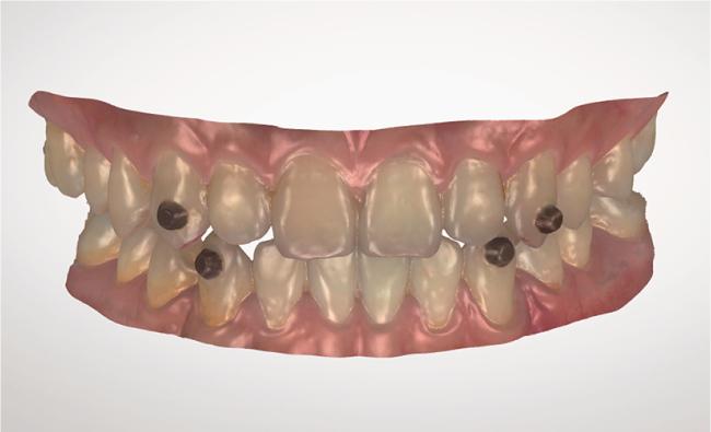
于受试者双侧上、下颌尖牙唇面中1/3处,在非酸蚀情况下使用RelyX U200树脂粘接剂(3M ESPE,美国)粘接标志物后(图 1),使用口内扫描仪(Trios 4,v20.1.2,3Shape A/S,丹麦)获得上、下颌数字牙列模型,嘱患者在牙尖交错位(intercuspal position,ICP)使用习惯性咬合力咬合,扫描获取颊面咬合配准数据。引导并训练受试者完成如下的下颌运动后,采集完整的动态咬合记录:(1)正前伸运动,要求下颌沿上颌切牙舌面向正前方滑动到上、下颌切牙切端相对;(2)侧前伸运动,要求下颌沿上颌切牙舌面向侧前方滑动到上、下颌切牙切端相对。口内扫描完成后,在扫描软件内检查运动轨迹是否连续、完整,检查是否有明显的偏移或穿透。确认无误后使用洁治器去除标志物并清除粘接剂,导出数字牙列模型数据(.stl)和包含动态咬合数据的口内扫描订单数据(.3oxz)。
图1 粘接标志物

Figure 1 Adhesive bonding of artificial landmarks

1.3 测量分析

所有指标均由同一名熟练使用相关软件的口腔医生按照方案进行标准化测量。除正中咬合接触面积由软件自动计算而未进行重复测量外,其余定量指标均在2周后重新测量,以2次测量的平均值作为最终结果,并计算组内相关系数(intraclass correlation coefficient,ICC)以评价复测信度。

1.3.1 前伸和正中咬合接触的测量分析

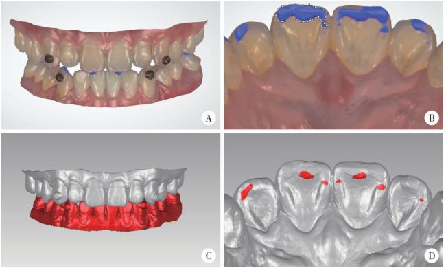
将获取的包含动态咬合数据的口内扫描订单数据(.3oxz)导入CAD软件(3Shape Dental System,丹麦),使用“特定于患者的下颌运动”功能显示前伸运动中的咬合接触区域作为引导部位,调整视图使其垂直于上颌切牙舌面,截取图像后导入图像分析软件(ImageJ Version 1.53q,美国国立卫生研究院),按照上颌切牙舌面的解剖结构(切嵴、近中边缘嵴、远中边缘嵴、舌窝及舌隆突)记录其位置分布,测量每个牙位引导部位的面积并计算其与四个切牙引导总面积的比值,作为该牙位的引导面积占比(图 2)。将获取的三维牙列模型数据(.stl)导入逆向工程软件,使用“抽壳”功能将下颌牙列模型向牙合方增厚200 μm,使用布尔运算(Boolean operation)获取上颌切牙舌面与下颌牙列相交的部分作为该牙位的咬合接触区域,按照上颌切牙舌面的解剖结构(切嵴、近中边缘嵴、远中边缘嵴、舌窝及舌隆突)记录其位置分布,测量每个牙位咬合接触区域的曲面面积并计算其与四个切牙咬合接触总面积的比值,作为该牙位的正中咬合接触面积占比(图 2)。
图2 前伸和正中咬合接触的测量

Figure 2 Measurement of the occlusal contacts in protrusive movement and intercuspal position

A: Reproduce the mandibular protrusive movement by the patient-specific motion (PSM) function within the computer-aided design software (3Shape Dental System). B: Measure the area of the guiding locations (blue) during protrusive movement within the image analysis software (ImageJ Version 1.53q). C: Thicken the mandibular model occlusally by 200 μm by the Shell function within the reverse engineering software (Geomagic Wrap 2021); Obtain the intersection areas between the maxillary incisors and the mandibular dentition by the Boolean operation function, which was defined as the occlusal contact areas in intercuspal position (ICP). D: Measure the area of the occlusal contact areas (red) in ICP.

1.3.2 切导相关解剖特征的测量分析

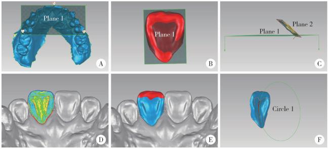
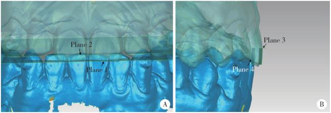
将获取的三维牙列模型数据(.stl)导入逆向工程软件,选取上颌牙列模型上的三个点确定牙合平面(上颌中切牙近中切角点与双侧上颌第一磨牙近中颊尖点)。分别按照以下步骤对切导相关舌面特征(图 3)和覆牙合、覆盖(图 4)进行测量分析:(1)舌面倾斜度:选取整个舌面,使用“最佳拟合”功能生成平面,计算其与参考平面(牙合平面)的夹角。(2)边缘嵴与切嵴面积占比:使用“绘制曲线”功能绘制目标牙位舌面边界后,选取其舌面生成新对象;在舌面上依据“曲率图”绘制切嵴、近中边缘嵴与远中边缘嵴的边界;使用“计算面积”功能分别计算以上解剖结构与该牙位舌面的曲面面积,并计算其相对于舌面的面积占比。(3)舌面中央曲率半径:使用“直线选择工具”选取舌面近远中方向上的中央部分自切嵴龈方至舌隆突上最突点的连线,拟合生成圆形特征,记录该圆的半径。(4)覆牙合:平行移动牙合平面,使其与目标牙位的切缘相切,得到平面1;平行移动牙合平面,使其与目标牙位对应的下颌切牙切缘相切,得到平面2;计算平面1与平面2之间的距离。(5)覆盖:选取目标牙位的唇面切1/3的区域,通过“最佳拟合”功能获得与唇面相切的平面3;平行移动平面3,使其与目标牙位对应的下颌切牙唇面相切,得到平面4;计算平面3与平面4之间的距离。
图3 切导相关舌面特征的测量

Figure 3 Measurement of lingual surface characteristics related to incisal guidance

A: Generate the occlusal plane (plane 1) within Geomagic wrap 2021. B: Use the maxillary right central incisor as an example; Select the lingual surface and generate Plane 2 by the Best-Fit function. C: calculate the angle between Plane 2 and Plane 1, defined as the lingual surface inclination. D: Plot the curve based on the Curvature Map. E: Convert the curve into the boundary and calculate the areas of the marginal ridges and the incisal ridge. F: Select the central portion of the lingual surface, generate Circle 1 by the Best-Fit function, and calculate its radius.

图4 覆牙合覆盖的测量

Figure 4 Measurement of overbite and overjet

A: Use the maxillary right central incisor as an example within Geomagic Wrap 2021; Move the occlusal plane until it is tangent to the incisal edge, generating Plane 1; Move Plane 1 until it is tangent to the incisal edge of the corresponding mandibular incisor, generating Plane 2; Calculate the distance between Plane 1 and Plane 2, defined as the overbite. B: Select the incisal third region of the labial surface and generate Plane 3 by the Best-Fit function, which is tangent to the labial surface; Translate Plane 3 parallel until it is tangent to the labial surface of the corresponding mandibular incisor, generating Plane 4; Calculate the distance between Plane 3 and Plane 4, defined as the overjet.

1.4 统计分析

使用SPSS 26.0(IBM v24.0,美国)软件进行统计分析。对引导部位面积占比、正中咬合接触面积占比、边缘嵴与切嵴面积占比、舌面中央曲率半径、舌面倾斜度和覆牙合、覆盖等定量指标使用Shapiro-Wilk检验进行正态性检验,数据均符合正态分布,采用均数±标准差表示,使用重复测量方差分析(repeated measures analysis of variance,RM-ANOVA)比较四个牙位是否存在整体差异,当整体差异有统计学意义时,采用Bonferroni法进行事后两两比较以控制多重比较的一类错误。使用配对t检验比较中切牙与侧切牙或双侧同名牙是否存在差异。对于引导部位与正中咬合接触部位的分布,使用McNemar检验进行比较。使用ICC评价定量指标的复测信度,以双侧检验P < 0.05为差异有统计学意义。

2 结果

本研究纳入的30例正常受试者中男性13例,女性17例,平均年龄(24.8±1.7)岁。各指标的复测信度均较高,引导部位面积占比ICC为0.991,边缘嵴与切嵴面积占比ICC为0.908,舌面中央曲率半径ICC为0.869,舌面倾斜度ICC为0.888,覆牙合、覆盖ICC为0.951。

2.1 前伸和正中咬合接触特征

前伸运动中,中切牙的引导部位面积占比显著高于侧切牙(73.4%±12.3% vs. 26.6%±12.3%,t=10.390,P < 0.001),双侧同名牙之间差异无统计学意义(P>0.05,表 1);各牙位中,切嵴、近中边缘嵴与远中边缘嵴发生引导的频率显著高于舌窝与舌隆突(P < 0.05,表 2)。牙尖交错位时,中切牙和侧切牙的咬合接触面积占比差异无统计学意义(48.8%±20.0% vs. 51.2%±20.0%, t=-0.312,P=0.758),双侧同名牙之间差异亦无统计学意义(P>0.05,表 1);各牙位中,近中边缘嵴与远中边缘嵴发生咬合接触的频率显著高于切嵴、舌窝与舌隆突(P < 0.05,表 2)。
表1 上颌切牙的前伸和正中咬合接触面积占比(%)

Table 1 Occlusal contact area proportion (%) of maxillary incisors in protrusive movement and intercuspal position

Items Central incisor Lateral incisor
Left Right t P Left Right t P
Protrusive movement (n=30) 39.3±10.1 34.1±11.2 -1.632 0.113 15.0±7.5 11.7±8.0 -1.953 0.061
Intercuspal position (n=28) 26.2±14.8 22.6±14.5 -0.896 0.378 29.2±15.2 22.0±15.2 -1.332 0.194

Data are expressed as ${\bar x}$±s.

表2 舌面解剖结构上的咬合接触分布频率(%,n=30)

Table 2 Frequency distribution (%) of occlusal contacts across the anatomical structures of the lingual surface (n=30)

Items Central incisor Lateral incisor
Left Right Left Right
Protrusive movement
  Mesial marginal ridge 86.2 79.3 86.2 79.3
  Distal marginal ridge 75.9 62.1 69.0 44.8
  Incisal ridge 93.1 89.7 86.2 69.0
  Lingual fossa 24.1 24.1 3.5 6.9
  Lingual cingulum 0 3.5 0 0
  No occlusal contact 0 0 3.5 3.5
Intercuspal position
  Mesial marginal ridge 80.0 76.7 73.3 76.7
  Distal marginal ridge 70.0 56.7 53.3 40
  Incisal ridge 13.3 10.0 30.0 26.7
  Lingual fossa 20.0 20.0 13.3 3.3
  Lingual cingulum 3.3 0 0 0
  No occlusal contact 10.0 13.3 13.3 16.7
以上结果表明,在前伸运动中以上颌中切牙引导为主,近远中边缘嵴与切嵴是主要的引导部位;而在牙尖交错位,上颌中切牙和侧切牙表现出相似的咬合接触分布特点与面积占比。

2.2 切导相关解剖特征

中切牙的近远中边缘嵴面积占比显著小于侧切牙(P < 0.05),切嵴面积占比差异无统计学意义(P>0.05),双侧同名牙之间各结构面积占比差异均无统计学意义(P>0.05)。中切牙的覆牙合、覆盖显著大于侧切牙(P < 0.05),双侧同名牙之间覆牙合、覆盖差异无统计学意义(P>0.05)。各牙位的舌面中央曲率半径与舌面倾斜度差异均无统计学意义(P>0.05,表 3)。
表3 切导相关上颌切牙的解剖特征(n=30)

Table 3 Anatomical features of maxillary incisors related to incisal guidance (n=30)

Items Central incisor Lateral incisor F P
Left Right Left Right
Area proportion/%
  Mesial marginal ridge 20.2±3.0 20.2±2.8 25.3±4.3 25.9±4.4 28.263 < 0.001
  Distal marginal ridge 21.5±2.8 20.6±2.7 24.3±4.3 22.9±3.8 9.990 < 0.001
  Incisal ridge 20.6±3.1 21.5±3.2 21.8±3.8 21.8±2.5 1.390 0.252
Lingual surface inclination/(°) 41.87±5.87 41.36±5.87 41.23±6.92 41.76±7.62 0.214 0.886
Radius of curvature of lingual surface/mm 6.97±2.28 7.34±2.70 6.16±2.04 6.40±1.49 2.135 0.102
Overbite/mm 3.05±0.92 3.18±0.84 2.63±0.78 2.60±0.85 10.378 < 0.001
Overjet/mm 3.12±1.06 3.33±1.12 2.66±1.04 2.90±0.91 9.671 < 0.001

3 讨论

本研究采用数字化测量咬合接触面积占比来比较不同牙位所承担的咬合功能。在既往前伸运动的引导部位研究中,Lulla等[8]计算出上颌中切牙正中咬合接触点与切缘的直线距离为3~7 mm;Bauer等[6]在石膏模型上利用咬合纸标记了切牙引导区域,并对引导距离进行测量,得出上颌中切牙的引导区域分布最多,且在前伸运动中有着最长引导距离,左右均值分别为2.4 mm与2.3 mm;Kulmer等[7]在石膏模型上测量出上颌中切牙和侧切牙引导部位的平均长度分别为4.5 mm和3.9 mm。然而咬合接触的分布并非单一的、线性的,引导距离难以真实反映上颌切牙实际参与引导的区域,石膏模型与口内真实的咬合情况也有差异。
数字化咬合分析便捷可靠,咬合接触面积与咬合力有较强相关性,可用于精准定量评估其在咀嚼运动中的功能[26]。但目前尚缺乏切导相关的数字化咬合分析研究,且既往研究未见关于前伸运动中上颌切牙咬合接触面积和部位的个性化、精确的测量数据。本研究通过改良动态咬合记录法获取受试者的个性化前伸运动数据,数字化定量测量了切牙前伸运动和正中咬合的咬合接触面积,可为临床上前牙修复体的咬合接触设计提供关键数据参考。本研究结果显示,上颌中切牙的引导面积占比约为侧切牙的3倍(均值分别为73.4%和26.6%),相较于既往研究,更直观地展现出上颌中切牙在前伸运动中所发挥的重要引导功能。同时本研究还显示上颌中切牙和侧切牙有相似的正中咬合接触面积占比。上述结果表明,在进行上颌切牙修复体的咬合设计时,应使上颌切牙的正中咬合接触面积大致相同,且前伸咬合时应增大上颌中切牙的咬合接触面积至接近侧切牙的3倍。
正常情况下,由于上颌切牙舌面略凹,在其引导下,下颌切牙在切割食物过程中呈略凸向唇侧的弧线形[3]。Donegan等[12]曾使用半径量规测量上颌切牙石膏模型的舌面中央曲率半径,均值为5.2~6.5 mm,与本研究测量结果相似(6.16~7.34 mm)。同时本研究得出不同牙位的舌面中央曲率半径差异无统计学意义。上述结果表明,上颌切牙修复体的舌面中央应设计为具有适当曲率的弧线形,且应参考其余切牙的弯曲程度。
覆牙合和覆盖是切导的影响因素,切牙覆牙合加深或覆盖减小会增加下颌前伸运动的向下运动幅度。本研究结果显示, 上颌切牙平均覆牙合为2.60~3.18 mm,平均覆盖为2.66~3.33 mm。正常情况下,前牙区的覆牙合、覆盖均不超过3 mm[27]。Bauer等[6]通过石膏模型测得的中切牙平均覆牙合为3.3 mm,平均覆盖为2.7 mm。Hasegawa等[14]使用显微计算机断层扫描系统(Micro-CT System)测量了上颌切牙各牙位的覆牙合、覆盖。相比于上述传统测量手段,本研究提供了一种数字化定量测量各牙位覆牙合和覆盖的方法,精准、便捷且具有良好的可重复性。覆牙合和覆盖是描述咬合关系的重要基础数据,随着口内扫描仪的广泛使用,本方法可进一步推广应用于各类临床诊疗和数字化临床研究中。
本研究定性记录了咬合接触在舌面解剖结构上的分布,结果显示前伸运动时咬合接触主要位于近远中边缘嵴与切嵴,正中咬合时咬合接触主要位于近远中边缘嵴,这与既往通过咬合纸记录的结果一致[27]。通过定量测量解剖结构的面积,本研究发现近远中边缘嵴与切嵴的舌面面积占比总和约为60%~70%,中切牙的近远中边缘嵴面积占比显著小于侧切牙。这一定量结果结合上述定性分布规律,表明近远中边缘嵴和切嵴在咀嚼运动中承担重要功能,在修复体设计时应将咬合接触位置控制在近远中边缘嵴和切嵴上,并参考其余切牙尽可能恢复其天然形态和各结构的面积占比。
目前临床上最常用的切导设计方法是使用实体机械牙合架或数字牙合架,通过将髁导斜度、Bennett角等基础参数设置为平均值来调整咬合,而牙合架的咬合关系与口内实际情况存在误差,再经过临床调牙合后容易将舌面调整为平板状,丧失了天然的功能结构和生理曲度,难以恢复其曲线引导路径[4, 28]。切导恢复不良容易造成咬合干扰、后牙咬合分离不足,从而影响咀嚼效率[3-4, 28]。这进一步表明,在前牙修复体咬合设计和临床调牙合时,不应仅以消除咬合干扰为目的,而是应当恢复理想的切导相关功能结构并建立精准适宜的咬合接触分布。本团队前期通过虚拟模型实验[24]和临床预试验[23]证明了本研究所采用的改良动态咬合记录法可用于个性化切导的记录和设计。因而,通过改良动态咬合记录法记录患者的前伸运动数据并辅助调牙合,参考本研究的解剖特征规律构建修复体仿生的舌面形态,依据切牙咬合接触规律优化和辅助判断咬合设计,可建立个性化、精准的切导设计流程,为实现口颌系统健康协调提供保障。
本研究存在以下不足之处。首先,本研究的样本量较少,难以排除个体间差异带来的影响。其次,本研究纳入的均为咬合无明显异常且牙齿无明显磨耗的年轻健康受试者,而随着年龄增长,往往会发生牙齿磨耗和位移,咬合接触关系和牙齿解剖特征会发生改变。后续可扩大样本量并纳入中老年人、磨耗较重及其余咬合类型的不同人群,丰富切导功能性数据,以进一步探索不同的前牙接触类型(如深覆牙合)与全口牙列咬合类型(如Angle分类)及运动状态(如正前伸、侧前伸和侧方运动等)的相关内容。最后,本研究所采用的数字化测量方法,部分步骤仍需要测量者手动选点和勾勒轮廓,尽管本研究已经对同一位操作者的复测信度进行了评价,且结果显示可信度较高,但仍缺乏对不同操作者间测量误差的评估,后续可根据需要进一步纳入2~3名经过标准化培训的操作者,开展不同操作者间的测量一致性评估,以更全面地验证本研究测量方法的可靠性。
综上所述,本研究通过数字化方法系统性地测量并分析了切导相关上颌切牙功能数据,结果显示其咬合接触与解剖特征表现出左右对称性,中切牙与侧切牙有显著差异。前伸运动时以上颌中切牙引导为主,中切牙的引导面积约为侧切牙的3倍,正中咬合时上颌中切牙与侧切牙具有相似的咬合接触面积。本研究所获取的舌面解剖特征数据可作为修复体形态构建的参考,咬合接触数据可指导咬合设计并辅助判断咬合接触是否合理,为优化上颌前牙修复体的切导设计提供了参考。随着人工智能与口腔医学的深度融合,智能化的咬合接触设计与分析展现出广阔的应用前景[29]。本研究测得的结果数据和测量方法可进一步用于人工智能模型的学习训练,有助于建立更加精准、个性化的智能切导设计方案,使修复体切导更加符合口腔生理功能。

利益冲突  所有作者均声明不存在利益冲突。

作者贡献声明  邵梁:设计研究方案,招募受试者,收集、分析、整理数据,撰写论文;马雯洁:招募受试者,收集数据;陈莹:收集、分析、整理数据;丁茜、张磊:提出研究思路,总体把关和审定论文。所有作者均参与论文修改,并对最终文稿进行审读和确认。

1
The glossary of prosthodontic terms 2023: Tenth edition[J]. J Prosthet Dent, 2023, 130(4 Suppl 1): e7-e126.

2
谢秋菲. 临床牙合学——成功修复指导[M]. 北京: 科学出版社, 2012.

3
王美青. 牙合学[M]. 4版 北京: 人民卫生出版社, 2020.

4
Feng Y , Sun X , Li J , et al. Is dynamic occlusal design necessary for anterior guidance recovery in the computer-aided design process? An in vitro study[J]. J Dent, 2024, 142, 104833.

DOI

5
Lepidi L , Grande F , Kim BC , et al. Dynamic modelling of the anterior guidance slope using a jaw tracking system in a fully digi-tal CAD-CAM workflow[J]. J Prosthet Dent, 2025, S0022-S3913 (25): 00373- 7.

6
Bauer W , van den Hoven F , Diedrich P . Wear in the upper and lower incisors in relation to incisal and condylar guidance[J]. J Orofac Orthop, 1997, 58 (6): 306- 319.

DOI

7
Kulmer S , Ruzicka B , Niederwanger A , et al. Incline and length of guiding elements in untreated naturally grown dentition[J]. J Oral Rehabil, 1999, 26 (8): 650- 660.

DOI

8
Lulla SS , Adaki R , Samant A , et al. Evaluation of influence of incisal guidance on the force exerted at the maximum possible intercuspal position and in protrusion: An in vivo study[J]. J Pharm Bioallied Sci, 2023, 15 (Suppl 2): S931- S933.

DOI

9
Çakmak G , Cho JH , Choi J , et al. Can deep learning-designed anterior tooth-borne crown fulfill morphologic, aesthetic, and functional criteria in clinical practice?[J]. J Dent, 2024, 150, 105368.

DOI

10
Tiwari S , Santosh A , Ramegowda S , et al. An analysis of the palatal surface anatomy of maxillary central incisors: Complexity behind the simplicity[J]. Int J Esthet Dent, 2021, 16 (4): 570- 578.

11
Botzenhart UU , Henningsen A , Quaas S , et al. 3D assisted morphological analysis of lingual upper central and lateral incisor surfaces[J]. Biomed Tech (Berl), 2012, 57 (1): 71- 77.

DOI

12
Donegan SJ , Knap FJ . A study of anterior guidance[J]. J Pros-thodont, 1995, 4 (4): 226- 232.

13
Knösel M , Jung K , Gripp-Rudolph L , et al. Changes in incisor third-order inclination resulting from vertical variation in lingual bracket placement[J]. Angle Orthod, 2009, 79 (4): 747- 754.

DOI

14
Hasegawa Y , Ezura A , Nomintsetseg B . The relationship between the incisor position and lingual surface morphology in normal occlusion[J]. Odontology, 2017, 105 (1): 84- 90.

DOI

15
Chughtai AM , Jeelani W , Ahmed M , et al. Correlation between faciolingual inclination of maxillary central incisors and molars with maxillary and mandibular morphology[J]. J Ayub Med Coll Abbottabad, 2024, 36 (1): 29- 34.

DOI

16
张瑞, 薛绯, 张勇, 等. 口腔扫描数字化测量法在评估膜龈手术疗效中的可靠性及有效性研究[J]. 实用口腔医学杂志, 2021, 37 (3): 357- 361.

17
金春晓, 娄梦伟, 蔡新杰, 等. 美学区牙齿二维口内照片与三维数字模型的测量对比研究[J]. 中华口腔医学杂志, 2024, 59 (6): 565- 570.

18
Nishine M , Kobayashi H , Ito K , et al. Comparison between interocclusal registration using silicone bite registration material and intraoral scanner on clenching strength[J]. Int J Prosthodont, 2024, 37 (4): 457- 459.

DOI

19
Rovira-Lastra B , Khoury-Ribas L , Flores-Orozco EI , et al. Accuracy of digital and conventional systems in locating occlusal contacts: A clinical study[J]. J Prosthet Dent, 2024, 132 (1): 115- 122.

DOI

20
Lee YC , Lee C , Shim JS , et al. Comparison between occlusal errors of single posterior crowns adjusted using patient specific motion or conventional methods[J]. Appl Sci (Basel), 2020, 10, 9140.

DOI

21
Jirajariyavej B , Ounvorawong P , Pornprasertsuk-Damrongsri S , et al. Accuracy of individual mandibular motion records using intra-oral scanner for fixed implant- supported prosthesis designs: A comparative study[J]. BMC Oral Health, 2025, 25 (1): 878.

DOI

22
Li L , Chen H , Li W , et al. Design of wear facets of mandibular first molar crowns by using patient-specific motion with an intraoral scanner: A clinical study[J]. J Prosthet Dent, 2023, 129 (5): 710- 717.

DOI

23
Wang H , Shao L , Sun J , et al. A fully digital workflow to design anterior guidance for an implant-supported single crown using a modified patient-specific motion technique[J]. J Prosthet Dent, 2025, 134 (4): 947- 951.

DOI

24
李穗, 马雯洁, 王时敏, 等. 上前牙种植单冠修复体切导的数字化设计正确度[J]. 北京大学学报(医学版), 2024, 56 (1): 81- 87.

DOI

25
Tu Y , Zhao X , Zhao G , et al. Assessment of physiological posterior-tooth displacement under habitual occlusal force by intraoral scanning using implant-supported crowns as the reference[J]. J Prosthet Dent, 2025, 133 (5): 1260- 1267.

DOI

26
Zhao Z , Wang Q , Li J , et al. Occlusal contact characteristics of molar teeth with food impaction: Insights from a new digital technique[J]. J Dent, 2024, 147, 105133.

DOI

27
谢秋菲, 张磊. 牙体解剖与口腔生理学[M]. 3版 北京: 北京大学医学出版社, 2021.

28
Lee W , Lim YJ , Kim MJ , et al. Occlusal consequence of using average condylar guidance settings: An in vitro study[J]. J Prosthet Dent, 2017, 117 (4): 532- 538.

DOI

29
王富, 牛丽娜, 陈吉华. 数字化咬合分析的方案与效能[J]. 中华口腔医学杂志, 2025, 60 (8): 822- 828.

Outlines

/