北京大学学报(医学版) ›› 2024, Vol. 56 ›› Issue (1): 190-195. doi: 10.19723/j.issn.1671-167X.2024.01.030
• 病例报告 • 上一篇
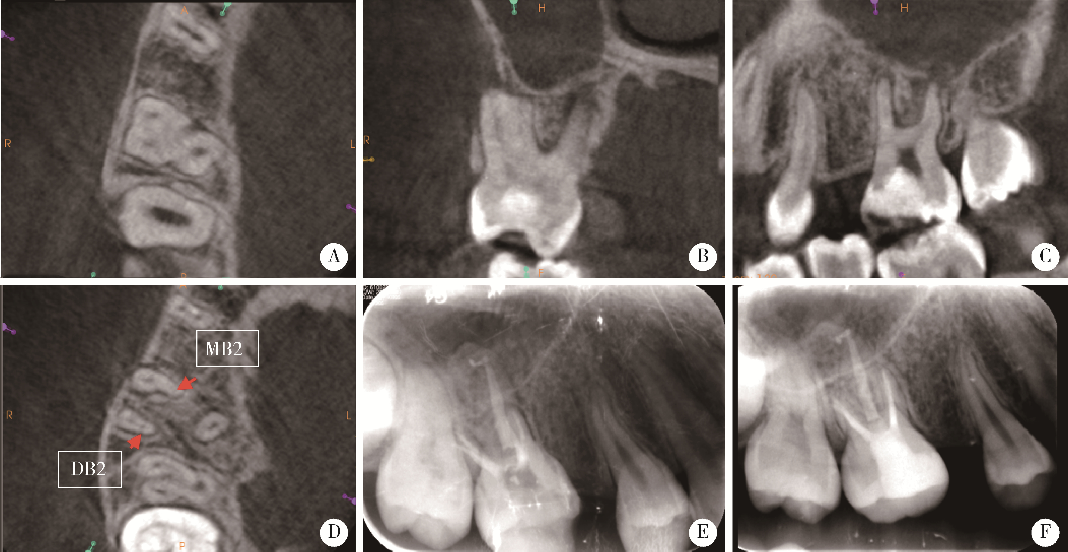
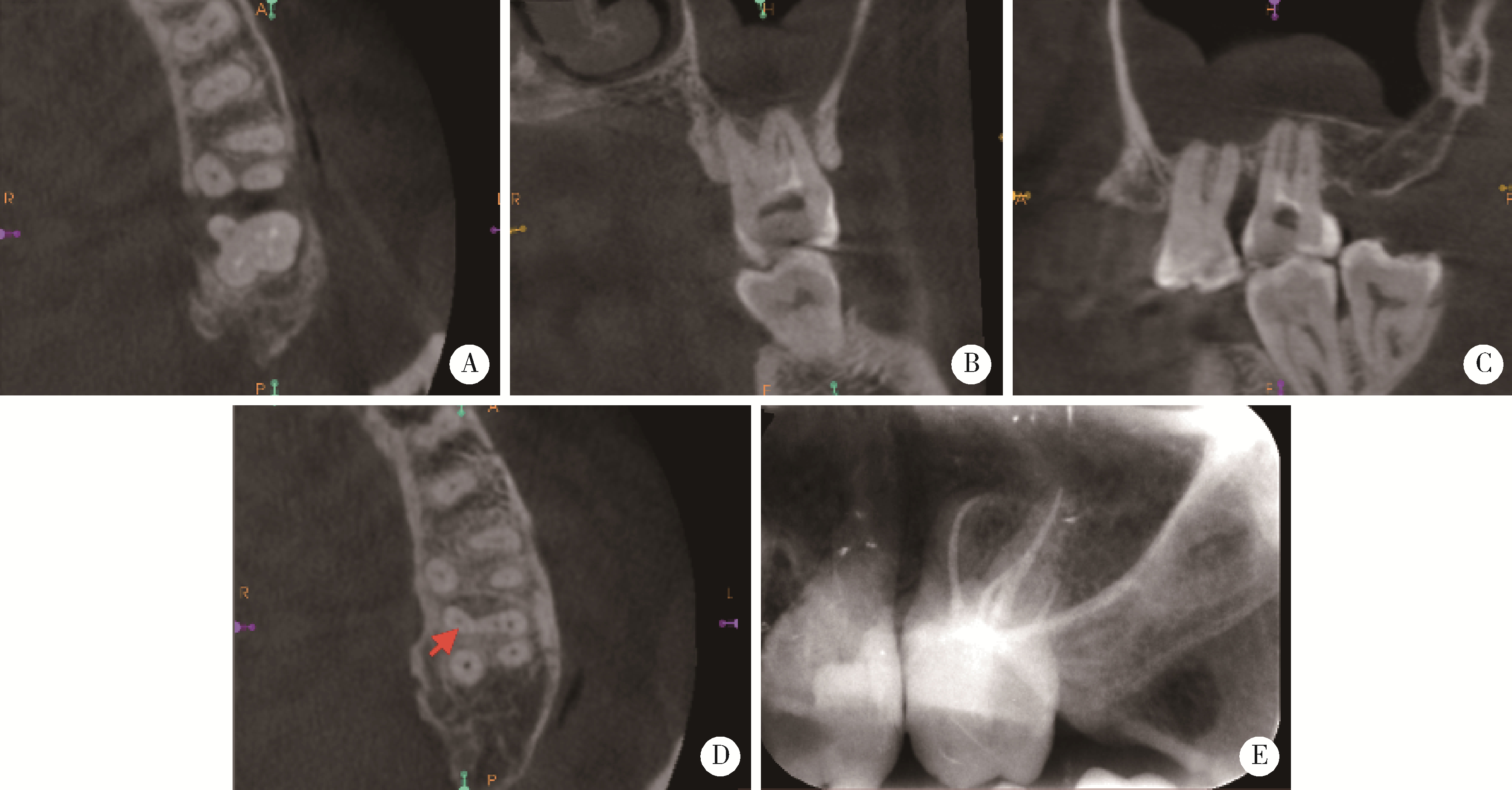
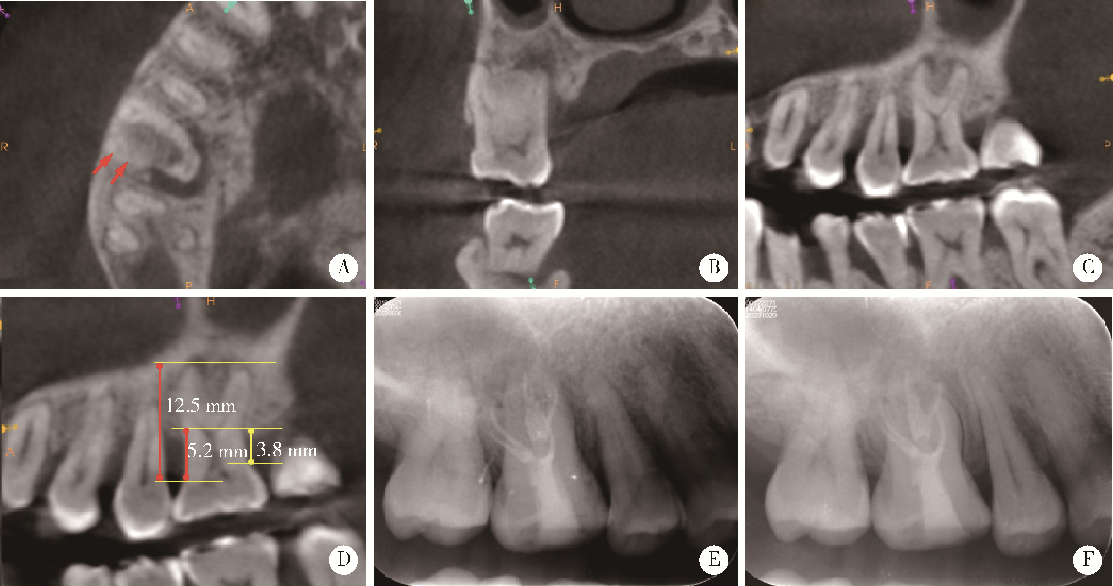
复杂根管上颌磨牙的根管治疗3例
陈晨1,梁宇红2,*( )
)
- 1. 北京大学口腔医学院·口腔医院门诊部, 国家口腔医学中心, 国家口腔疾病临床医学研究中心, 口腔生物材料和数字诊疗装备国家工程研究中心, 口腔数字医学北京市重点实验室, 国家卫生健康委员会口腔医学计算机应用工程技术研究中心, 国家药品监督管理局口腔生物材料重点实验室, 北京 100034
2. 北京大学口腔医学院·口腔医院急诊科, 北京 100081
Root canal therapy of maxillary molars with atypical canals: A report of three cases
Chen CHEN1,Yuhong LIANG2,*()
- 1. First Clinical Division, Peking University School and Hospital of Stomatology & National Center of Stomatology & National Clinical Research Center for Oral Diseases & National Engineering Research Center of Oral Biomaterials and Digital Medical Devices & Beijing Key Laboratory of Digital Stomatology & NHC Research Center of Engineering and Technology for Computerized Dentistry & NMPA Key Laboratory for Dental Materials, Beijing 100034, China
2. Department of Oral Emergency, Peking University School and Hospital of Stomatology, Beijing 100081, China
中图分类号:
- R781.05

| 1 |
Peters OA . Current challenges and concepts in the preparation of root canal systems: A review[J]. J Endod, 2004, 30 (8): 559- 567.
doi: 10.1097/01.DON.0000129039.59003.9D |
| 2 | 高学军, 岳林. 牙体牙髓病学[M]. 2版 北京: 北京大学医学出版社, 2013: 431. |
| 3 |
Tabassum S , Khan FR . Failure of endodontic treatment: The usual suspects[J]. Eur J Dent, 2016, 10 (1): 144- 147.
doi: 10.4103/1305-7456.175682 |
| 4 | Cohen S , Hargreaves KM . Pathways of the pulp[M]. 9th ed St. Louis, Mo: Mosby Elsevier, 2006: 230- 235. |
| 5 |
Cleghorn BM , Christie WH , Dong CC . Root and root canal morphology of the human permanent maxillary first molar: A literature review[J]. J Endod, 2006, 32 (9): 813- 821.
doi: 10.1016/j.joen.2006.04.014 |
| 6 |
Zhang R , Yang H , Yu X , et al. Use of CBCT to identify the morphology of maxillary permanent molar teeth in a Chinese subpopulation[J]. Int Endod J, 2011, 44 (2): 162- 169.
doi: 10.1111/j.1365-2591.2010.01826.x |
| 7 |
Neelakantan P , Subbarao C , Ahuja R , et al. Cone-beam computed tomography study of root and canal morphology of maxillary first and second molars in an Indian population[J]. J Endod, 2010, 36 (10): 1622- 1627.
doi: 10.1016/j.joen.2010.07.006 |
| 8 |
Kim Y , Lee SJ , Woo J . Morphology of maxillary first and second molars analyzed by cone-beam computed tomography in a korean population: variations in the number of roots and canals and the incidence of fusion[J]. J Endod, 2012, 38 (8): 1063- 1068.
doi: 10.1016/j.joen.2012.04.025 |
| 9 |
Alavi AM , Opasanon A , Ng YL , et al. Root and canal morphology of Thai maxillary molars[J]. Int Endod J, 2002, 35 (5): 478- 485.
doi: 10.1046/j.1365-2591.2002.00511.x |
| 10 |
Martins JN , Quaresma S , Quaresma MC , et al. C-shaped maxillary permanent first molar: A case report and literature review[J]. J Endod, 2013, 39 (12): 1649- 1653.
doi: 10.1016/j.joen.2013.06.032 |
| 11 |
Qian Y , Li Y , Song J , et al. Evaluation of C-shaped canals in maxillary molars in a Chinese population using CBCT[J]. BMC Med Imaging, 2022, 22 (1): 104.
doi: 10.1186/s12880-022-00831-4 |
| 12 |
Jabali AH , Chourasia HR , Wasli AS , et al. Taurodontism in maxillary and mandibular molars using cone beam computed tomography in a dental center in Saudi Arabia[J]. Ann Saudi Med, 2021, 41 (4): 232- 237.
doi: 10.5144/0256-4947.2021.232 |
| 13 |
Nair R , Khasnis S , Patil JD . Bilateral taurodontism in permanent maxillary first molar[J]. Indian J Dent Res, 2019, 30 (2): 314- 317.
doi: 10.4103/ijdr.IJDR_770_17 |
| 14 |
叶佳学, 梁宇红. 牙髓专科医师应用锥形束CT的现况调查[J]. 北京大学学报(医学版), 2023, 55 (1): 114- 119.
doi: 10.19723/j.issn.1671-167X.2023.01.017 |
| 15 | Mirza MB , Gufran K , Alhabib O , et al. CBCT based study to analyze and classify root canal morphology of maxillary molars: A retrospective study[J]. Eur Rev Med Pharmacol Sci, 2022, 26 (18): 6550- 6560. |
| 16 |
Tian XM , Yang XW , Qian L , et al. Analysis of the root and canal morphologies in maxillary first and second molars in a Chinese population using cone-beam computed tomography[J]. J Endod, 2016, 42 (5): 696- 701.
doi: 10.1016/j.joen.2016.01.017 |
| 17 |
Felsypremila G , Vinothkumar TS , Kandaswamy D . Anatomic symmetry of root and root canal morphology of posterior teeth in Indian subpopulation using cone beam computed tomography: A retrospective study[J]. Eur J Dent, 2015, 9 (4): 500- 507.
doi: 10.4103/1305-7456.172623 |
| 18 |
Tzeng LT , Chang MC , Chang SH , et al. Analysis of root canal system of maxillary first and second molars and their correlations by cone beam computed tomography[J]. J Formos Med Assoc, 2020, 119 (5): 968- 973.
doi: 10.1016/j.jfma.2019.09.012 |
| 19 |
de Moor RJ . C-shaped root canal configuration in maxillary first molars[J]. Int Endod J, 2002, 35 (2): 200- 208.
doi: 10.1046/j.1365-2591.2002.00461.x |
| 20 |
Newton CW , McDonald S . A C-shaped canal configuration in a maxillary first molar[J]. J Endod, 1984, 10 (8): 397- 399.
doi: 10.1016/S0099-2399(84)80162-4 |
| 21 |
Dankner E , Friedman S , Stabholz A . Bilateral C shape configuration in maxillary first molars[J]. J Endod, 1990, 16 (12): 601- 603.
doi: 10.1016/S0099-2399(07)80204-4 |
| 22 |
Yilmaz Z , Tuncel B , Serper A , et al. C-shaped root canal in a maxillary first molar: A case report[J]. Int Endod J, 2006, 39 (2): 162- 166.
doi: 10.1111/j.1365-2591.2006.01069.x |
| 23 |
Kottoor J , Velmurugan N , Ballal S , et al. Four-rooted maxillary first molar having C-shaped palatal root canal morphology evaluated using cone-beam computerized tomography: A case report[J]. Oral Surg Oral Med Oral Pathol Oral Radiol Endod, 2011, 111 (5): 41- 45.
doi: 10.1016/j.tripleo.2010.12.009 |
| 24 | Joshi C , Joshi S . C-shaped canal in maxillary first molars: A case report[J]. J Dent (Tehran), 2014, 11 (1): 111- 117. |
| 25 | Paksefat S , Rahimi S . Root canal treatment of a two-rooted C-shaped maxillary first molar: A case report[J]. Iran Endod J, 2014, 9 (4): 301- 303. |
| 26 | Kharouf N , Haïkel Y , Mancino D . Unusual maxillary first molars with C-shaped morphology on the same patient: Variation in root canal anatomy[J]. Case Rep Dent, 2019, 2019, 1857289. |
| 27 | Wahane KD , Bansod AV , Mattigatti S , et al. Cone-beam com-puted tomography (CBCT) analysis of an unusual configuration of the upper first molar with a C-shaped canal with apically fused roots: A case report[J]. Cureus, 2023, 15 (3): e36474. |
| 28 |
Chaintiou Piorno R , Consoli Lizzi EP , Gualtieri AF , et al. C-shaped canal system in maxillary molars evaluated by cone-beam computed tomography in an Argentine subpopulation[J]. Acta Odontol Latinoam, 2022, 35 (3): 164- 170.
doi: 10.54589/aol.35/3/164 |
| 29 |
Shifman A , Chanannel I . Prevalence of taurodontism found in radiographic dental examination of 1 200 young adult Israeli patients[J]. Community Dent Oral Epidemiol, 1978, 6 (4): 200- 203.
doi: 10.1111/j.1600-0528.1978.tb01150.x |
| 30 | Keene HJ . A morphologic and biometric study of taurodontism in a contemporary population[J]. Am J Phys Anthrop, 1966, 25, 208- 209. |
| [1] | 赵菡,卫彦,张学慧,杨小平,蔡晴,宁成云,徐明明,刘雯雯,黄颖,何颖,郭亚茹,江圣杰,白云洋,吴宇佳,郭雨思,郑晓娜,李文静,邓旭亮. 口腔硬组织修复材料仿生设计制备和临床转化[J]. 北京大学学报(医学版), 2024, 56(1): 4-8. |
| [2] | 雍颹,钱锟,朱文昊,赵晓一,刘畅,潘洁. 成年恒牙牙髓切断后牙髓钙化的X线片评价[J]. 北京大学学报(医学版), 2023, 55(1): 88-93. |
| [3] | 彭俐,王祖华,孙玉春,渠薇,韩扬,梁宇红. 根尖切除手术导板的计算机辅助设计及三维打印[J]. 北京大学学报(医学版), 2018, 50(5): 905-910. |
| [4] | 谭京,魏秀霞,张庆辉,周永胜. 使用改良半固定桥修复单个缺失后牙的3年临床观察[J]. 北京大学学报(医学版), 2018, 50(2): 314-317. |
| [5] | 焦洋, 王继德, 邓久鹏. 不同表面处理对氧化锆晶相结构及性能的影响[J]. 北京大学学报(医学版), 2018, 50(1): 49-52. |
| [6] | 廖宇,刘晓强,陈立,周建锋,谭建国. 不同表面处理方法对氧化锆与树脂水门汀粘接强度的影响[J]. 北京大学学报(医学版), 2018, 50(1): 53-57. |
| [7] | 张皓羽,姜婷,程明轩,张玉玮. 类瓷树脂及玻璃陶瓷牙合贴面疲劳实验前后的磨耗及表面粗糙度的变化[J]. 北京大学学报(医学版), 2018, 50(1): 73-77. |
| [8] | 唐琳,张一,李皓,刘玉华,周永胜,李博文,吴唯伊,王思雯. 不同浓度的碳二亚胺乙醇溶液处理对牙本质自酸蚀粘接剂粘接强度的影响[J]. 北京大学学报(医学版), 2017, 49(6): 1055-1059. |
| [9] | 李皓,刘玉华,罗志强. 生物活性玻璃用于缓解活髓牙全冠预备后敏感的效果评价[J]. 北京大学学报(医学版), 2017, 49(4): 709-713. |
| [10] | 赵一姣,刘怡,孙玉春,王勇. 一种基于曲率连续算法的冠、根三维数据融合方法[J]. 北京大学学报(医学版), 2017, 49(4): 719-723. |
|
