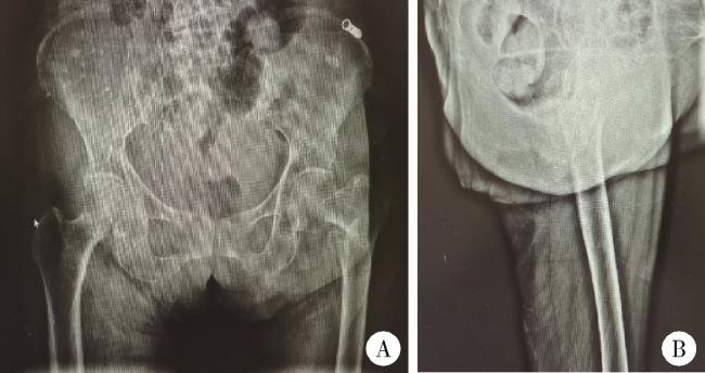
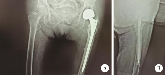
病例报告

股骨粗隆间骨折术后同侧股骨颈骨折1例

  • 宁太国 ,
  • 潘利平 ,
  • 叶一林 , Δ ,
  • 曹永平
展开
  • 北京大学第一医院骨科,北京 100034

* These authors contributed equally to this work

收稿日期: 2022-07-27

  网络出版日期: 2025-06-13

版权

版权所有,未经授权,不得转载。

Ipsilateral femoral neck fracture after fixation of intertrochanteric fracture by InterTAN intramedullary nail: A case report

  • Taiguo NING ,
  • Liping PAN ,
  • Yilin YE , Δ ,
  • Yongping CAO
Expand
  • Department of Orthopaedics, Peking University First Hospital, Beijing 100034, China
YE Yilin, e-mail,

Received date: 2022-07-27

  Online published: 2025-06-13

Copyright

All rights reserved. Unauthorized reproduction is prohibited.

本文引用格式

宁太国 , 潘利平 , 叶一林 , 曹永平 . 股骨粗隆间骨折术后同侧股骨颈骨折1例[J]. 北京大学学报(医学版), 2025 , 57(3) : 610 -613 . DOI: 10.19723/j.issn.1671-167X.2025.03.028

Abstract

Hip fractures are common in elderly patients and are associated with significant morbidity and mortality, often referred to as the "last fracture of life". These fractures frequently result in a loss of functional independence. Evidence suggests that early surgical intervention can reduce mortality. The selection of treatment modality should take into account factors such as the type of fracture, the patient' s age, and overall health status. This case report discusses an 88-year-old female patient who sustained an unstable intertrochanteric fracture of the left femur following a fall. She underwent closed reduction and internal fixation using an InterTAN intramedullary nail, resulting in a satisfactory postoperative recovery. Sixteen months following the surgical procedure, the patient presented with progressive pain in the left hip and ambulatory difficulties, absent from any evident trauma. Radiographic analysis identified a fracture of the left femoral neck accompanied by some degree of acetabular bone degradation attributable to the implant. Subsequently, the patient underwent removal of the internal fixation device and received a hemiarthroplasty. The postoperative course was uneventful, with marked improvements in both pain levels and functional capacity. This case underscored the intricate nature of femoral neck fractures following the internal fixation of intertrochanteric fractures. Contributing factors may include advanced age, osteoporosis, and stress shielding induced by the implant. In patients presenting with hip pain or gait disturbances months to years post-intertrochanteric fracture surgery, the potential for a new fracture should be consi- dered, even in the absence of an explicit traumatic incident. Radiographic imaging is imperative to exclude the presence of a fracture, particularly in individuals with high-risk factors such as advanced age, osteoporosis, alcohol abuse, and a history of hormone therapy. Management of such cases may necessitate the removal of internal fixation devices and the implementation of hemiarthroplasty or total hip arthroplasty, contingent upon the patient ' s surgical tolerance. Crucially, anti-osteoporosis therapy serves as a vital preventive strategy. Considering the high-risk profile of elderly patients with hip fractures, diligent follow-up and timely intervention are paramount to mitigating complications and mortality, thereby enhancing the quality of life for these patients. This case highlights the critical need for increased vigilance and comprehensive management of elderly patients with hip fractures to enhance treatment outcomes and improve prognosis.

随着全球人口的老龄化,髋部骨折患者的数量正在逐渐增加。据估计,到2025年将有260万人遭受髋部骨折,到2050年将达到700万到2 100万人[1]。手术是老年髋部骨折患者的主要治疗方法,保守治疗仅用于骨折前不能行走或不能耐受手术的患者。股骨粗隆间骨折常采用闭合复位内固定的手术方式,影响手术效果的因素包括骨骼强度、复位程度、内固定物的选择、术后疼痛和康复的管理等。术后常见并发症包括骨折不愈合、内植物切出、假体周围骨折、股骨头坏死等[2]。当患者骨折愈合后出现髋部疼痛、步态异常等表现时需警惕相关并发症的发生。现将北京大学第一医院收治的1例股骨粗隆间骨折术后同侧股骨颈骨折病例报道如下。

1 病例资料

老年女性患者,88岁,患者2020年10月26日步行时不慎摔倒,左髋着地。于北京大学第一医院急诊查双髋X线片示左侧股骨粗隆间不稳定骨折(图 1)。查体:生命体征平稳。左髋强迫外展外旋位,左下肢短缩约2 cm。左髋局部压痛,轴向叩击痛,活动度拒查。双下肢感觉无异常,双侧足背动脉搏动可。入院后行左股骨粗隆间骨折闭合复位InterTAN髓内钉固定术,术中复位满意,过程顺利(图 2),术后复查髋关节X线片示骨折复位及内固定物位置满意(图 3)。术后次日开始骨化三醇、碳酸钙D3抗骨质疏松治疗,术后1周加用地舒单抗。术后1个半月部分负重,术后3个月可下地行走,无明显不适。2021年10月7日(术后12个月)无明显诱因出现左髋疼痛,进行性加重,逐渐出现行走困难。2022年2月28日(术后16个月)于北京大学第一医院门诊复查双髋正位X线片考虑左侧股骨颈骨折,内固定物对髋臼侧骨质有一定破坏(图 4),为行治疗收治入院。既往有高血压、腔隙性脑梗、房颤病史。查体:生命体征平稳,左髋关节周围压痛明显,无红肿,关节活动受限,疼痛明显,查体配合差。入院查血常规:白细胞计数7.1×10⁹/L,中性粒细胞计数6.5×109/L,中性粒细胞百分比92.3%,超敏C反应蛋白4.27 mg/L,降钙素原0.02 μg/L,除外感染后行左髋关节内固定物取出加半髋关节置换术,术中未见浑浊关节液及组织液,既往股骨粗隆间骨折部位已完全愈合,新发股骨颈骨折(图 5),髋臼外上方有一处头颈螺钉切割出的缺损区,直径约1.5 cm,内有纤维瘢痕组织填充,取局部组织留取病理,送咽拭子细菌培养,培养结果阴性。术中采用骨水泥型11号股骨假体柄(长度200 mm)和外径44 mm的双极金属股骨头行半髋关节置换术,术后复查髋关节X线片(图 6),可见髋臼侧骨质密度减低。术后预防性抗感染治疗,术后1周出院,院外继续抗骨质疏松治疗。术后3个月患者反馈左髋疼痛及功能明显改善,患者及家属对治疗满意。
图1 患者第一次入院前髋关节X线片

Figure 1 Hip joint X-ray of patient before initial admission

A, radiographic examinations, including X-ray imaging of anteroposterior view of hips; B, lateral view of left hip, indicate the presence of an intertrochanteric fracture in the left femur.
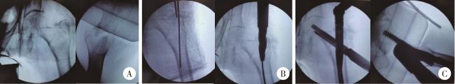
图2 患者股骨粗隆间骨折闭合复位内固定术术中透视X线片

Figure 2 X-ray in closed reduction and internal fixation of femur intertrochanteric fracture of paitient

A, following closed reduction, the X-ray of the left hip confirmed a well-reduced fracture; B, after inserting the guide pin into the femoral medullary cavity, reaming was performed; C, subsequently, the proximal femoral intramedullary nail and lag screw were inserted, and an X-ray of the femur was taken.
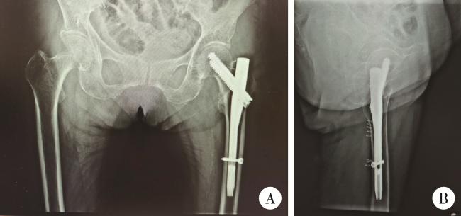
图3 患者左股骨粗隆间骨折闭合复位髓内钉内固定术后X线片

Figure 3 X-ray after closed reduction and intramedullary nail fixation of left femoral intertrochanteric fracture of patient

A, post-operative anteroposterior view of hips; B, lateral view of left hip, showed that the fracture reduction and internal fixation was satisfactory.
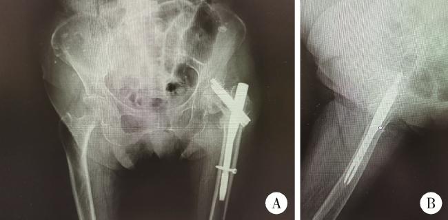
图4 患者术后16个月髋部X线片

Figure 4 Radiographic examination of the hip of patient 16 months postoperatively

A, sixteen months post-operation, X-ray imaging of anteroposterior view of hips; B, lateral view of left hip, revealed a fracture in the left femoral neck and a cut-out screw.
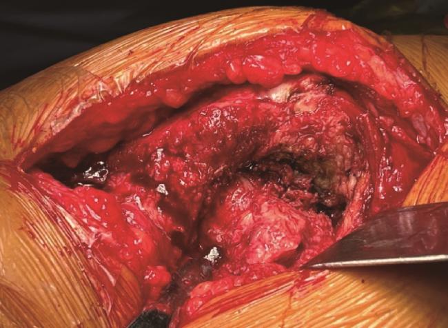
图5 术中所见,患者原股骨粗隆间骨折已愈合,新发股骨颈骨折

Figure 5 During the operation, the patient' s original intertrochanteric femur fracture had healed, revealing a femoral neck fracture

图6 患者左侧半髋关节置换术后X线片

Figure 6 X-ray films of patient after left hemiarthroplasty

A, X-ray films of both hips; B, left hip lateral view, post-left hip hemiarthroplasty.

2 讨论

髋部骨折是一种老年患者常见骨折,具有较高的发病率和死亡率,被称为人生最后一次骨折,骨折后30%~50%的患者失去功能独立性[3]。较早的手术干预有益于降低死亡率[4],治疗的方式取决于骨折类型、患者年龄、一般情况等多种因素。动态髋螺钉(dynamic hip screw,DHS)适用于大多数股骨粗隆间骨折,尤其是稳定性骨折,对于不稳定性骨折,股骨近端髓内钉具有更好的适应性和固定效果[5]。本例患者第一次骨折属于不稳定性骨折,手术方式选择闭合复位髓内钉固定术,复位后在正位和侧位上、前方皮质和内侧皮质均无移位,符合最佳的复位标准[6],术后早期恢复顺利,可下地正常活动。本病例中患者术后早期使用地舒单抗抗骨质疏松治疗,地舒单抗是目前可用于人体治疗的活性最强的核因子-κB受体活化因子配体(receptor activator of nuclear factor-kappa B ligand, RANKL)抑制剂,通过与RANKL结合抑制破骨细胞的生成与功能, 从而减少骨吸收、增加骨量、改善骨强度[7]。在一项随机、双盲、安慰剂对照的Ⅲ期临床试验中,因非椎体骨折接受手术治疗的患者未发现地舒单抗影响骨折的愈合[8]
股骨粗隆间骨折内固定后股骨颈骨折(femoral neck fractures after internal fixation of trochanteric fractures with the implant in situ,FNFAIFTFWIIS)的病因较为复杂,尚不明确,已有的病例报道中提出了众多可能的危险因素,总的来说可以分为两大类,一类是生物因素,主要是骨质疏松症,以及能够影响骨骼强度的危险因素,如高龄、绝经后女性、酗酒、激素治疗史、佩吉特(Paget)骨病、糖尿病、维生素D缺乏、股骨头和股骨颈尺寸较小、股骨颈基底部骨折、粗隆间骨折不愈合、亚临床慢性骨髓炎和继发性股骨头缺血性坏死等[9];另一类是生物力学相关的因素,如手术中骨折复位的情况、固定装置类型、植入物在股骨颈和股骨头的位置不合适、内固定物产生的应力遮挡或者股骨粗隆间骨折后长期的负重受限或部分负重减轻导致局部骨质疏松、进入股骨颈和股骨头的拉力螺钉不稳定、螺钉或钉子回退、滑动髋螺钉失效、颈-干角内翻、严重创伤等[9]。与大多数FNFAIFTFWIIS患者一样,本例患者二次住院前并无明确外伤史,高龄、合并骨质疏松、植入物产生应力遮挡等是其主要危险因素。此外,文献报道我国成人股骨头直径为(46.5±3.5) mm,其中男、女股骨头直径分别为(48.9±2.1) mm和(43.4±2.2) mm,差异有统计学意义[10-11]。本研究中该患者股骨头直径约44.0 mm,女性股骨头相对男性较小,对于较小股骨头和较短股骨颈的患者,髓内钉螺纹附近骨骼承受的应力要高得多,更容易发生FNFAIFTFWIIS。
大多数病例报告推测,局部骨质疏松和应力集中效应是造成FNFAIFTFWIIS的原因[12-13]。Tronzo[13]认为当髓内钉过短,仅到达股骨颈与股骨头交界处时,钉子尖端的重复性微动会导致疲劳性或应力性骨折。Cameron等[12]对此观点持有争议,他们在类似的骨折患者中没有发现小梁应力性骨折,也没有发现皮质骨改变,他们认为该骨折是由于金属和骨骼的刚度不同,在植入物尖端存在应力集中,弹性模量的突然变化会导致骨折,也称为杨氏模量骨折。
有研究者建议手术中将螺钉置于软骨下5~10 mm处,以避免FNFAIFTFWIIS的发生[14],但并不能将FNFAIFTFWIIS的发生归因于此。本研究中患者的螺钉尖端在正位片上距离关节面9 mm,尖顶距(正、侧位X线片上螺钉尖部到股骨头中心的距离之和[15])20 mm。与本病例类似,在大多数病例报告中,尽管软骨下植入物放置正确,但仍会发生FNFAIFTFWIIS,且股骨颈骨折部位远低于钉尖[11, 16]。在大多数情况下,优化手术技术可能无法预防FNFAIFTFWIIS的发生,因此,应该正确预防和处理可干预的危险因素,如骨质疏松[9]
Barquet等[9]总结了104例股骨粗隆间骨折后股骨颈骨折病例,发现该类型的骨折有以下特点:股骨颈骨折发生时间平均在股骨粗隆间骨折后9个月(1~132个月);99%的患者骨折是由低能量创伤所致或自发的骨折;几乎所有该类型股骨颈骨折都是头下型骨折,其中约80%伴有移位;骨折后的主要表现为髋部疼痛或髋部疼痛伴跛行,少数患者无法行走或无明显不适;通过X线片能够诊断,少数患者需要完善髋部CT或磁共振协助诊断;治疗上对于能够耐受手术的患者采取半髋或全髋关节置换。
由于高龄、活动不便、术后死亡率较高等原因导致髋部骨折患者术后随访率低,实际FNFAIFTFWIIS的发生率并没有报道的那么少见[9]。对于股骨粗隆间骨折术后数月到数年内出现髋关节疼痛、跛行的患者都应该引起医师的重视,即使没有明确的外伤史,也应该考虑到骨折的可能,接诊时有必要行影像学检查除外新发骨折,尤其是对有高龄、骨质疏松、酗酒、接受激素治疗等高危因素的患者。考虑到骨折患者大都高龄,对功能要求不高,股骨粗隆间骨折术后再次股骨颈骨折治疗方式可选择内固定物取出加半髋或全髋关节置换,坚持抗骨质疏松治疗是重要的预防措施。

利益冲突  所有作者均声明不存在利益冲突。

作者贡献声明  宁太国:收集、分析、整理数据,撰写论文;潘利平:收集、分析、整理数据;叶一林、曹永平:总体把关和审定论文。

1
Gullberg B , Johnell O , Kanis JA . World-wide projections for hip fracture[J]. Osteoporos Int, 1997, 7 (5): 407- 413.

DOI

2
Tosounidis TH , Castillo R , Kanakaris NK , et al. Common complications in hip fracture surgery: Tips/tricks and solutions to avoid them[J]. Injury, 2015, 46 (Suppl 5): S3- S11.

3
Sing CW , Lin TC , Bartholomew S , et al. Global epidemiology of hip fractures: A study protocol using a common analytical platform among multiple countries[J]. BMJ Open, 2021, 11 (7): e047258.

DOI

4
Blanco JF , da Casa C , Pablos-Hernández C , et al. 30-day morta-lity after hip fracture surgery: Influence of postoperative factors[J]. PLoS One, 2021, 16 (2): e0246963.

DOI

5
Huang SG , Chen B , Zhang Y , et al. Comparison of the clinical effectiveness of PFNA, PFLCP, and DHS in treatment of unstable intertrochanteric femoral fracture[J]. Am J Ther, 2017, 24 (6): e659- e666.

DOI

6
Yoon YC , Oh CW , Sim JA , et al. Intraoperative assessment of reduction quality during nail fixation of intertrochanteric fractures[J]. Injury, 2020, 51 (2): 400- 406.

DOI

7
Lacey DL , Boyle WJ , Simonet WS , et al. Bench to bedside: Elucidation of the OPG-RANK-RANKL pathway and the development of denosumab[J]. Nat Rev Drug Discov, 2012, 11 (5): 401- 419.

DOI

8
Adami S , Libanati C , Boonen S , et al. Denosumab treatment in postmenopausal women with osteoporosis does not interfere with fracture-healing[J]. J Bone Joint Surg Am, 2012, 94 (23): 2113- 2119.

DOI

9
Barquet A , Giannoudis PV , Gelink A . Femoral neck fractures after internal fixation of trochanteric fractures with implants in situ in adults: A systematic review[J]. Injury, 2018, 49 (12): 2121- 2131.

DOI

10
薛磊, 李乾, 胡刚峰, 等. 成人股骨CT建模及解剖学参数三维自动测量[J]. 中华医学杂志, 2019, 99 (39): 3093- 3099.

11
Lung YT , Kam WL , Leung YF , et al. Subcapital femoral neck fracture following successful trochanteric fracture treatment with a dynamic hip screw: A report of five cases[J]. J Orthop Surg (Hong Kong), 2007, 15 (2): 238- 241.

DOI

12
Cameron HU , Pilliar RM , Hastings DE , et al. Iatrogenic subcapital fracture of the hip: A new complication of intertrochanteric fractures[J]. Clin Orthop Relat Res, 1975 (112): 218- 220.

13
Tronzo RG. Fractures of the hip in adults[M]//Tronzo RG. Surgery of the hip joint. Philadelphia: Lea & Febiger, 1973: 526.

14
Syed F , Nunag P , Mustafa A , et al. Ipsilateral intracapsular hip fracture 2 years after fixation of extracapsular fracture by dynamic hip screw[J]. J Orthop Case Rep, 2015, 5 (2): 18- 20.

15
Baumgaertner MR , Curtin SL , Lindskog DM , et al. The value of the tip-apex distance in predicting failure of fixation of peritrochanteric fractures of the hip[J]. J Bone Joint Surg Am, 1995, 77 (7): 1058- 1064.

DOI

16
Kaneko H , Matsuda K , Kim S , et al. Subcapital femoral neck fracture after fixation of an intertrochanteric fracture with a proximal femoral nail: A report of two cases[J]. J Orthop Surg (Hong Kong), 2009, 17 (3): 370- 373.

DOI

文章导航

/