
1 资料与方法
2 结果
表1 6例口腔黏膜嗜酸性溃疡患者临床资料Table 1 Clinical features of 6 cases with eosinophilic ulcer of the oral mucosa |
| Case | Gender | Age/years | Duration/weeks | Location | Range/(cm×cm) | Etiology | EO(×109/L) | Clinical impression | Management | Follow-up/months | Recurrence | Times of other ulcers* |
| 1 | M | 30 | 1 | The dorsum of the tongue | 2.0×1.5 | Stab | 1.66↑ | Malignant tumor? | Resection | 12 | No | 3 |
| 2 | F | 60 | 12 | Lateral margin of the tongue | 1.5×1.0 | Psychological | 0.03 | Malignant tumor | Biopsy | 24 | No | 2 |
| 3 | M | 10 | 4 | Buccal mucosa | 2.0×2.0 | None | - | Malignant tumor | Biopsy | 66 | No | 0 |
| 4 | F | 57 | 2 | Lateral margin of the tongue | 1.5×1.0 | None | - | Malignant tumor | Biopsy | 54 | No | 0 |
| 5 | M | 0.5 | 8 | The belly of the tongue | 1.5×1.0 | Incisor rubbing | 0.85↑ | Tumor? | Resection | 42 | No | 4 |
| 6 | M | 49 | 1.5 | Lateral margin of the tongue | 4.0×3.0 | Bite | 0.07 | Tumor or infection? | Biopsy | 11 | No | 0 |
M, male; F, female;EO, eosinophil. *Other ulcers appeared during the follow-up period. -, data are not collected. |
2.1 典型病例1
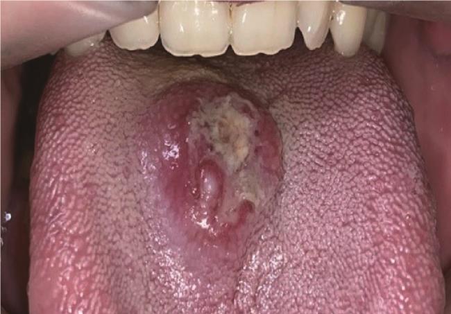
图1 病例1,舌背黏膜溃疡,表面覆盖假膜,周缘隆起Figure 1 Case 1, an ulcer on the dorsum of the tongue, which is covered with pseudomembrane and surrounded by elevated lesion |
2.2 典型病例2
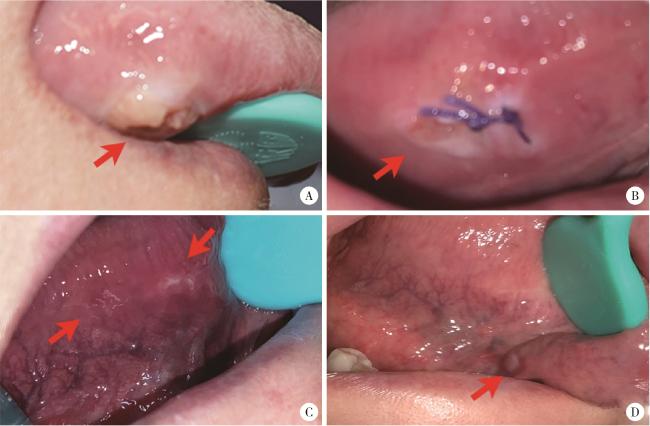
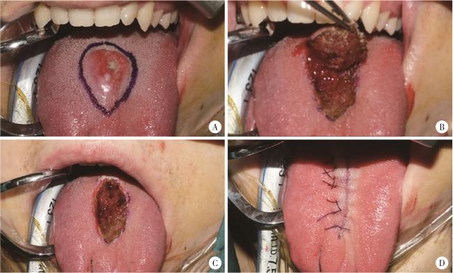
图5 病例2,右舌侧缘溃疡Figure 5 Case 2, an ulcer on the lateral margin of the right tongue A, appearance of ulcer at the initial diagnosis; B, one week after biopsy, the lesion is smaller than before; C, one month later, the ulcer in the anterior part of the right lingual margin disappeared, and an ulcer appeared in the posterior part of the right lingual margin with rough surface; D, one year later, a mung bean-sized ulcer appeared(arrowhead). |
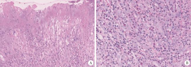
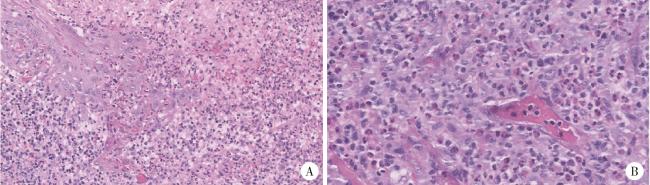
图6 病例2的组织病理学表现Figure 6 Histopathological findings of case 2 A, ulcer on the surface of mucosa with epithelial necrosis, inflammatory cells infiltration in the underlying submucosa (HE ×20); B, mixed chronic inflammatory cell infiltration in the submucosa and muscle,note the presence of many eosinophilic cells (HE ×40). |
