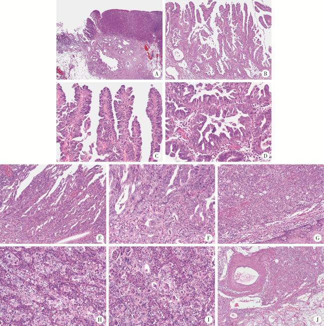
病例报告

食管胃结合部具有显著空泡状核特征的神经内分泌癌1例

  • 侯卫华 1, * ,
  • 宋书杰 2, * ,
  • 石中月 3 ,
  • 刘露 2 ,
  • 金木兰 , 3, *
展开
  • 1. 襄城县人民医院病理科, 河南许昌 461700
  • 2. 解放军联勤保障部队第九八九医院消化内科, 河南平顶山 467099
  • 3. 首都医科大学附属北京朝阳医院病理科, 北京 100020

* These authors contributed equally to this work

收稿日期: 2023-03-15

  网络出版日期: 2025-09-16

基金资助

国家自然科学基金(62176168)

中国消化道早癌医师共同成长计划科研专项基金(GTCZ-2023-HN-01)

版权

版权所有,未经授权,不得转载。

Neuroendocrine carcinoma with significantly vacuolar nucleus at the esophagogastric junction: A case report

  • Weihua HOU 1 ,
  • Shujie SONG 2 ,
  • Zhongyue SHI 3 ,
  • Lu LIU 2 ,
  • Mulan JIN , 3, *
Expand
  • 1. Department of Pathology, Xiangcheng County People's Hospital, Xuchang 461700, Henan, China
  • 2. Department of Gastroenterology, 989 Hospital of Joint Logistic Support Force, Pingdingshan 467099, Henan, China
  • 3. Department of Pathology, Beijing Chaoyang Hospital, Capital Medical University, Beijing 100020, China
JIN Mulan, e-mail,

Received date: 2023-03-15

  Online published: 2025-09-16

Supported by

the National Natural Science Foundation of China(62176168)

Scientific Research Special Fund for the Joint Growth Plan of Chinese Early Gastrointestinal Cancer Physicians(GTCZ-2023-HN-01)

Copyright

All rights reserved. Unauthorized reproduction is prohibited.

摘要

以肿瘤细胞中神经内分泌分化标记物的表达为特征的肿瘤被定义为神经内分泌肿瘤。本文报道了1例在食管胃交界处伴有少量乳头状腺癌成分的具有显著空泡核特征的神经内分泌癌(neuroendocrine carcinomas,NECs)。患者为男性,77岁,吞咽不畅1周。内镜检查提示食管胃结合部早期癌,活检诊断为低分化癌,遂行内镜黏膜下剥离术。组织学上,肿瘤呈现出由衬覆靴钉样细胞特征的典型分枝的乳头状结构(高分化),到乳头的衬覆上皮增生形成筛孔状结构(中分化),再到缺乏乳头结构的实性区(低分化)的连续性过程;在此过程中肿瘤细胞核内空泡呈现由轻微到明显,再到显著空泡特征的连续性变化。肿瘤主要由实性区构成(约占95%),细胞单一,细胞体积大,核圆形或卵圆形至不规则形,核呈显著空泡状,空泡较大的核呈圆圈状,空泡内可见少许稀薄的弱嗜碱性或弱嗜酸细颗粒,空泡边缘不光滑。空泡外侧核染色质细颗粒状,核分裂多见(20~30个/mm2),可见非典型核分裂,核仁易见,细胞质弱嗜酸性,细胞的界线不清楚。细胞排列呈巢状、梁索状或弥漫片状,部分排列呈腺管状。黏膜下层静脉内见癌栓;肿瘤内富含毛细血管的间质中伴有大量中性粒细胞浸润。免疫组织化学染色肿瘤实性区细胞突触素(synaptophysin,Syn)和嗜铬粒蛋白A(chromogranin A,CgA)阳性,而乳头状腺癌阴性。黏蛋白5AC(mucin 5AC,MUC5AC)乳头状腺癌弥漫阳性,而肿瘤实性区细胞阳性比例约为10%~15%。总之,该病例展示了NECs空泡状核特征的极端情况,极为罕见,从某种意义上说,扩展了NECs形态谱的边界; 了解这种核的极端空泡状特征有助于做出正确的病理诊断。

本文引用格式

侯卫华 , 宋书杰 , 石中月 , 刘露 , 金木兰 . 食管胃结合部具有显著空泡状核特征的神经内分泌癌1例[J]. 北京大学学报(医学版), 2025 , 57(5) : 1005 -1009 . DOI: 10.19723/j.issn.1671-167X.2025.05.029

Abstract

Neoplasms characterized by the expression of markers of neuroendocrine differentiation in neoplastic cells are defined as neuroendocrine neoplasms. A case of neuroendocrine carcinomas (NECs) with a small amount of papillary adenocarcinoma and significantly vacuolar nucleus at the esophagogastric junction was reported in this article. A 77-year-old male had dysphagia for one week. Endoscopy revealed early-stage esophagogastric junction carcinoma, and biopsy was diagnosed as poorly differentiated carcinoma. Endoscopic submucosal dissection was performed. Histologically, the papillary adenocarcinoma progresses from typically branching papillary structures (well-differentiated) to hyperplasia of the lining epithelium of the papilla to form a cribriform structure (moderately differentiated), to solid area lacking papillary structures (poorly differentiated). There was a continuous process, and during this process, the vacuoles in the nuclei of tumor cells showed progressive changes from mild to obvious and finally to significant vacuoles. The tumor was mainly composed of solid areas (about 95%), with single cell, large cell, round or oval to irregular nuclei, and significantly vacuolar nuclei, nuclei with larger vacuoles appeared in a loop, a few thin weakly basophilic or weakly eosinophilic fine particles could be seen in the vacuoles, and the vacuoles had rough edges. The nucleus chromatin at the outer edge of the vacuoles was fine particles, and mitosis was common (20-30/mm2), atypical mitosis could be seen, and nucleoli could be seen easily, the cytoplasm was weakly eosinophilic, and the boundaries of cells were unclear. The cells were arranged in a nested, trabecular, or diffuse sheet shape, with some arranged in a glandular tube shape. Tumor thrombus was found in the vein of submucosa; the interstitial tissue rich in capillaries within the tumor was accompanied by a large number of neutrophil infiltration. Immunohistochemical staining showed that the solid area of the tumor was positive for synaptophysin (Syn) and chromogranin A (CgA), while papillary adenocarcinoma was negative. Mucin 5AC (MUC5AC) was diffusely positive in papillary adenocarcinoma, while the proportion of positive cells in the solid area of the tumor was about 10% to 15%. In a word, this case showed the extreme situation of the vacuolar nuclear characteristics of NECs, extremely rare, in a sense, this case expanded the boundary of the morphological spectrum of NECs. Understanding the extreme vacuolar features of this nucleus is helpful to make a correct pathological diagnosis.

以肿瘤细胞中神经内分泌分化标记物的表达为特征的肿瘤被定义为神经内分泌肿瘤(neuroendocrine neoplasms,NENs)[1]。世界卫生组织最近的分类系统将消化道NENs分为高分化神经内分泌瘤(neuroendocrine tumors,NETs)、低分化神经内分泌癌(neuroendocrine carcinomas,NECs)(小细胞和大细胞)和混合性神经内分泌-非神经内分泌肿瘤(mixed neuroendocrine-nonneuroendocrine neoplasms,MiNENs)[2]。根据定义,MiNENs是一个独特的类型,结合了神经内分泌和非神经内分泌成分,在形态学和免疫组织化学上都具有可识别的离散成分,构成肿瘤的每种成分均>30%[2]。大多数MiNENs具有分化不良的NECs成分[3];在超过90%的病例中,非神经内分泌成分由腺癌组成[3-6]。这些混合肿瘤曾被归类为混合性腺神经内分泌癌(mixed adeno-neuroendocrine carcinomas,MANECs)[2]。MANECs预后较差(与单纯NECs重叠),与非神经内分泌成分无关[2-3, 5, 7]。大细胞NECs细胞体积大,核染色空淡,染色质分布不均,核仁明显,胞质嗜酸,排列成器官样结构[2]。NECs的有丝分裂率通常大于20个/mm2,Ki-67阳性指数通常大于60%~70%[2]。本文报道1例食管胃结合部伴有少量乳头状腺癌成分的具有显著空泡状核特征的NECs。

1 病例报告

患者男性,77岁,因吞咽不畅1周就诊。1周前无明显诱因出现吞咽不畅,尤以进食固体食物为著,伴胸闷、乏力和上腹部胀满。门诊内镜检查提示食管胃结合部早期癌,活检诊断为低分化癌,遂入住联勤保障部队第九八九医院。既往有高血压病史40余年。无肿瘤病史,无特殊个人史及家族史。
内镜下于食管胃结合部前侧壁有一菜花样隆起,亚蒂,表面充血、糜烂。胸部CT检查显示食管下段及贲门部管壁不规则增厚,局部可见软组织影充填,管腔明显变窄,周围境界尚清,未见肿大淋巴结。
临床经过:通过内镜观察诊断为食管胃结合部早期癌,癌浸润黏膜下层,巴黎分型0~Ⅰs型(扁平隆起型),无溃疡。判断病变为内镜黏膜下剥离术(endoscopic submucosal dissection,ESD)的相对适应证,结合患者意愿并签署知情同意书后实施ESD治疗。
病理检查:大体观察为不规则黏膜组织一块,面积4.6 cm×4.0 cm,中央区域有一灰红色亚蒂肿物,肿物大小2.5 cm×2.4 cm×1.2 cm,切面实性,灰白色,质地较脆。镜下检查:超低倍观察肿瘤隆起于黏膜表面,局部浸润黏膜下层。肿瘤组织顶部黏膜缺失伴少量坏死,肿瘤周围部分覆盖正常鳞状上皮,部分覆有胃黏膜(图 1)。肿瘤肛侧黏膜内见少量乳头状腺癌成分,表现为有典型分枝的乳头状结构(图 1A~C),乳头表面衬覆靴钉样、柱状和多边形细胞,细胞核大,圆形至不规则形,染色质细颗粒状,部分细胞核呈空泡状,可见核仁及核分裂,核极向紊乱,胞质嗜中性,细胞密集,排列呈单层至多层(图 1CD)。部分乳头的衬覆上皮呈簇状、条索状或假乳头状增生并融合形成筛状结构(图 1EF)。肿瘤的主体呈实性,细胞形态单一,细胞体积大,核圆形或卵圆形至不规则形,核染色质细颗粒状,核呈显著空泡状改变,空泡较大者核呈圈套状,空泡内可见少许稀薄的弱嗜碱性或弱嗜酸细颗粒,空泡边缘不光滑,核分裂多见(20~30个/mm2),可见非典型核分裂,核仁易见,细胞质弱嗜酸性,细胞膜和细胞边界不清。细胞排列呈巢状、梁索状或弥漫片状,部分排列呈腺管状(图 1G~J)。黏膜下层静脉内见癌栓,肿瘤间质内有丰富的毛细血管网伴大量中性粒细胞(图 1HI)。周围鳞状上皮黏膜内有少量炎细胞浸润;胃黏膜内有较多淋巴细胞、浆细胞浸润,固有腺无明显减少,个别腺体有肠上皮化生,局灶腺体呈结节状陷入黏膜下层伴囊性扩张及炎细胞浸润(图 1A);未见幽门螺杆菌。
图1 食管胃结合部具有显著空泡状核特征的神经内分泌癌的形态学特征(HE染色)

Figure 1 Morphological characteristics of neuroendocrine carcinoma with significantly vacuolar nucleus at the esophagogastric junction (HE staining)

A, the hierarchical structure of mucosa and submucosa existed; abnormal branched glands were seen in the tumor area, some were solid, and nodular glands and cystic expansion were seen in the submucosa; B, intramucosal tumor with branching papillary structure; C, D, the surface of the papillary and the glandular was lined with boot-like or cuboidal cells, and a few nuclei were slightly vacuolated; E, F, the lining epithelium of some papillae and glandular cord hyperplasia fused to form a cribriform structure; the nucleus was obviously vacuolated; G, the main body of the tumor is solid, with diffuse arrangement of cells and no involvement of the surface squamous epithelium; H, I, the cells of the solid area were structured with nests, cord, and glandular tubes, with significantly vacuolar changes in nuclei, atypical mitoses, and a large capillary network with a large number of neutrophils in the tumor stroma; J, cancer cell mass seen in submucosa veins. A, 20×; B, G, 100×; C-F, H, I, 400×; J, 40×. HE, hematoxylin and eosin.

病理辅助检查:免疫组织化学染色显示,肿瘤实性区细胞突触素(synaptophysin,Syn)、嗜铬粒蛋白A(chromogranin A,CgA)(图 2AB)和CD56阳性,而乳头状腺癌阴性,胃黏膜内未见神经内分泌细胞异型增生。广谱细胞角蛋白(cytokeratin,CK) 表达于肿瘤细胞的细胞质和细胞膜,而细胞核及核内空泡阴性(图 2C)。p16(图 2DE)、MLH1、MSH2、MSH6和PMS2均阳性。黏蛋白(mucin,MUC)5AC乳头状腺癌弥漫阳性,而肿瘤实性区细胞阳性比例约为10%~ 15%(图 2FG);MUC6、MUC2、CD10、胃蛋白酶原Ⅰ和H+/K+-ATPase均阴性。单纯疱疹病毒(herpes simplex virus,HSV)-1、HSV-2、人类疱疹病毒(human herpesvirus,HHV)-8、腺病毒、S-100、CK14、CK5/6、P40、P63、甲状腺转录因子-1(thyroid transcription factor-1,TTF-1)、程序性死亡配体-1(programmed death-ligand 1,PD-L1)和波形蛋白(vimentin)均阴性,Ki-67阳性指数约80%~90%,p53蛋白呈弥漫阳性的错义突变表达模式(图 2H),HER2(0+)。D2-40显示淋巴管,CD31显示血管,结蛋白(desmin)显示黏膜肌。组织化学EVG染色(elastica van Gieson stain)显示血管壁弹力纤维层。原位杂交技术检测EB病毒编码的小RNA(Epstein-Barr virus-encoded small RNA,EBER),结果为阴性。采用聚合酶链反应-荧光探针法检测人乳头瘤病毒16种常见型别基因,结果均为阴性。
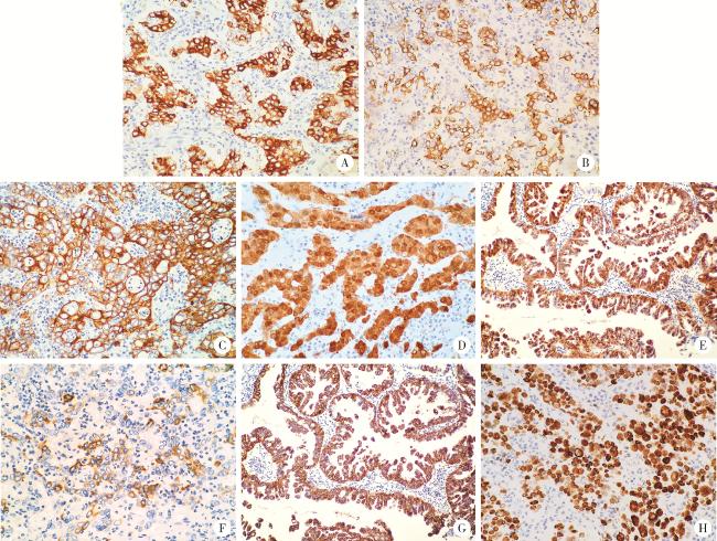
图2 食管胃结合部具有显著空泡状核特征的神经内分泌癌的免疫表型特征(IHC染色)

Figure 2 Immunophenotypic characteristics of neuroendocrine carcinoma with significantly vacuolar nucleus at the esophagogastric junction (IHC staining)

A, the cells of the solid area showed positive staining for chromogranin A (CgA); B, the cells of the solid area showed positive staining for synaptophy-sin (Syn); C, the tumor cells showed cytokeratin-pan (CKpan) positive for cytoplasmic membrane and negative for nucleus and intranuclear vacuoles; D, the cells of the solid area showed diffusely positive staining for P16; E, the papillary adenocarcinoma component showed diffusely positive staining for P16; F, the cells of the solid area showed partially positive for MUC5AC; G, the papillary adenocarcinoma component showed diffusely positive staining for MUC5AC; H, the cells of the solid area showed diffusely positive staining for p53. A, B, E, G, 200×; C, D, F, H, 400×. IHC, immunohistochemical.

病理诊断:食管胃结合部伴有少量乳头状腺癌成分(约5%)的具有显著空泡状核特征的NECs(约95%),肿瘤最大直径2.5 cm,癌浸润模式为浸润式,并浸润破坏黏膜肌进入黏膜下层(浸润深度12 mm,浸润宽度9 mm),静脉内有癌栓,未见淋巴管浸润,水平切缘和垂直切缘阴性(癌距垂直切缘最近距离约200 μm)。肿瘤口侧鳞状上皮黏膜慢性炎,肛侧黏膜慢性浅表性胃炎伴个别腺体肠上皮化生及局灶深在囊性胃炎,幽门螺杆菌阴性。
随访:ESD术后患者拒绝追加外科手术及放疗和化疗。术后56个月内镜和CT检查,未见复发与转移。

2 讨论

食管胃结合部乳头状腺癌是一种分化良好的外生性腺癌,由表面被覆柱状或立方状上皮、纤维血管结缔组织轴心支撑的细长指状突起构成[8]。本例患者乳头状腺癌从典型分枝的乳头状结构(高分化),到乳头的衬覆上皮增生形成筛孔状结构(中分化),再到缺乏乳头结构的实性区(低分化),即存在由高分化腺癌到低分化癌的连续性过程。此过程中可以观察到核内空泡由轻微到明显,再到具有显著空泡特征的连续性变化。肿瘤主要由低分化癌构成,且低分化癌的几乎所有细胞均存在突出的核内空泡,富含毛细血管的间质中伴有大量中性粒细胞浸润。高分化乳头状腺癌的纤细血管结缔组织轴心表面被覆有明显的靴钉样细胞,因此形态学特征十分独特。需要指出的是,核的极端空泡状特征会给肿瘤组织学类型的鉴别诊断带来困难。
在免疫表型方面,MUC5AC由高中分化乳头状腺癌的弥漫表达到低分化癌阳性细胞仅占15%,表明肿瘤细胞出现了胃型黏蛋白的表达丢失;而MUC6、MUC2、CD10、胃蛋白酶原Ⅰ和H+/K+-ATPase阴性进一步支持该肿瘤具有小凹上皮表型或可能起源于小凹上皮,两个区域共同表达p16蛋白是支持二者同源性的又一辅助证据。高中分化乳头状腺癌无神经内分泌表型,p53呈野生型表达,而低分化癌(包括其中的一些腺管样结构)不但表达了神经内分泌标志物,还出现了p53的突变型表达和胃型黏蛋白表型的丢失,表明低分化癌是NECs[9-10]TP53是胃NECs和MiNENs中经常突变的单个基因,类似于胃腺癌的突变谱,而在分化良好的NET中并不常见[2, 5, 10]。目前的普遍观点是,MiNENs是来自单个多能干细胞的克隆性肿瘤[6, 11-12]。一些学者认为MiNENs可能从非NENs开始,然后在某些时候进行反向分化以形成侵袭性NECs成分[5-6],虽然本病例在免疫表型和组织形态学方面均支持这一观点,但由于在该肿瘤中乳头状腺癌成分占比较少(5%),因此尚不能诊断MiNENs或MANECs [2]
NECs的肿瘤细胞可排列呈腺管样结构,因此使用神经内分泌标志物进行免疫组织化学染色对于NENs的诊断至关重要。Jepsen等[13]评估了在食管胃结合部肿瘤中常规进行神经内分泌免疫组织化学染色的必要性,结果发现在262例食管胃结合部腺癌和低分化癌的常规切片中仅有9.2%的病例诊断了NENs,如果没有在所有食管胃结合部肿瘤中常规进行神经内分泌染色,高达22.7%的食管胃结合部NENs将被遗漏。另外,本例患者肿瘤细胞核内空泡角蛋白不着色,因此不支持核内空泡为假包涵体的证据,同时由于EB病毒、单纯疱疹病毒等阴性,可排除病毒感染。本例患者属于早期癌,在ESD术后近5年的随访中尚未见复发和转移,但其更长期的预后还不清楚。本研究的主要不足是缺少基因水平的克隆性肿瘤进化分析,但这并不影响该病例的临床价值。
总之,本病例展示了NECs空泡状核特征的极端情况,从某种意义上说,本例扩展了NECs形态谱的边界,其形态学特征十分独特,极为罕见。了解这种核的极端空泡状特征有助于做出正确的病理诊断,进而指导临床医生选择更恰当的治疗方案。

利益冲突  所有作者均声明不存在利益冲突。

作者贡献声明  侯卫华:资料收集、病理诊断和论文撰写;宋书杰、石中月和刘露:病例讨论和论文修改;金木兰:指导病理诊断,总体把关和审定论文; 所有作者均对最终文稿进行审读并确认。

1
Inzani F , Rindi G . Introduction to neuroendocrine neoplasms of the digestive system: Definition and classification[J]. Pathologica, 2021, 113 (1): 1- 4.

DOI

2
Klimstra DS, Kloppel G, La Rosa S, et al. Classification of neuroendocrine neoplasms of the digestive system[M]//WHO classification of tumours editorial board. WHO classification of tumours, digestive system tumours. 5th ed. Lyon, France: IARC Press, 2019: 16-19.

3
Jacob A , Raj R , Allison DB , et al. An update on the management of mixed neuroendocrine-non-neuroendocrine neoplasms (MiNEN)[J]. Curr Treat Options Oncol, 2022, 23 (5): 721- 735.

DOI

4
La Rosa S , Sessa F , Uccella S . Mixed neuroendocrine-nonneuroendocrine neoplasms (MiNENs): Unifying the concept of a heterogeneous group of neoplasms[J]. Endocr Pathol, 2016, 27 (4): 284- 311.

DOI

5
Frizziero M , Chakrabarty B , Nagy B , et al. Mixed neuroendocrine non-neuroendocrine neoplasms: A systematic review of a controversial and underestimated diagnosis[J]. J Clin Med, 2020, 9 (1): 273.

DOI

6
Toor D , Loree JM , Gao ZH , et al. Mixed neuroendocrine-non-neuroendocrine neoplasms of the digestive system: A mini-review[J]. World J Gastroenterol, 2022, 28 (19): 2076- 2087.

DOI

7
Elpek GO . Mixed neuroendocrine-nonneuroendocrine neoplasms of the gastrointestinal system: An update[J]. World J Gastroenterol, 2022, 28 (8): 794- 810.

DOI

8
Lam AK, Kumarasinghe MP. Adenocarcinoma of the oesophagus and oesophagus and oesophagogastric junction NOS[M]//WHO classification of tumours editorial board. WHO classification of tumours, digestive system tumours. 5th ed. Lyon, France: IARC Press, 2019: 38-43.

9
Tomita Y , Seki H , Matsuzono E , et al. Early gastric mixed neuroendocrine-non-neuroendocrine neoplasm with early poor prognosis after endoscopic submucosal dissection: A case report[J]. DEN Open, 2021, 2 (1): e10.

10
Ishida S , Akita M , Fujikura K , et al. Neuroendocrine carcinoma and mixed neuroendocrine-non-neuroendocrine neoplasm of the stomach: A clinicopathological and exome sequencing study[J]. Hum Pathol, 2021, 110, 1- 10.

DOI

11
La RS . Challenges in high-grade neuroendocrine neoplasms and mixed neuroendocrine/non-neuroendocrine neoplasms[J]. Endocr Pathol, 2021, 32 (2): 245- 257.

DOI

12
Guerrera LP , Suarato G , Napolitano R , et al. Mixed neuroendocrine non-neuroendocrine neoplasms of the gastrointestinal tract: A case series[J]. Healthcare (Basel), 2022, 10 (4): 708.

13
Jepsen DNM , Fiehn AK , Garbyal RS , et al. Immunohistochemical staining with neuroendocrine markers is essential in the diagnosis of neuroendocrine neoplasms of the esophagogastric junction[J]. Appl Immunohistochem Mol Morphol, 2021, 29 (6): 454- 461.

DOI

文章导航

/