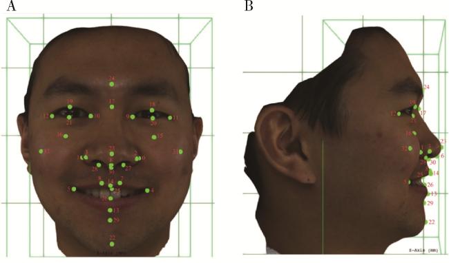
论著

姿势性微笑的三维形态学研究

  • 肖宇嘉 ,
  • 毛渤淳 ,
  • 周彦恒 , *
展开
  • 北京大学口腔医学院·口腔医院正畸科,国家口腔医学中心,国家口腔疾病临床医学研究中心,口腔生物材料和数字诊疗装备国家工程研究中心,口腔数字医学北京市重点实验室,国家卫生健康委员会口腔医学计算机应用工程技术研究中心,国家药品监督管理局口腔生物材料重点实验室,北京 100081

收稿日期: 2022-10-08

  网络出版日期: 2024-04-03

基金资助

国家自然科学基金(62076011)

北京大学口腔医学院青年科研基金(PKUSS20200114)

版权

版权所有,未经授权,不得转载。

Three-dimensional morphological analysis of posed smile

  • Yujia XIAO ,
  • Bochun MAO ,
  • Yanheng ZHOU , *
Expand
  • Department of Orthodontics, Peking University School and Hospital of Stomatology & National Center of Stomatology & National Clinical Research Center for Oral Diseases & National Engineering Research Center of Oral Biomaterials and Digital Medical Devices & Beijing Key Laboratory of Digital Stomatology & NHC Research Center of Engineering and Technology for Computerized Dentistry & NMPA Key Laboratory for Dental Materials, Beijing 100081, China
ZHOU Yanheng, e-mail,

Received date: 2022-10-08

  Online published: 2024-04-03

Supported by

the National Natural Science Foundations of China(62076011)

the Research Foundation of Peking University School and Hospital of Stomatology(PKUSS20200114)

Copyright

All rights reserved. Unauthorized reproduction is prohibited.

摘要

目的: 研究姿势性微笑时三维面部软组织变化和对称性,并分析姿势性微笑的性别差异,同时验证姿势性微笑的一致性。方法: 应用光学面部三维扫描设备获取41名成年志愿者每一张休息位和两张姿势位的面部软组织图像,其中男性16人,女性25人,年龄(26.76±2.70)岁。将面部图像数据导入三维分析软件进行模型定位后应用三维可变模型(3-dimensional morphable face model method, 3DMM)标定软组织特征点,选取眼部、面颊部、鼻部及口周测量指标进行软组织分析,比较两种表情状态下的面中下部软组织变化情况和对称性,并分析男女差异,同时对两次姿势性微笑的测量结果进行统计学检验。结果: 与休息位相比,除鼻唇角变化量(1.45°±7.65°)差异无统计学意义外,姿势性微笑时其余软组织测量值均有改变,且眼部区域也有显著变化(P < 0.001)。面下部软组织主要表现为鼻基底变宽,上下唇区域后向运动,颏部前移,唇红变窄变薄,颏唇沟变浅;姿势性微笑时不对称性以口角点[2.78 (1.73,3.49) mm]、眶下中点[2.36 (1.22, 3.27) mm]和外眼角点[2.31(1.29,2.80) mm]最为显著,另外与休息位相比,除口角点和鼻翼基部外不对称性变化差异无统计学意义(P>0.05);姿势性微笑时,男性右眼高和下唇红深度变化量大于女性(P < 0.05),外眼角点和脸颊点的不对称性增加程度较女性大(P < 0.05);两次姿势性微笑的一致性较好。结论: 姿势性微笑时眼部、面颊部、鼻部及口周软组织存在不同程度的变化,且口角和鼻翼基部不对称性较休息位增加;另外,姿势性微笑时面部软组织的一致性较好,可为临床面部微笑美学研究提供参考。

本文引用格式

肖宇嘉 , 毛渤淳 , 周彦恒 . 姿势性微笑的三维形态学研究[J]. 北京大学学报(医学版), 2025 , 57(5) : 989 -995 . DOI: 10.19723/j.issn.1671-167X.2025.05.026

Abstract

Objective: To investigate the changes and symmetry of facial soft tissue during posed smile, to analyze the feature of posed smile in different gender, and verify the reproducibility of posed smile. Methods: Three-dimensional (3D) facial images of 41 adults (16 males and 25 females with an average age of 26.76±2.70 years) which were taken by FaceScan three-dimensional sensor, including one rest position and two posed smile images. Then these images were imported into 3D soft tissue analysis software for model repositioning. 3D morphable model method (3DMM) was carried out to automatic landmarks setting. After that, the measurement of the eyes, cheeks, nose and perioral area were carried out for 3D soft tissue analysis. Finally, the changes and symmetry of the soft tissues between the two expression states and the gender differences during the posed smiles were compared. Meanwhile, the reproducibility of posed smile was statistically tested. Results: Compared with the rest position, except for nasolabial angle (1.45°±7.65°), the measurements of 3D soft tissue in other region were changed in posed smile (P < 0.001). It should be noted that the eye region was also significantly changed (P < 0.001). Furthermore, the prominent feature of posed smile was that the alar base length became longer, the upper and lower vermilions were narrow and thin, and the mentolabial furrows became shallow. Meanwhile the chin extended anteriorly while the mouth retracted; During posed smile, the labial fissure asymmetry [2.78 (1.73, 3.49) mm], mid-infraorbital asymmetry [2.36 (1.22, 3.27) mm] and outercanthal asymmetry [2.31(1.29, 2.80) mm] were most apparent. Compared with the rest position, the asymmetry was not significantly increased except for cheilion and alar curvature points during the posed smile (P>0.05). In the posed smile, the changes of the right palpebral fissure height and the thickness of lower vermilion (|Li-Stoi| z) of males were greater than those of females (P < 0.05), and asymmetry of exocanthion and cheekbone increased more than that of females (P < 0.05). There was no obvious difference between the two posed smiles. Conclusion: In this study, during the posed smile the soft tissues of the eyes, cheeks, nose, lips and chin changed in different degrees, and the asymmetry of cheilion and alar curvature point was greater than that of the rest position; In addition, the reproducibility of posed smile was excellent, which can be a reference for clinical aesthetics and functional research of smile.

微笑是一种具有美感的面部表情,在人与人的交往中常传达愉快、积极的情绪,或是作为社交场合下的一种礼仪。而姿势性微笑,又称为社会性微笑具有较高的再现性[1-2],在微笑美学研究、临床辅助诊断中比较常用[3-5],可以为口腔正畸和修复治疗提供参考。另外,对于因炎症、肿瘤、创伤或先天性疾病导致的面部表情异常或面瘫患者,研究姿势性微笑时面部软组织的变化规律,通过面部拟态指导术后功能恢复也具有重要意义[6-7]。以往关于姿势性微笑的研究,多是通过二维照片测量唇齿关系,聚焦于上下唇口周组织[8],且少有关于面中下区域软组织的线角测量。随着三维立体摄影系统的出现,在更方便捕捉稳定的姿势性微笑的同时,也提供了面部更多的三维信息。并且,通过三维可变模型(three-dimensional morphable face model method, 3DMM)实现对目标人脸的三维定点方式[9],已经被证实具有高于手动定点的精确性,但目前该方法较少用于面部三维微笑的研究。本研究旨在尝试运用3DMM的方法探究姿势性微笑时三维面部软组织的变化规律及对称性,同时分析姿势性微笑的性别差异,并验证其一致性。

1 资料与方法

1.1 研究对象

2021年9月至2022年5月在北京大学口腔医学院研究生中招募志愿者,共获取41名研究生志愿者每人的1张休息位和2张姿势性微笑位的三维面扫图像,其中男性16人,女性25人,年龄25~36岁,平均(26.76±2.70)岁。本研究开始前已经北京大学口腔医院医学伦理委员会审查批准(PKUSSIRB-202058135),所有志愿者均签署知情同意书,个别研究对象(图 12)取得了肖像应用的本人书面许可。
图1 三维面部软组织特征点标定

Figure 1 Landmarks of 3D facial soft tissue

A, front view of 3D facial landmarks; B, lateral profile of 3D facial landmarks. 0, alar curvature point (L); 1, alar curvature point (R); 2, alare (L); 3, alare (R); 4, cheilion (L); 5, cheilion (R); 6, columella; 7, crista philtri (L); 8, crista philtri (R); 9, endocanthion (L); 10, endocanthion (R); 11, exocanthion (L); 12, exocanthion (R); 13, labrale inferius (Li); 14, labrale superius (Ls); 15, mid-infraorbital (L); 16, mid-infraorbital (R); 17, nasion; 18, palpebrale superius (L); 19, palpebrale superius (R); 20, palpebrale inferius (L); 21, palpebrale inferius (R); 22, pogonion (Pg); 23, pronasale (Prn); 24, glabella (Gl); 25, stomion superius (Stos); 26, stomion inferius (Stoi); 27, subalare (L); 28, subalare (R); 29, sublabial (Sl); 30, subnasale (Sn); 31, cheekbone (L); 32, cheekbone (R).

图2 面部对称性测量示意图

Figure 2 Schematic of facial symmetry measurement

Measurement items of symmetry in posed smile: 1, endocanthion; 2, exocanthion; 3, mid-infraorbital; 4, cheekbone; 5, alar curvature point; 6, alare; 7, subalare; 8, crista philtri; 9, cheilion.

纳入标准:年龄≥18岁;面型协调,软组织无异常,无正颌外科、整形外科治疗史;牙弓形态基本正常,牙齿排列基本整齐,磨牙中性关系,前牙覆牙合覆盖正常;无严重错位、间隙、前牙缺失等。
排除标准:颌面部软组织发育缺陷或软组织不对称超过5 mm者;面部神经肌肉系统疾病者;不能保持拍摄时面部固定及表情稳定者;佩戴唇侧固定矫治器及隐形矫治器者;严重的错牙合畸形者。

1.2 面部休息位和姿势性微笑位信息获取

采用面部扫描设备FaceScan (3DShape, Erlangen, Germany) 获取三维数据,流程及拍摄要求如下:受试者端坐于仪器正中前方,距离相机组件约2 m;受试者拍摄时暴露额头及双耳不能有眼镜、饰品等阻挡;受试者于休息位时,嘴部放松,无紧咬牙及张口等,眼睛平视前方镜头,头部无偏转;嘱受试者对着镜子自然微笑或发“E”音辅助训练[10],训练至表情达到稳定后,间隔5 min重复拍摄两张。将拍摄获取的数据导入3D viewer1.56软件(3Dshape, Erlangen, Germany)进行过滤平滑处理,最终以. obj格式导出。利用三维数据处理分析软件Cliniface 5.2.1(Unlocking Facial Clubs, Australia),对开源非刚性配准程序MeshMonk中的标准人脸模型进行配准[11],即应用一个由7 160个点与14 050个三角面片组成的三维可变模型,模型自动具有三维坐标信息,以过所有面部的中点定为正中矢状面,以眶耳平面为水平面,以垂直于两平面定为冠状面,通过非刚性变形映射于目标人脸进行精细配准及模型定位,并自动标定33个特征点(图 1),最后对有偏差的标志点进行人工修正。上述所有标志点及测量项目均由同一位正畸医生完成。

1.3 三维测量项目

姿势性微笑的三维软组织改变、对称性及一致性:当三维模型重定位及标志点选定后,利用Clinifac5.2.1软件的“Measurements Brower”及“Add Calliper Measurements”功能,对眼部、面颊部、鼻部和口周软组织进行测量(表 1)。对休息位及两次姿势位微笑的三维面像数据分别进行测量,选取9组标志点,以正中矢状面为对称平面,比较对应标志点的三维坐标偏差量以衡量左右面部的不对称性(图 2)。比较两次姿势性微笑的软组织测量值,评估姿势性微笑的一致性。
表1 休息位和姿势性微笑三维软组织测量项目定义

Table 1 Definitions of 3D soft tissue measurements of rest position and posed smile

Measurement items Definition
Palpebral fissure length (L/R)/mm Horizontal distance between the exocanthion and endocanthion
Palpebral fissure height (L/R)/mm Vertical distance between the upper and lower eyelids
Facial convexity/(°) Angle at Sn subtended by side Gl-Pg
Cheekbone width/mm Distance between the L/R cheekbone
Outer canthal nasal angel/(°) Angle at Sn subtended by the outer canthi of the eyes
Nasal width/mm Horizontal width of the nose at the base of the nasal alare
Nasolabial angle /(°) Angle at Sn subtended by side Prn-Ls
Philtral length/mm Distance between the Sn and Ls
Philtral width/mm Distance between the philtral ridges,measured just above the vermilion border
Labial width/mm Distance between L/R cheilion
Upper vermilion height/mm Vertical distance between Sn and Stos
Upper lip height/mm Vertical distance between Sn and Ls
Lower vermilion height/mm Vertical distance between Sl and Li
Lower lip height/mm Vertical distance between Sl and Li
Upper lip protrusion(|Prn-Ls|z)/mm Sagittal distance between Prn and Ls
Lower lip protrusion(|Prn-Li|z)/mm Sagittal distance between Prn and Li
Mentolabial furrows depth(|Ls-Sl|z)/mm Sagittal distance between Li and Sl
Thickness of upper vermilion (|Ls-Stos|z)/mm Sagittal distance between Ls and Stos
Thickness of lower vermilion (|Li-Stoi|z)/mm Sagittal distance between Li and Stoi

L, left; R, right.

姿势性微笑的性别差异:计算不同性别受试者第一次姿势性微笑与休息位的软组织测量指标(图 2)和对称性的差值,以排除自身软组织不对称性的影响,评估姿势性微笑过程中软组织变化情况和对称性改变的男女差异。

1.4 统计学分析

使用SPSS 26.0软件进行分析,计量数据若符合正态分布,以均数±标准差表示;若成偏态分布,以M(P25P75)表示。随机选取姿势性微笑和休息位各5幅图像,间隔一周重复定点,一致性结果ICC均大于0.8。对休息位和第一次姿势性微笑位时的数据进行软组织变化和对称性比较,将两次姿势性微笑的软组织测量结果进行对比,若数据为正态分布,采用配对样本t检验,若数据呈偏态分布,采用Wilcoxon配对样本秩和检验;另外,进行软组织变化情况和对称性的性别评估时,若数据呈正态分布,采用独立样本t检验;若数据呈偏态分布,则采用Mann-Whitney U检验,P < 0.05认为差异有统计学意义。

2 结果

姿势性微笑的软组织改变: 与休息位相比,姿势性微笑时眼部、面颊部、鼻部和口周区域的软组织具有不同程度的变化(表 2)。眼高、上唇部高度、下唇红高、颏唇沟深度在姿势性微笑时均减小,差值具有统计学意义(P < 0.05), 而鼻唇角在不同面部表情状态下差异无统计学意义(P=0.231),其余测量值如上下唇区域的深度,面突角,眼部、脸颊、鼻翼基部和唇周组织的宽度,在姿势性微笑时均大于休息位,差异具有统计学意义(P < 0.05),面中下软组织整体呈向后向外运动趋势。不同性别的受试者在姿势性微笑时,软组织变化值的增减趋势基本一致,仅在右侧眼高变化量和下唇红深度变化量上男性大于女性(P < 0.05),其余软组织变化量在不同性别之间差异均无统计学意义(P>0.05, 表 3)。
表2 姿势性微笑与休息位三维软组织测量结果

Table 2 Results of 3D soft tissue measurements in posed smile and rest position

Measurement items Rest (n=41) Posed smile (n=41) Deviation t P
Palpebral fissure length (L)/mm 31.28±1.66 31.97±1.90 0.69±1.16 3.79 < 0.001
Palpebral fissure height (R)/mm 31.86±1.58 32.80±2.06 0.94±1.39 4.33 < 0.001
Palpebral fissure height (L)/mm 10.29±1.46 8.47±1.39 -1.81±1.45 -8.00 < 0.001
Palpebral fissure height (R)/mm 10.48±1.42 8.52±1.35 -1.96±1.37 -9.11 < 0.001
Facial convexity /(°) 169.64±5.27 171.19±4.93 1.54±1.68 5.88 < 0.001
Cheekbone width/mm 107.46±3.73 112.75±4.11 5.30±1.95 17.37 < 0.001
Outer canthal nasal angel/(°) 93.76±3.64 96.42±3.93 2.66±1.77 9.62 < 0.001
Nasal width/mm 34.58±2.32 36.72±2.33 2.14±0.89 15.43 < 0.001
Nasolabial angle/(°) 98.30±8.46 99.75±7.95 1.45±7.65 1.22 0.231
Philtral length/mm 15.17±1.74 12.82±1.77 -2.35±1.34 -11.27 < 0.001
Philtral width/mm 13.52±1.39 14.71±1.37 1.19±1.33 5.74 < 0.001
Labial width/mm 49.26±4.36 59.65±5.24 10.39±3.51 18.98 < 0.001
Upper vermilion height/mm 7.50±1.11 5.51±1.19 -1.99±1.12 -11.32 < 0.001
Upper lip height/mm 14.52±1.70 12.57±1.66 -1.96±1.40 -8.93 < 0.001
Lower vermilion height/mm 8.90±1.54 8.42±1.67 -0.48±1.51 -2.05 0.047
Lower lip height/mm 7.00±1.84 7.92±1.92 0.92±1.40 4.21 < 0.001
Upper lip protrusion(|Prn-Ls|z)/mm 8.70±2.24 11.33±2.34 2.63±1.16 14.51 < 0.001
Lower lip protrusion(|Prn-Li|z)/mm 12.17±3.56 14.13±3.39 1.96±1.28 9.82 < 0.001
Mentolabial furrows depth(|Ls-Sl|z)/mm 5.45±1.45 4.18±1.75 -1.27±1.07 -7.64 < 0.001
Thickness of upper vermilion (|Ls-Stos|z)/mm 4.43±1.30 5.59±1.85 1.16±1.81 4.11 < 0.001
Thickness of lower vermilion (|Li-Stoi|z)/mm 1.26±1.29 3.62±1.81 2.36±1.91 7.89 < 0.001

Data are $\bar x \pm s$. L, left; R, right.

表3 不同性别受试者姿势性微笑时三维软组织变化量的比较

Table 3 Comparison of 3D soft tissue changes in posed smile of different gender

Deviation (posed smile-rest) Female (n=25) Male (n=16) t P
Palpebral fissure length (L)/mm 0.77±1.04 0.56±1.35 -0.52 0.610
Palpebral fissure height (R)/mm 0.80±1.38 1.16±1.42 0.80 0.431
Palpebral fissure height (L)/mm -1.33±1.20 -2.57±1.53 -2.76 0.100
Palpebral fissure height (R)/mm -1.38±1.03 -2.86±1.38 -3.69 0.010#
Facial convexity /(°) 1.53±1.59 1.57±1.87 0.09 0.932
Cheekbone width /mm 5.22±1.76 5.43±2.28 0.32 0.750
Outer canthal nasal angel/(°) 2.54±1.85 2.84±1.68 0.54 0.593
Nasal width/mm 2.23±1.00 2.00±0.69 -0.85 0.399
Nasolabial angle /(°) 0.96±6.60 2.23±9.24 0.48 0.638
Philtral length/mm -2.19±1.13 -2.61±1.62 -0.90 0.377
Philtral width/mm 1.33±1.39 0.97±1.25 -0.87 0.392
Labial Width/mm 11.09±3.66 9.29±3.04 -1.70 0.097
Upper vermilion height/mm -1.83±0.87 -2.23±1.43 -1.02 0.321
Upper lip height/mm -1.77±1.10 -2.26±1.78 -0.99 0.332
Lower vermilion height/mm -0.81±1.39 0.04±1.57 1.78 0.086
Lower lip height /mm 0.75±1.36 1.18±1.46 0.94 0.356
Upper lip protrusion(|Prn-Ls|z)/mm 2.53±0.86 2.78±1.53 0.60 0.556
Lower lip protrusion(|Prn-Li|z)/mm 2.06±1.29 1.81±1.29 0.60 0.552
Mentolabial furrows depth(|Ls-Sl|z)/mm -1.44±1.08 -1.01±1.01 1.30 0.205
Thickness of upper vermilion (|Ls-Stos|z)/mm 1.02±1.33 1.39±2.42 0.59 0.583
Thickness of lower vermilion (|Li-Stoi|z)/mm 1.67±1.82 3.45±1.56 3.33 0.002#

Data are $\bar x \pm s$. L, left; R, right. # P < 0.05.

姿势性微笑的不对称性变化: 姿势性微笑时口角点不对称性最为明显[2.78 (1.73,3.49) mm]。另外,在姿势性微笑时口角点(Z=3.226,P=0.001)和鼻翼基部(Z=2.035,P=0.042)的不对称性较休息位显著增加,余标志点除鼻下点外不对称性均有所增加但差异无统计学意义(P>0.05,表 4)。对于不同性别的受试者,男性在姿势性微笑时外眼角点和脸颊点处不对称性增加程度较女性大,且差异具有统计学意义(P < 0.05,表 5)。
表4 姿势性微笑与休息位时左右面部对称性

Table 4 Symmetry in posed smile and rest position /mm

Symmetry Rest(n=41) Posed smile (n=41) Deviation Z P
Endocanthion 1.35 (0.95,1.70) 1.51 (0.95,1.77) 0.01 (-0.53,0.61) 0.60 0.551
Exocanthion 2.25 (1.42,2.93) 2.31 (1.29,2.80) -0.05 (-0.80,0.52) 0.51 0.613
Mid-infraorbital 2.25 (1.18,2.91) 2.36 (1.22,3.27) 0.26 (-0.74,0.89) 0.98 0.330
Cheekbone 1.16 (0.74,1.30) 1.25 (0.81,1.60) 0.23 (-0.29,0.71) 1.72 0.085
Alar curvature point 1.08 (0.74,1.45) 1.27 (0.60,1.77) 0.15 (-0.17,0.56) 2.04 0.042
Alare 1.12 (0.68,1.32) 1.19 (0.78,1.64) 0.07 (-0.22,0.35) 1.08 0.282
Subalare 1.21 (0.78,1.58) 1.14 (0.71,1.54) -0.21 (-0.41,0.30) 0.87 0.385
Crista philtri 1.12 (0.72,1.26) 1.18 (0.66,1.41) 0.00 (-0.43,0.55) 0.28 0.778
Cheilion 1.93 (1.13,2.42) 2.78 (1.73,3.49) 1.07 (-0.10,2.03) 3.23 0.001

Data are M(P25, P75).

表5 不同性别受试者姿势性微笑时对称性变化的比较

Table 5 The change of symmetry in posed smile of different gender /mm

Symmetry (posed smile-rest) Female(n=25) Male(n=16) Z P
Endocanthion 0.01 (-0.37,0.87) -0.45 (-0.73,0.53) -1.22 0.227
Exocanthion -0.48 (-1.08,0.15) 0.20 (-0.13,0.85) -2.38 0.017
Mid-infraorbital 0.28 (-0.67,0.83) 0.03 (-0.76,1.06) -0.04 0.968
Cheekbone 0.05 (-0.47,0.35) 0.49 (-0.15,1.11) -2.13 0.032
Alar curvature point 0.14 (-0.14,0.45) 0.35 (-0.38,0.76) -0.83 0.419
Alare 0.07 (0.07,0.29) 0.20 (-0.30,0.54) -0.98 0.333
Subalare -0.27 (-0.49,0.16) -0.12 (-0.30,0.43) -1.67 0.095
Crista philtri 0.10 (-0.33,0.61) -0.41 (-0.66,0.47) -1.74 0.085
Cheilion 1.30 (0.04,2.03) 0.62 (-0.53,2.02) -0.54 0.606

Data are M(P25, P75).

姿势性微笑的一致性:对两次姿势性微笑的软组织测量值进行比较(表 6),所有的测量指标在两组间差异均无统计学意义(P>0.05)。姿势性微笑在眼部、面颊部、鼻部及口周区域的一致性均较好。
表6 姿势性微笑的一致性

Table 6 Reproducibility of posed smile

Measurement items Posed smile 1 (n=41) Posed smile 2 (n=41) Deviation t P
Palpebral fissure length (L)/mm 31.97±1.90 31.75±1.79 -0.22±1.06 -1.31 0.197
Palpebral fissure height (R)/mm 32.80±2.06 32.59±1.67 -0.21±1.01 -1.31 0.197
Palpebral fissure height (L)/mm 8.47±1.39 8.54±1.29 0.07±0.75 0.56 0.581
Palpebral fissure height (R)/mm 8.52±1.35 8.65±1.41 0.12±0.74 1.00 0.325
Facial convexity/(°) 171.19±4.93 171.16±5.00 -0.03±0.82 -0.26 0.799
Cheekbone width/mm 112.75±4.11 112.77±4.15 -0.02±1.35 0.10 0.932
Outer canthal nasal angel/(°) -96.42±3.93 -96.04±3.67 0.38±1.55 1.56 0.126
Nasal width/mm 36.72±2.33 36.77±2.30 0.05±0.56 0.59 0.560
Nasolabial angle /(°) 99.75±7.95 99.93±7.05 0.18±4.65 0.24 0.809
Philtral length/mm 12.82±1.77 12.75±1.96 -0.07±1.10 -0.43 0.668
Philtral width/mm 14.71±1.37 14.47±1.23 -0.23±0.95 -1.56 0.127
Labial width/mm 59.65±5.24 59.45±5.46 -0.20±2.41 -0.96 0.600
Upper vermilion height/mm 5.51±1.19 5.45±1.10 -0.06±0.67 -0.59 0.556
Upper lip height/mm 12.57±1.66 12.53±1.89 -0.03±1.12 -0.20 0.847
Lower vermilion height/mm 8.42±1.67 8.37±1.74 -0.05±0.68 -0.48 0.635
Lower lip height/mm 7.92±1.92 8.23±1.83 0.27±1.05 1.67 0.104
Upper lip protrusion (|Prn-Ls|z)/mm 11.33±2.34 11.31±1.92 -0.02±0.83 -0.13 0.896
Lower lip protrusion (|Prn-Li|z)/mm 14.13±3.39 14.26±3.45 0.13±0.97 0.83 0.412
Mentolabial furrows depth (|Ls-Sl|z)/mm 4.18±1.75 4.10±1.61 -0.08±0.79 -0.66 0.512
Thickness of upper vermilion (|Ls-Stos|z)/mm 5.59±1.85 5.72±1.93 0.13±1.86 0.45 0.658
Thickness of lower vermilion (|Li-Stoi|z)/mm 3.62±1.81 3.59±1.17 -0.04±1.61 -0.14 0.888

Data are $\bar x \pm s$.

3 讨论

姿势性微笑的一致性: 姿势性微笑较高的一致性是微笑研究的重要基础,是探究得到可靠的姿势性微笑时面部软组织变化规律的重要前提。既往研究利用二维照片[3-4],聚焦于姿势性微笑口周区域的美学研究及软组织变化规律探索,而本研究通过收集三维面部扫描数据,嘱受试者多次训练达到表情稳定后获取质量较高的图像,在三维层面上初步验证了姿势性微笑于口周、眼周、鼻部和面颊部较好的一致性。因此本研究在数据分析时只纳入第一次姿势性微笑的数据进行软组织变化的评价、不对称性分析和性别差异分析。
姿势性微笑的软组织变化:既往有研究显示,姿势性微笑并不会存在眼轮匝肌的活动,并认为此特征是区别于自发性微笑的重要标志,即杜氏特征(Duchenne marker)之一[12]。姿势性微笑是社交场合或摄影状态下出于礼貌而产生的微笑[13-14],这种微笑与情绪状态无关,主要是人为控制唇周肌肉收缩所形成的特殊面部表情。而自发性微笑常被认为是一种与情绪状态相关的微笑[15],是一种无意识的面部表情。学者们发现[16-19],与自发性微笑相比,姿势性微笑往往总持续时间较长,但消退时间更短,运动幅度更小,嘴角不对称性更大。目前越来越多的研究证实,姿势性微笑在个体中存在不同程度和形式[14, 20],当微笑强度增大时也可能出现眼部的变化。Schmidt等[16]发现,姿势性微笑与自发性微笑时眼轮匝肌的活动差异无统计学意义,姿势性微笑可以模仿自发性微笑的表情状态[21]。甚至在Gunnery等[22]的研究中,71%的姿势性微笑存在杜氏标志。本研究姿势性微笑时眼部软组织发生了改变,也印证了这一观点。在本研究中,姿势性微笑时鼻唇角(99.75°±7.95°)相较于休息位时(98.30°±8.46°)并未发生明显变化(P=0.231),与Parra等[23]研究结果相同,该研究认为,降鼻中隔肌起于上颌骨切牙窝,肌纤维向上附着于鼻翼和鼻中隔,是引起鼻尖下降和鼻唇角减小的重要肌群之一。而Rubin等[24]通过尸体解剖研究得出,姿势性微笑的形成主要是通过提上唇肌、颧肌和颊肌升支收缩引起上唇的向上向后牵拉,加深鼻唇沟口角上翘产生微笑,所以降鼻中隔肌在微笑状态下与其他肌肉的相互作用上可能并不明显。另外,姿势性微笑时上下唇软组织向后向外移动,厚度减小,与Freytag等[25]结果类似,且面突角较休息位时平均增加1.54°±1.68°,颏唇沟变浅,说明颏部后向运动幅度可能比上下唇组织小。
姿势性微笑的不对称性: 姿势性微笑的不对称性在口角点、眶下中点、外眼角点较大,一方面休息位时这三个部位本身存在较大的不对称性,另一方面是因为姿势性微笑时右侧大脑半球中的相关神经元被激活,而这部分神经元控制左侧的面部组织,从而两侧表情的表达差异增加了姿势性微笑时的不对称性[26]。相比于休息位,只有口角和鼻翼基部的不对称性在姿势性微笑时有显著增加,可能与鼻唇沟的解剖结构和微笑时口角鼻翼部位软组织变化量大有关[27]。另外,姿势性微笑时男性外眼角点和脸颊点的不对称性增加程度高于女性,可能与颊部和上下眼睑的软组织变化量有关。本研究男性眼部变化量和脸颊宽度增加量较女性多,后者没有统计学差异,并且与Freytag等[25]的研究相一致,微笑状态下男性面中部皮肤向前额和太阳穴的位移大于女性。
综上所述,本研究通过三维面扫图像探索姿势性微笑时的软组织变化规律,对于各种原因导致的面部表情异常者,可为其术后表情恢复训练提供指导,同时也可为正畸治疗前后的微笑美学指标评估提供参考。

利益冲突  所有作者均声明不存在利益冲突。

作者贡献声明  肖宇嘉:收集、分析和整理数据,论文撰写;毛渤淳:数据分析指导;周彦恒:提出研究思路,总体把关和审核论文。所有作者均参与文章修改。

感谢李晶在本篇文章的论文选题、基金支持、初稿修改等方面的指导工作,感谢柳大为在本论文修改中的指导意见。

1
Ackerman JL , Ackerman MB , Brensinger CM , et al. A morphometric analysis of the posed smile[J]. Clin Orthod Res, 1998, 1 (1): 2- 11.

DOI

2
Dobreva D , Gkantidis N , Halazonetis D , et al. Smile reproducibility and its relationship to self-perceived smile attractiveness[J]. Biology (Basel), 2022, 11 (5): 719.

3
Koidou VP , Chatzopoulos GS , Rosenstiel SF . Quantification of facial and smile esthetics[J]. J Prosthet Dent, 2018, 119 (2): 270- 277.

DOI

4
Armalaite J , Jarutiene M , Vasiliauskas A , et al. Smile aesthetics as perceived by dental students: A cross-sectional study[J]. BMC Oral Health, 2018, 18 (1): 225.

DOI

5
Dym H , Pierre R 2nd . Diagnosis and treatment approaches to a "gummy smile"[J]. Dent Clin North Am, 2020, 64 (2): 341- 349.

DOI

6
Alagha MA , Ju X , Morley S , et al. Reproducibility of the dynamics of facial expressions in unilateral facial palsy[J]. Int J Oral Maxillofac Surg, 2018, 47 (2): 268- 275.

DOI

7
Pucciarelli V , Gibelli D , Barni L , et al. Assessing normal smiling function through 3D-3D surfaces registration: An innovative method for the assessment of facial mimicry[J]. Aesthetic Plast Surg, 2018, 42 (2): 456- 463.

DOI

8
Rao A , Badavannavar A , Acharya A . An orthodontic analysis of the smile dynamics with videography[J]. J Oral Biol Craniofac Res, 2021, 11 (2): 174- 179.

DOI

9
Claes P , Walters M , Shriver MD , et al. Sexual dimorphism in multiple aspects of 3D facial symmetry and asymmetry defined by spatially dense geometric morphometrics[J]. J Anat, 2012, 221 (2): 97- 114.

DOI

10
邱淑婷, 朱玉佳, 王时敏, 等. 姿势微笑位口唇对称参考平面的数字化构建及初步应用验证[J]. 北京大学学报(医学版), 2022, 54 (1): 193- 199.

DOI

11
White JD , Ortega-Castrillón A , Matthews H , et al. MeshMonk: Open-source large-scale intensive 3D phenotyping[J]. Sci Rep, 2019, 9 (1): 6085.

DOI

12
Ekman P . Darwin, deception, and facial expression[J]. Ann N Y Acad Sci, 2003, 1000, 205- 221.

DOI

13
Rigsbee OH 3rd , Sperry TP , BeGole EA . The influence of facial animation on smile characteristics[J]. Int J Adult Orthodon Orthognath Surg, 1988, 3 (4): 233- 239.

14
Maharjan S , Rajbhandari A , Pradhan R , et al. Average type smile in posed smile of individuals visiting department of orthodontics of a tertiary care hospital: A descriptive cross-sectional study[J]. JNMA J Nepal Med Assoc, 2022, 60 (247): 286- 289.

DOI

15
Lee K , Lee EC . Siamese architecture-based 3D densenet with person-specific normalization using neutral expression for spontaneous and posed smile classification[J]. Sensors (Basel), 2020, 20 (24): 7184.

DOI

16
Schmidt KL , Ambadar Z , Cohn JF , et al. Movement differences between deliberate and spontaneous facial expressions: Zygoma-ticus major action in smiling[J]. J Nonverbal Behav, 2006, 30 (1): 37- 52.

DOI

17
Park S , Lee K , Lim JA , et al. Differences in facial expressions between spontaneous and posed smiles: Automated method by action units and three-dimensional facial landmarks[J]. Sensors (Basel), 2020, 20 (4): 1199.

DOI

18
Kawulok M , Nalepa J , Kawulok J , et al. Dynamics of facial actions for assessing smile genuineness[J]. PLoS One, 2021, 16 (1): e0244647.

DOI

19
Guo H , Zhang XH , Liang J , et al. The dynamic features of lip corners in genuine and posed smiles[J]. Front Psychol, 2018, 9, 202.

DOI

20
Yasuda K , Nakano H , Yamada T , et al. Identifying differences between a straight face and a posed smile using the homologous modeling technique and the principal component analysis[J]. J Craniofac Surg, 2019, 30 (8): 2378- 2380.

DOI

21
Burstone CJ . Part 1 facial esthetics. Interview by ravindra Nanda[J]. J Clin Orthod, 2007, 41 (2): 79- 87.

22
Gunnery SD , Hall JA , Ruben MA . The deliberate Duchenne smile: Individual differences in expressive control[J]. J Nonverbal Behav, 2013, 37 (1): 29- 41.

DOI

23
Parra M , Pardo R , Haidar ZS , et al. Three-dimensional analysis of nasolabial soft tissues while smiling using stereophotogrammetry (3dMDTM)[J]. Int J Morphol, 2019, 37 (1): 232- 236.

DOI

24
Rubin LR , Mishriki Y , Lee G . Anatomy of the nasolabial fold: The keystone of the smiling mechanism[J]. Plast Reconstr Surg, 1989, 83 (1): 1- 10.

DOI

25
Freytag DL , Alfertshofer MG , Frank K , et al. The difference in facial movement between the medial and the lateral midface: A 3-dimensional skin surface vector analysis[J]. Aesthet Surg J, 2022, 42 (1): 1- 9.

DOI

26
Lindell A . Lateralization of the expression of facial emotion in humans[J]. Prog Brain Res, 2018, 238, 249- 270.

27
Tanikawa C , Takada K . Test-retest reliability of smile tasks using three-dimensional facial topography[J]. Angle Orthod, 2018, 88 (3): 319- 328.

DOI

文章导航

/