
1 病例资料
1.1 一般资料
1.2 诊治过程
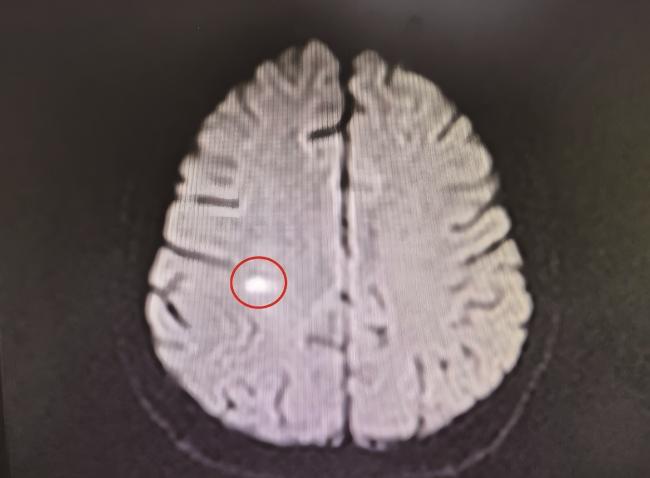
图1 2024年7月30日患者头颅MRIFigure 1 Cranial MRI of patient on July 30, 2024 Patchy abnormal signal is observed in the right parietal subcortical white matter (marked with a red box). T1WI shows isointense signal, T2WI and T2FLAIR show hyperintense signal, DWI shows hyperintense signal, and ADC shows hypointense signal, suggesting an acute inflammatory lesion (possible infection or demyelination). T1WI, T1-weighted imaging; T2WI, T2-weighted imaging; T2FLAIR, T2-fluid attenuated inversion recovery; DWI, diffusion-weighted imaging; ADC, apparent diffusion coefficient. |
 唑片抗感染治疗。2024年8月2日患者发热症状较前缓解,行腰椎穿刺,见清亮脑脊液,测压力为185 mmH2O(1 mmH2O= 9.81 Pa),留取标本。患者家属诉患者苏醒后,意识清晰,问话可答,追溯病史,患者数十日前因食剩饭腹痛、腹泻1周。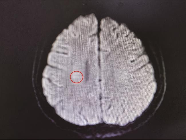
图2 2024年8月14日患者头颅MRIFigure 2 Cranial MRI of patient on August 14, 2024 The previously abnormal signal area in the subcortical white matter of the right parietal lobe (marked with a red box) has significantly reduced compared to before, with decreased signal intensity (T2 FLAIR high signal weakened, DWI high signal disappeared), indicating lesion absorption consistent with effective anti-infective treatment, and NPSLE is excluded (NPSLE lesions are mostly non-infectious inflammation; without adjusting the immunotherapy regimen, lesions are unlikely to absorb in the short term). T2FLAIR, T2-fluid attenuated inversion recovery; DWI, diffusion-weighted imaging; NPSLE, neuropsychiatric systemic lupus erythematosus. |



