技术方法

肾透明细胞癌FABP6基因长转录本的表达及意义

  • 殷昊明 1, * ,
  • 王子杰 1, * ,
  • 舒帆 1 ,
  • 张展奕 1 ,
  • 梁会 , 2, * ,
  • 张树栋 , 1, *
展开
  • 1. 北京大学第三医院泌尿外科,北京 100191
  • 2. 北京大学基础医学院病理学系,北京 100191

*These authors contributed equally to this work

收稿日期: 2024-03-15

  网络出版日期: 2026-01-12

基金资助

国家自然科学基金(82273389)

北京市自然科学基金(7232212)

版权

版权所有,未经授权,不得转载。

Expression and significance of the FABP6 long transcript in clear cell renal cell carcinoma

  • Haoming YIN 1 ,
  • Zijie WANG 1 ,
  • Fan SHU 1 ,
  • Zhanyi ZHANG 1 ,
  • Hui LIANG , 2, * ,
  • Shudong ZHANG , 1, *
Expand
  • 1. Department of Urology, Peking University Third Hospital, Beijing 100191, China
  • 2. Department of Pathology, Peking University School of Basic Medical Sciences, Beijing 100191, China
LIANG Hui, e-mail,
ZHANG Shudong, e-mail,

Received date: 2024-03-15

  Online published: 2026-01-12

Supported by

the National Natural Science Foundation of China(82273389)

Beijing Natural Science Foundation(7232212)

Copyright

All rights reserved. Unauthorized reproduction is prohibited.

摘要

目的: 探讨脂肪酸结合蛋白6(fatty acid binding protein 6,FABP6)基因长转录本在肾透明细胞癌(clear cell renal cell carcinoma,ccRCC)中的表达情况及其与肿瘤生物学行为的关系,进一步分析其作为潜在生物标志物和治疗靶点的可能性。方法: 通过基因表达汇编(Gene Expression Omnibus,GEO)和癌症基因组图谱(The Cancer Genome Atlas,TCGA)数据库进行生物信息学分析后,筛选得到与ccRCC发生发展和预后相关的FABP6基因,并通过实验进一步确认其长短转录本的存在情况及表达模式。利用实时荧光定量逆转录PCR(reverse transcription quantitative real-time PCR,RT-qPCR)和蛋白免疫印迹实验检测FABP6基因长短转录本在ccRCC细胞系和组织样本中的表达水平差异。构建FABP6基因长转录本过表达和敲低的ccRCC细胞系,并分别通过5-乙炔基-2’-脱氧尿苷(5-ethynyl-2’-deoxyuridine, EdU)增殖实验和克隆形成实验,评估FABP6基因长转录本对ccRCC细胞增殖能力的影响。结果: 生物信息学数据库分析显示,FABP6基因在ccRCC细胞系和组织样本中的表达高于其正常对照细胞和组织样本(P=0.02),并且其长转录本为主要表达形式(P=0.02)。RT-qPCR和蛋白免疫印迹实验结果进一步证实,FABP6基因长转录本在ccRCC细胞系769P、A498、CAKI1、OSRC2、786O等中的表达水平高于短转录本。体外功能实验中,过表达FABP6基因长转录本促进ccRCC细胞的增殖能力;相反,敲低FABP6基因长转录本显著抑制ccRCC细胞的增殖能力,提示FABP6基因长转录本可能通过驱动细胞周期进程或调控相关增殖信号通路,在ccRCC的发生发展中扮演了癌基因样的促进作用。结论: 系统报道了FABP6基因长转录本在ccRCC中特异性高表达,并通过功能增益和功能缺失实验证实了其促进ccRCC细胞增殖的关键作用,揭示了FABP6基因,尤其是其长转录本在ccRCC恶性进展中的重要新功能。

本文引用格式

殷昊明 , 王子杰 , 舒帆 , 张展奕 , 梁会 , 张树栋 . 肾透明细胞癌FABP6基因长转录本的表达及意义[J]. 北京大学学报(医学版), 2026 , 58(2) : 393 -398 . DOI: 10.19723/j.issn.1671-167X.2026.02.026

Abstract

Objective: To investigate the expression of the fatty acid binding protein 6 (FABP6) long transcript in clear cell renal cell carcinoma (ccRCC), its correlation with tumor biological behavior, and further analyze its potential as a biomarker and therapeutic target. Methods: Following bioinformatics analysis of the Gene Expression Omnibus (GEO) and The Cancer Genome Atlas (TCGA) databases, the FABP6 gene associated with ccRCC development and prognosis was screened. The existence and expression patterns of FABP6 long and short transcripts were further confirmed experimentally. In the experimental section, reverse transcription quantitative real-time PCR (RT-qPCR) and Western blot were used to detect the differential expression levels of the FABP6 long and short transcripts in ccRCC cell lines and tissue samples. ccRCC cell lines with overexpression and knockdown of the FABP6 long transcript were constructed. The impact of the FABP6 long transcript on the proliferation capacity of ccRCC cells was assessed using the 5-ethynyl-2'-deoxyuridine proliferation assay and the colony formation assay, respectively. Results: Bioinformatics database analysis revealed that the expression of the FABP6 gene was higher in ccRCC cell lines and tissue samples compared with their normal counterparts (P=0.02), with FABP6 long transcript being the predominant form (P=0.02). RT-qPCR and Western blot results further confirmed that the expression level of the FABP6 long transcript was higher than that of the FABP6 short transcript in ccRCC cell lines such as 769P, A498, CAKI1, OSRC2, and 786O. In in vitro functional experiments, overexpression of the FABP6 long transcript promoted the proliferation of ccRCC cells. Conversely, knockdown of the FABP6 long transcript significantly inhibited the proliferation of ccRCC cells. This suggested that the FABP6 long transcript might play an oncogenic role in the development and progression of ccRCC, potentially by driving cell cycle progression or regulating related proli-ferative signaling pathways. Conclusion: This study systematically reports the specific high expression of the FABP6 long transcript in ccRCC. Gain-of-function and loss-of-function experiments confirmed its crucial role in promoting ccRCC cell proliferation. This reveals an important new function of the FABP6 gene, particularly FABP6 long transcript, in the malignant progression of ccRCC.

肾透明细胞癌(clear cell renal cell carcinoma,ccRCC)是成人中最常见的肾脏原发性肿瘤之一,在肾脏恶性肿瘤中约占85%[1]。尽管在肾脏恶性肿瘤的发病中占据主导地位,ccRCC发生发展的分子生物学基础尚未完全阐明。我们在基因表达汇编(Gene Expression Omnibus,GEO)数据库中对GSE53757、GSE36895、GSE16449、GSE16441、GSE40435的数据和癌症基因组图谱(The Cancer Genome Atlas,TCGA)数据库中的ccRCC数据进行生物信息学分析,得到了一个在ccRCC中高表达且影响预后的脂肪酸结合蛋白6(fatty acid binding protein 6,FABP6)基因。FABP6基因编码的是一种细胞内脂肪酸运载蛋白,在正常生理条件下主要在肠道中表达,参与胆汁酸的转运和代谢[2-3]。然而,近年来的研究表明,FABP6基因不仅在肠道组织中表达,在多种肿瘤中也呈现异常表达,并且与肿瘤的发生发展及预后密切相关[4-7]。在ccRCC中,FABP6基因也被发现具有高表达的特征,虽然有研究显示,FABP6基因在ccRCC组织中的表达水平可能与肿瘤的发生发展相关,且可作为ccRCC的生物标志物,但相关机制尚未完全阐明[8]FABP6基因编码2个蛋白,其表达量在不同组织中略有差别,发挥的作用也不同,但目前少有研究将其具体区分开来[2-3]。因此,本研究深入探讨FABP6基因在ccRCC中的表达情况及其可能的作用机制,为ccRCC的诊断、治疗和预后评估提供新的生物学标志物。

1 资料与方法

本研究获得北京大学第三医院医学科学研究伦理委员会批准(批准号:M2017147),研究对象均签署知情同意书。

1.1 主要试剂和仪器

杜氏改良伊格尔培养基(Dulbecco’ s modified Eagle medium,DMEM,PM150210)购自中国Procell公司,罗斯韦尔公园纪念研究所(Roswell Park Memorial Institute,RPMI)1640培养基(PM150110)购自美国Gibco公司,4’,6-二脒基-2-苯基吲哚(4’,6-diamidino-2-phenylindole,DAPI,C1002)购自中国Beyotime公司,细胞/组织裂解液(PC101)购自美国Epizyme公司,特级胎牛血清(164210)购自中国Procell公司,青霉素-链霉素-两性霉素B溶液(PB18012)购自中国Procell公司,FABP6多克隆抗体(NBP1-32482)购自美国Novus公司。

1.2 细胞培养

肾癌细胞系769P、A498、ACHN、CAKI1、CAKI2、786O、OSRC2和正常肾小管细胞系HK2购自美国菌种保藏中心(American Type Culture Collection,ATCC)。将细胞放入含10%(体积分数)特级胎牛血清的RPMI1640培养基中,置于37 ℃、5%(体积分数)CO2的细胞孵育箱中培养。

1.3 实时荧光定量逆转录PCR

从培养皿中收集细胞,使用Trizol方法提取总RNA。从ccRCC组织中提取RNA时先将黄豆大小的组织在Trizol中剪碎(ccRCC组织样本来源于北京大学第三医院泌尿外科2023年2月到8月住院诊治的肾癌患者40例),再和细胞RNA提取步骤一样,使用Trizol方法提取总RNA。检测RNA的浓度和纯度后,使用逆转录试剂盒(G592,ABM公司,美国)将RNA逆转录为cDNA。设计特异性引物,用于实时荧光定量逆转录PCR(reverse transcription quantitative real-time PCR,RT-qPCR)检测目标基因的表达水平,引物序列见表 1
表1 实时荧光定量逆转录PCR所用引物序列

Table 1 Nucleotide sequences of primers used in RT-qPCR

Genes Primer
FABP6S Forward:5′-CCACCCATTCTCCTCATCCCTCTGCTC-3′
Reverse:5′-ACCAAGTGAAGTCCTGCCCATCCTG-3′
FABP6L Forward:5′-ACATGGGTGAGCCGGAAAGGAGAC-3′
Reverse:5′-CCGGAGTAGTGCTGGGACCAAGTGAAGT-3′

RT-qPCR, reverse transcription quantitative real-time PCR; FABP6S, fatty acid binding protein 6 short transcript; FABP6L, fatty acid binding protein 6 long transcript.

1.4 蛋白免疫印迹实验

从培养皿中收集细胞,或者从收集的ccRCC组织样本(组织样本来源于北京大学第三医院泌尿外科2023年2月到8月住院诊治的肾癌患者40例)中使用放射免疫沉淀法(radio immunoprecipitation assay, RIPA)裂解液提取细胞或组织中的总蛋白,并检测蛋白浓度,将蛋白样品与十二烷基硫酸钠-聚丙烯酰胺(sodium dodecyl sulfate-polyacrylamide gel electrophoresis, SDS-PAGE)蛋白上样缓冲液(P0015,Beyotime公司,中国)混合,进行蛋白变性处理,随后将蛋白样品加入SDS-PAGE凝胶槽中,根据蛋白的大小和分离需求,进行SDS-PAGE凝胶电泳分离。将SDS-PAGE凝胶中的蛋白迁移到聚丙烯酰胺膜(polyvinylidene fluoride,PVDF)上,蛋白转印条件为200 mA,3 h。使用5%(体积分数)脱脂奶粉(P0216,Beyotime公司,中国)阻塞膜上的非特异性结合位点,使用增强型化学发光试剂(enhanced chemiluminescence, ECL, 36222ES60,Yeasen公司,中国)进行显色反应。

1.5 5-乙炔基-2’-脱氧尿苷增殖实验

将细胞在含有10 μm 5-乙炔基-2’-脱氧尿苷(5-ethynyl-2’-deoxyuridine, EdU)的培养基中处理2 h。使用4%(体积分数)的多聚甲醛对细胞进行固定处理,使用EdU法细胞增殖成像分析试剂盒(KTA2031,Abbkine公司,中国)进行EdU标记,并记录图像。

1.6 克隆形成实验

将ccRCC细胞以每一个孔板500个细胞的数量接种于6孔板中,定期观察并记录克隆的形成情况,大约2周后,使用吉姆萨(Giemsa)染色方法进行克隆形成的检测和计数。

1.7 统计学分析

采用SPSS 25.0和R语言(版本4.2.1)进行数据分析。正态分布的计数资料采用Student t检验或Fisher’ s检验。肾组织FABP6长转录本(FABP6 long transcript, FABP6L)和FABP6短转录本(FABP6 short transcript, FABP6S)表达情况采用秩和检验。P<0.05为差异有统计学意义。

2 结果

2.1 FABP6基因在数据库中的信息及其不同转录形式

通过对GEO数据中的GSE53757、GSE40435、GSE16441、GSE16449和GSE36895进行单独的差异表达分析,获得在ccRCC中显著高表达的基因集(|Log2FC|>1, P<0.01),之后再取交集得到186个基因,然后将这些高表达的基因集做生存分析,并按照Log-rank P值进行排序,得到对患者生存情况有影响的基因,并注意到其中FABP6基因与生存情况关联最强。进一步在基于基因表达水平值的交互式分析平台(Gene Expression Profiling Interactive Ana-lysis,GEPIA)数据库(http://gepia.cancer-pku.cn)中进行检索确认,发现FABP6基因在ccRCC中高表达,其表达水平与分期相关,且其高表达与ccRCC患者的不良预后相关。但这些数据对FABP6基因的不同转录本不做区分,而实际上在美国国家生物技术信息中心(National Center for Biotechnology Information,NCBI)官网(https://www.ncbi.nlm.nih.gov/)上,可以查询到FABP6基因有3个转录本,分别为NM_001040442.1、NM_001130958.2和NM_001445.3,其翻译后对应的蛋白有2个,分别为128个氨基酸的FABP6S(CCDS4349.1)和177个氨基酸的FABP6L(CCDS43393.1),其中FABP6L蛋白在FABP6S蛋白的N端多出49个氨基酸。

2.2 FABP6在肾癌细胞系中的表达情况

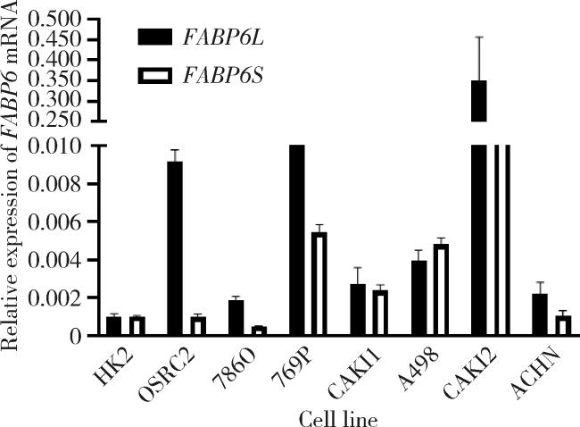
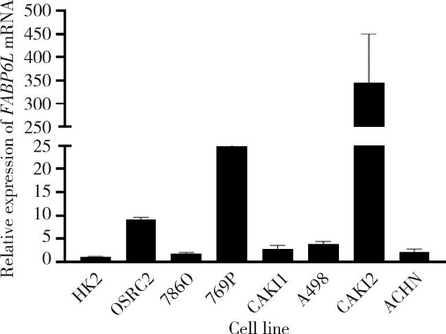
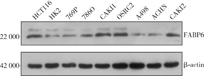
为了检测FABP6基因编码的两个蛋白及其对应的转录本在肾癌和正常肾组织中的表达情况,分别通过RT-qPCR检测了肾癌细胞系769P、A498、ACHN、CAKI1、CAKI2、786O、OSRC2和正常肾小管细胞HK2中FABP6基因FABP6LFABP6S的表达情况,结果显示多数肾癌细胞系中FABP6L水平显著高于FABP6S水平(图 1),并且肾癌细胞系的FABP6L水平高于正常肾小管细胞(图 2),该现象也体现在蛋白免疫印迹实验上(图 3),HCT116为已报道的FABP6高表达阳性对照细胞。
图1 正常肾小管细胞系和肾癌细胞系中FABP6基因mRNA的表达水平

Figure 1 mRNA expression profile of FABP6 gene isoforms in the normal renal tubular cell line and renal cancer cell lines

FABP6L, fatty acid binding protein 6 long transcript; FABP6S, fatty acid binding protein 6 short transcript.

图2 正常肾小管细胞和肾癌细胞系中FABP6L mRNA的表达情况

Figure 2 Expression of FABP6L mRNA in normal renal tubular cell line and renal cancer cell lines

FABP6L, fatty acid binding protein 6 long transcript.

图3 正常肾小管细胞和肾癌细胞系和中FABP6蛋白的表达情况,HCT116为已报道的FABP6高表达阳性对照细胞

Figure 3 Expression of FABP6 protein in renal cancer cell lines and normal renal tubular cell line HK2, HCT116 as a reported high FABP6 protein expression cell line

FABP6, fatty acid binding protein 6.

2.3 FABP6在ccRCC组织中的表达情况

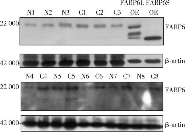
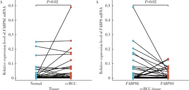
通过构建FABP6基因过表达质粒,感染293T细胞,并同时检测ccRCC组织和癌旁正常肾组织的FABP6两种蛋白的表达情况,发现组织表达的FABP6蛋白主要是FABP6L,且其在ccRCC组织中的表达高于其癌旁正常肾组织(图 4)。通过RT-qPCR实验检测了40对ccRCC癌和癌旁配对正常肾组织,发现在ccRCC组织中FABP6L mRNA的表达水平高于癌旁正常肾组织,并且ccRCC组织中的FABP6LFABP6S高(图 5n=40,P<0.05)。
图4 癌旁正常肾组织和ccRCC组织中FABP6蛋白的表达情况

Figure 4 Expression of FABP6 protein in normal kidney tissue and ccRCC tissue

FABP6L, fatty acid binding protein 6 long transcript; FABP6S, fatty acid binding protein 6 short transcript; FABP6, fatty acid binding protein 6; OE, over expression; N, normal kidney tissue; C, ccRCC tissue; ccRCC, clear cell renal cell carcinoma.

图5 FABP6 mRNA在癌旁正常肾组织和ccRCC组织中的表达情况

Figure 5 mRNA expression profile of FABP6 in normal kidney tissue and ccRCC tissue

FABP6L, fatty acid binding protein 6 long transcript; FABP6S, fatty acid binding protein 6 short transcript; FABP6, fatty acid binding protein 6; ccRCC, clear cell renal cell carcinoma.

2.4 FABP6L促进ccRCC细胞的增殖

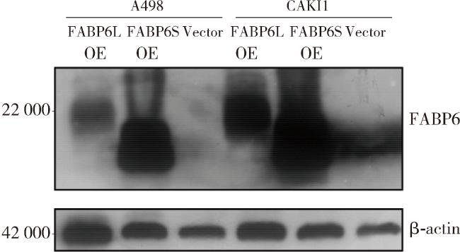
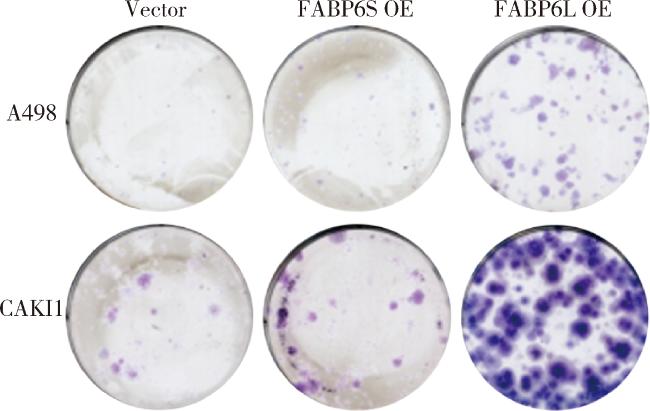
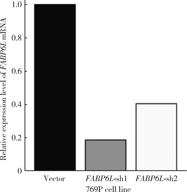
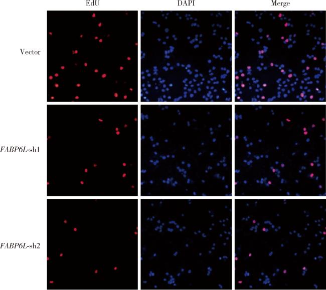
构建FABP6SFABP6L基因过表达的ccRCC细胞系A498和CAKI1,并通过蛋白质免疫印迹实验验证其过表达效率(图 6)。通过克隆形成实验发现FABP6L基因的过表达会促进A498和CAKI1细胞的增殖(图 7)。构建FABP6L基因敲低的ccRCC细胞系769P,通过RT-qPCR检验其敲低效率(图 8),发现在FABP6L基因敲低后,769P细胞增殖被抑制(图 9)。
图6 A498和CAKI1细胞系的过表达效率验证

Figure 6 A498 and CAKI1 cell line stable transfection efficiency validation

FABP6L, fatty acid binding protein 6 long transcript; FABP6S, fatty acid binding protein 6 short transcript; FABP6, fatty acid binding protein 6; OE, over expression; ccRCC, clear cell renal cell carcinoma.

图7 A498和CAKI1细胞系的克隆形成实验

Figure 7 Colony formation assay of A498 and CAKI1 cell line

FABP6S, fatty acid binding protein 6 short transcript; FABP6L, fatty acid binding protein 6 long transcript; OE, over expression.

图8 769P细胞系的敲低效率验证

Figure 8 Knockdown efficiency validation of 769P cell line

FABP6L, fatty acid binding protein 6 long transcript; sh, short hairpin RNA.

图9 769P细胞系的EdU增殖实验(×20)

Figure 9 EdU labeling of 769P cell line (×20)

EdU, 5-Ethynyl-2'-deoxyuridine; DAPI, 4', 6-diamidino-2-phenylindole; FABP6L, fatty acid binding protein 6 long transcript; sh, short hairpin RNA.

3 讨论

本研究旨在探讨FABP6基因在ccRCC中的表达水平以及其与ccRCC发生发展的联系。首先,我们观察到FABP6L相较于FABP6S在ccRCC细胞系中的表达显著增加,且ccRCC细胞系中FABP6L的表达水平高于正常肾小管细胞系,这一发现与之前的FABP6基因在结直肠癌中的研究结果较类似[2],表明FABP6L可能也在肾癌的发生和发展过程中发挥类似作用。
随后在FABP6L的过表达模型中,我们发现其高表达可能与ccRCC细胞的增殖相关联,因细胞和组织中其主要存在形式是FABP6L,故本研究主要聚焦于FABP6L对应的蛋白,但具体FABP6LFABP6S表达不一致的原因及其对表型影响的不一致性的具体机制,还有待进一步的分子生物学实验阐明。
总之,针对FABP6基因的治疗策略可能成为一种潜在的ccRCC治疗手段,通过抑制其表达或活性来改善患者的预后。
但是由于目前的FABP6蛋白抗体能同时识别FABP6L和FABP6S所对应的蛋白,缺乏特异性FABP6L蛋白的抗体,故目前我们在制作FABP6L的特异性抗体,以期在大批量组织样本中验证其表达情况。同时本研究也存在一些局限性,没有观察ccRCC细胞增殖之外的其他常见表型,且目前仅有体外实验结果。同时,没有关于FABP6基因对增殖影响的机制研究,之后我们计划使用动物模型和临床样本进行更深入的机制研究。
综上所述,FABP6基因在ccRCC中的表达水平与其发生发展相关,因此,FABP6基因可能成为一个潜在的ccRCC诊断、治疗和预后评估的生物标志物。然而,尚需进一步的临床和基础研究来验证这一假设,并深入探讨FABP6基因在ccRCC中的确切作用机制。

利益冲突  所有作者均声明不存在利益冲突。

作者贡献声明  殷昊明:撰写论文初稿,完成部分实验并分析,整理数据;王子杰:完成部分实验并分析,整理数据;舒帆:完成临床病例相关数据的收集;张展奕:完成临床病例相关数据的分析和整理;梁会:设计研究方案,指导论文修改;张树栋:提出研究思路,指导完成研究。所有作者均参与论文修改,并对最终文稿进行审读和确认。

1
Hu J , Wang SG , Hou Y , et al. Multi-omic profiling of clear cell renal cell carcinoma identifies metabolic reprogramming associated with disease progression[J]. Nat Genet, 2024, 56 (3): 442- 457.

DOI

2
Fang C , Dean J , Smith JW . A novel variant of ileal bile acid binding protein is up-regulated through nuclear factor-kappaB activation in colorectal adenocarcinoma[J]. Cancer Res, 2007, 67 (19): 9039- 9046.

DOI

3
D'Onofrio M , Zanzoni S , Munari F , et al. The long variant of human ileal bile acid-binding protein associated with colorectal cancer exhibits sub-cellular localization and lipid binding behaviour distinct from those of the common isoform[J]. Biochim Biophys Acta Gen Subj, 2017, 1861 (9): 2315- 2324.

DOI

4
Lian W , Wang Z , Ma Y , et al. FABP6 expression correlates with immune infiltration and immunogenicity in colorectal cancer cells[J]. J Immunol Res, 2022, 2022, 3129765.

5
Zhang Y , Zhao X , Deng L , et al. High expression of FABP4 and FABP6 in patients with colorectal cancer[J]. World J Surg Oncol, 2019, 17 (1): 171.

DOI

6
Zhang D , Zhao F , Liu H , et al. FABP6 serves as a new therapeutic target in esophageal tumor[J]. Aging, 2024, 16 (2): 1640- 1662.

DOI

7
Pai FC , Huang HW , Tsai YL , et al. Inhibition of FABP6 reduces tumor cell invasion and angiogenesis through the decrease in MMP-2 and VEGF in human glioblastoma cells[J]. Cells, 2021, 10 (10): 2782.

DOI

8
Zheng J , Li YZ , Ni WJ , et al. Comprehensive analysis of the roles of fatty acid transport related proteins in clear cell renal cell carcinoma[J]. Prostaglandins Other Lipid Mediat, 2023, 167, 106732.

DOI

文章导航

/